. 2009 May;59(3):181-90.
doi: 10.1007/s12576-009-0023-0. Epub 2009 Feb 25.

17beta-Estradiol-induced enhancement of estrogen receptor biosynthesis via MAPK pathway in mouse skeletal muscle myoblasts

Affiliations

17beta-Estradiol-induced enhancement of estrogen receptor biosynthesis via MAPK pathway in mouse skeletal muscle myoblasts

J Hatae et al. J Physiol Sci. 2009 May.

Abstract

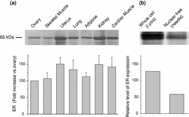
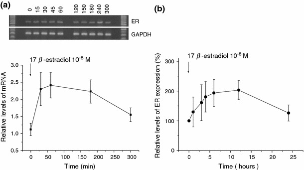
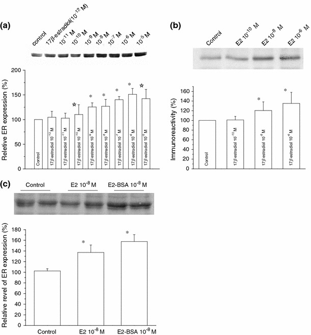
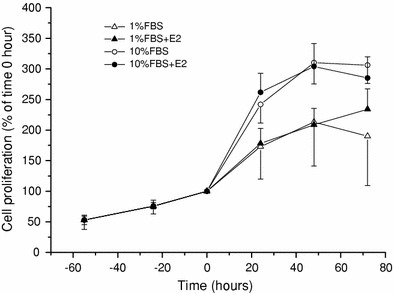
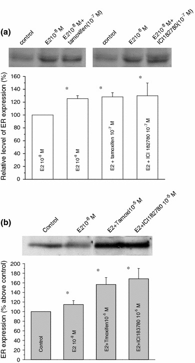
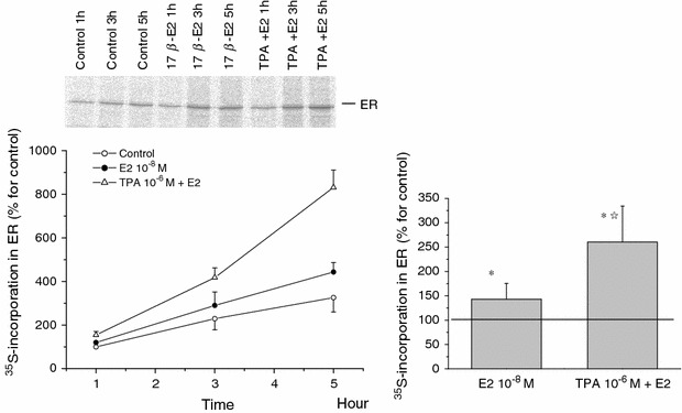
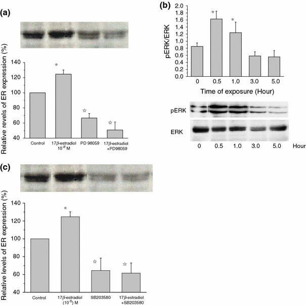
The skeletal muscle is one of the important target tissues for the actions of estrogen via both nuclear and extranuclear (non-genomic) pathways. However, there is a paucity of information about the receptor (ER) involved. The aim of this study was thus to explore the ER expression in skeletal muscle, and the influence of estrogen on it, by using C2C12 myoblasts derived from mouse skeletal muscle. Significant expression of a approximately 66-kD protein immunoreactive to ER type alpha (ERalpha) monoclonal antibody, which was comparable to that in ovary, was detected in the whole-cell (total) and nucleus-free (nonnuclear) fractions of C2C12 myoblasts. The expression level of these ER proteins increased in several hours with treatment with 17beta-estradiol (E2), which was preceded by the elevation of the ER mRNA level. This increase appeared to reflect the acceleration of de novo synthesis of ER protein, as proved by the (35)S-methionine immunoprecipitation method. A similar extent of fast increase in ER expression was also induced by a membrane-impermeable, BSA-conjugated estradiol (E2-BSA). Unexpectedly, the E2-induced increases in total and nonnuclear ER were further enhanced by the classic ER antagonists tamoxifen and ICI182,780 in a wide concentration range, implying some structural difference of the involved ER from the classical one. Treatment with the ERK1/2 inhibitor, PD98059 (10 microM), or the p38 MAPK-specific inhibitor, SB203580 (10 microM), greatly inhibited the E2-induced ER increase, while the protein kinase C (PKC) activator TPA (1 microM) enhanced it. These results collectively suggest that C2C12 skeletal myoblasts express a high level of ER, a considerable part of which is extranuclear. Further, the expression of ER in these cells may be significantly upregulated by estrogen itself via increased biosynthesis linked to membrane-bound ER and downstream MAPK-mediated signaling pathways.


Figures

Fig. 1
Fig. 1
Distribution of estrogen receptors in various organs and tissues of adult mouse. a Western blot analysis of whole-cell fractions with a specific monoclonal estrogen receptor (ER) antibody. Upper panel immunoreactive bands corresponding to the ER protein (66 kDa). The histogram (lower) indicates the results of densitometric analysis for a 66-kDa band (mean ± SE, n = 4). Relative expression level with respect to the ovary is shown in %. From left to right: skeletal muscle, uterus, lung, adipose tissue, kidney, and myocardium. b Relative expression (to ovary) of whole cell and nonnuclear ER proteins obtained from C2C12 mouse myoblasts (n = 3)
Fig. 2
Fig. 2
Time course of ER mRNA and protein expression during E2 exposure. a Representative of C2C12 mRNA transcripts for ER and GAPDH at several time points after exposure to E2 (10−8 M) (upper). The mRNA level of ER relative to GAPDH is averaged from five individual experiments and displayed as mean ± SE (statistically significant). At 0, 0.5, 1, 3, and 5 h (lower). b Proteins were extracted from nucleus-free C2C12 cells exposed to E2 (10−8 M) for varying periods of time (from left: 0, 1, 3, 4, 6, 12, and 24 h) and immunoblotted with the ER antibody. Immunodensity in a 66-kDa band is quantified by densitometry and presented as the % increase of control (time 0). Symbols and bars represent mean ± SE (n = 5); statistically significant
Fig. 3
Fig. 3
Dose–dependent effects of 17β-estradiol on the expression of total and nonnuclear ER. Representative immunoreactive bands for total ER in C2C12 myoblasts (upper panel) and the relative expression of total ER protein (a) or nonnuclear ER protein (b) quantified by densitometric analysis (lower panel); after treatment with either vehicle (control), different concentrations of 17β-estradiol (10−12–10−5 M) in a and (10−10–10−6 M) in b for 15 h. c Shows the relative expression of total ER protein after treatment with vehicle (control), 17β-estradiol (10−8 M), and BSA-conjugated E2 (10−8 M) for 15 h. Each bar is normalized to the control (far left) and expressed as %. Columns and bars indicate mean ± SE (n = 3 in a, n = 4 in b, and n = 5 in c). *P < 0.01, P < 0.05 compared with control values, determined using Student’s t test
Fig. 4
Fig. 4
Influence of 17-βestradiol on C2C12 proliferation. C2C12 cells were grown in the serum-free medium for 48 h, which was then changed to ones with or without E2 (10−8 M) in 1% FBS (open triangle, closed triangle) or 10% FBS (open circle, closed circle). The rate of cell growth is shown as the cell number at every 24 h relative to 0 h. Symbols and bars represent the mean ± SE (n = 5)
Fig. 5
Fig. 5
Effects of ER antagonists on E2-induced whole-cell and non-nuclear ER increase. C2C12 cells were treated with E2 (10−8 M) in the absence or presence of either tamoxifen or ICI 182,780 (10−7 M; a total ER) or (10−5 M; b non-nuclear fractions) for 15 h. Tamoxifen or ICI 182,780 was added to the culture medium 30 min prior to the administration of E2. Total and nonnuclear fractions of ER were prepared as described in “Materials and methods” and subjected to Western blotting. Histograms represent the extent of inhibition of E2-induced ER increase in total a and nonnuclear b fractions, which are expressed as the percentage of control (no stimulation), averaged from five separate experiments (mean ± SE). *P < 0.01, compared with control values, determined using Student’s t test
Fig. 6
Fig. 6
Effects of 17β-estradiol and/or TPA on ER de novo synthesis. In the absence (control) or presence of 17β-estradiol (10−8 M) and/or TPA (10−6 M), C2C12 cells were labeled with [35S] methionine. At the time points indicated in the figure, the total ER fractions were prepared and subjected to selective immunoprecipitation for ER. The immunoprecipitates were analyzed by SDS-PAGE and image scanning. Data are the mean ± SE from three separate experiments. Left panel time course of 35methionine incorporation in ER. Right panel rates of the increase at 5 h by the relative ratio of control. *P < 0.01, compared with control values, determined using Student’s t test. P < 0.01, compared with E2 values, determined using Student’s t test
Fig. 7
Fig. 7
E2-induced ER increase involves ERK1/2 and p38 pathways. Histograms indicate the relative density of ER immunoblots normalized to control (basal level). a From left to right, vehicle, E2 10−8, PD9809 alone, and E2 + PD98059. b Time course of ERK phosphorylation induced by E2. ERK phosphorylation assessed by pERK-specific antibody. c From left to right, vehicle, E2 (10−8 M), SB203580 (10 μM) alone, and E2 + SB203580. In a and c, PD98059 (10 μM) or SB203580 (10 μM) was added into the culture medium 30 min before addition of E2 (10−8 M). The bars show means ± SE of three independent experiments performed in duplicate. *P < 0.01, compared with control values, determined using Student’s t test. P < 0.01, compared with E2 values, determined using Student’s t test

References

    1. Nilsson S, Makela S, Treuter E, Tujague M, Thomsen J, Andersson G, Enmark E, Pettersson K, Warner M, Gustafsson JA. Mechanisms of estrogen action. Physiol Rev. 2001;81:1535–1565. - PubMed
    1. Nadal A, Diaz M, Valverde M. The estrogen trinity: membrane, cytosolic, and nuclear effects. News Physiol Sci. 2001;16:251–255. - PubMed
    1. Ho KJ, Liao JK. Nonnuclear action of estrogen. Arterioscler Thromb Vasc Biol. 2002;22:1952–1961. doi: 10.1161/01.ATV.0000041200.85946.4A. - DOI - PubMed
    1. Acconcia F, Ascenzi P, Fabozzi G, Visca P, Marino M. S-palmitoylation modulates human estrogen receptor-α functions. BBRC. 2004;316:878–883. - PubMed
    1. Boonyaratanakonkit V, Edwards DP. Receptor mechanisms mediating non-genomic actions of sex steroids. Semim Reprod Med. 2007;25(3):139–153. doi: 10.1055/s-2007-973427. - DOI - PubMed

MeSH terms