. 2009 May;59(3):157-63.
doi: 10.1007/s12576-009-0020-3. Epub 2009 Feb 6.

Abdominal expiratory muscle activity in anesthetized vagotomized neonatal rats

Affiliations

Abdominal expiratory muscle activity in anesthetized vagotomized neonatal rats

Makito Iizuka. J Physiol Sci. 2009 May.

Abstract

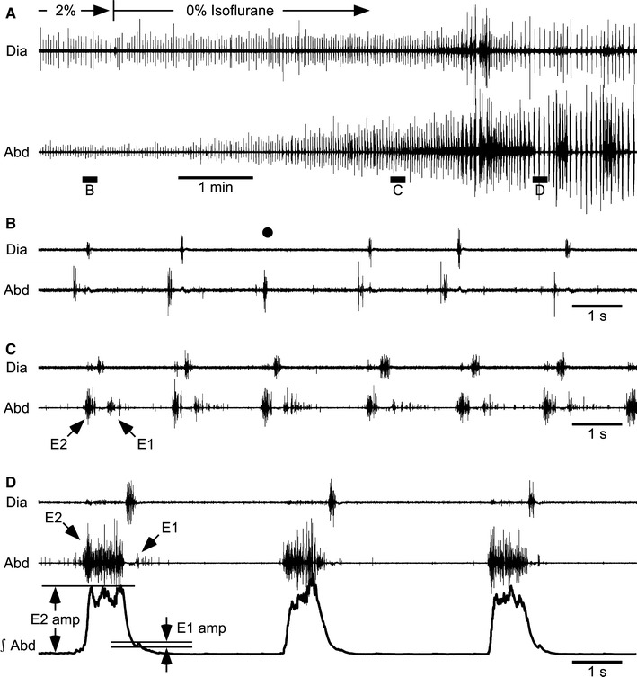
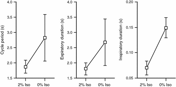
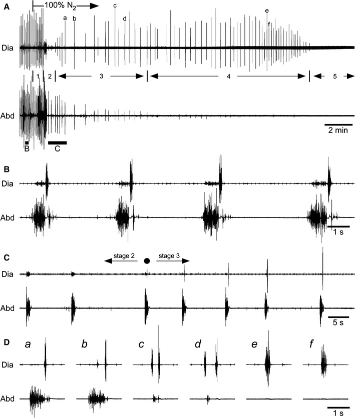
The pattern of respiratory activity in abdominal muscles was studied in anesthetized, spontaneously breathing, vagotomized neonatal rats at postnatal days 0-3. Anesthesia (2.0% isoflurane, 50% O(2)) depressed breathing and resulted in hypercapnia. Under this condition, abdominal muscles showed discharge late in the expiratory phase (E2 activity) in most rats. As the depth of anesthesia decreased, the amplitude of discharges in the diaphragm and abdominal muscles increased. A small additional burst frequently occurred in abdominal muscles just after the termination of diaphragmatic inspiratory activity (E1 or postinspiratory activity). Since this E1 activity is not often observed in adult rats, the abdominal respiratory pattern likely changes during postnatal development. Anoxia-induced gasping after periodic expiratory activity without inspiratory activity, and in most rats, abdominal expiratory activity disappeared before terminal apnea. These results suggest that a biphasic abdominal motor pattern (a combination of E2 and E1 activity) is a characteristic of vagotomized neonatal rats during normal respiration.


Figures

Fig. 1
Fig. 1
AD Respiratory activity in a vagotomized neonatal rat. Electromyograms were obtained from the diaphragm (Dia) and abdominal muscles (Abd). The integrated electromyogram of Abd (∫Abd, time constant 0.1 s) is also shown as the third trace in panel D. The concentration of isoflurane was decreased from 2 to 0% at the time indicated in panel A. BD Portions of recording A above the horizontal bars marked BD are shown on an expanded time scale. The vertical gain in Abd in B is higher by a factor of four compared with other records. As indicated by the black circle in B, the inspiratory burst was absent in some respiratory cycles under 2% isoflurane. The arrows indicate E1 and E2 activities. The amplitudes of E2 and E1 activities (E2 amp, E1 amp) were measured from the baseline or trough to the peak
Fig. 2
Fig. 2
Effects of the depth of anesthesia on respiratory parameters. The cycle period, expiratory duration, and inspiratory duration were measured under 2 and 0% isoflurane (Iso). For 0% isoflurane, data were obtained about 5 min after the concentration of isoflurane was switched from 2 to 0%. All three parameters were significantly prolonged by removal of isoflurane (P < 0.05, n = 8)
Fig. 3
Fig. 3
Effects of anoxia. AD Electromyograms obtained from the diaphragm (Dia) and abdominal muscles (Abd). Numbers 1–5 in A indicate the stages following anoxic stimulation (100% N2) referred to in the text. In B and C, the portions of recording A above the horizontal bars marked B and C are shown on an expanded time scale. D The portions of recording A indicated by the letters a–f are shown on an expanded time scale. After the inspired gas was switched to 100% N2, the inspiratory bursts in the diaphragm were depressed transiently and then increased gradually (C). The first inspiratory activity that occurred after the transient disappearance is indicated by a black circle, and this time was defined as the onset of gasping. The expiratory activity disappeared before terminal apnea (e and f in panel D)

Similar articles

Cited by

References

    1. Iscoe S. Control of abdominal muscles. Prog Neurobiol. 1998;56:433–506. doi: 10.1016/S0301-0082(98)00046-X. - DOI - PubMed
    1. Janczewski WA, Aoki M. Expiratory activity in the 1–4 day old rat. Jpn J Physiol. 1999;49(Suppl):S84.
    1. Janczewski WA, Aoki M. Expiratory muscle activity in neonatal rats. Soc Neurosci Abst. 1999;25:279.
    1. Janczewski WA, Onimaru H, Homma I, Feldman JL. Opioid-resistant respiratory pathway from the preinspiratory neurones to abdominal muscles: in vivo and in vitro study in the newborn rat. J Physiol. 2002;545:1017–1026. doi: 10.1113/jphysiol.2002.023408. - DOI - PMC - PubMed
    1. Iizuka M, Fregosi RF. Influence of hypercapnic acidosis and hypoxia on abdominal expiratory nerve activity in the rat. Respir Physiol Neurobiol. 2007;157:196–205. doi: 10.1016/j.resp.2007.01.004. - DOI - PubMed

Substances