Review
. 2009 Jan;59(1):3-21.
doi: 10.1007/s12576-008-0008-4. Epub 2008 Dec 9.

The maxi-anion channel: a classical channel playing novel roles through an unidentified molecular entity

Affiliations
Review

The maxi-anion channel: a classical channel playing novel roles through an unidentified molecular entity

Ravshan Z Sabirov et al. J Physiol Sci. 2009 Jan.

Abstract

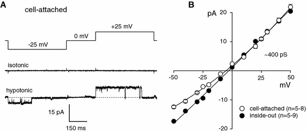
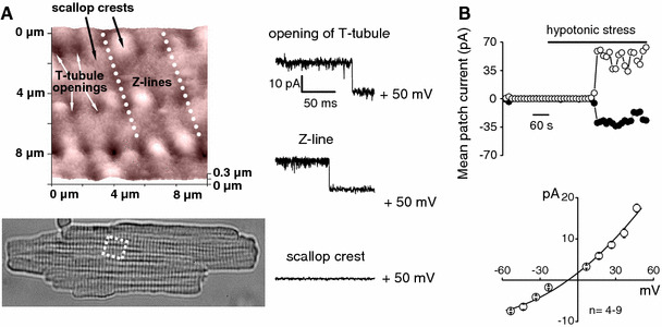
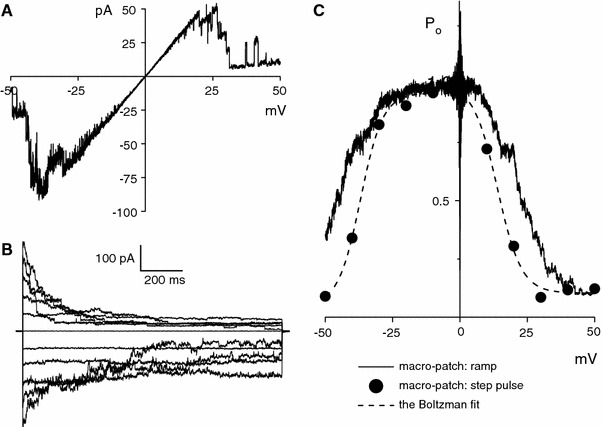
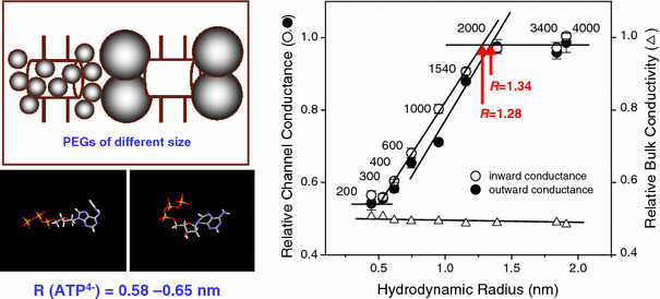
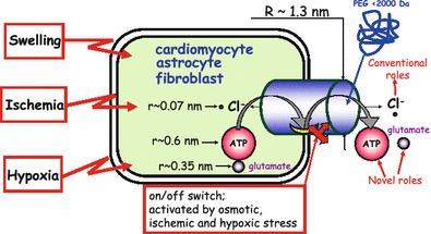
The maxi-anion channel is widely expressed and found in almost every part of the body. The channel is activated in response to osmotic cell swelling, to excision of the membrane patch, and also to some other physiologically and pathophysiologically relevant stimuli, such as salt stress in kidney macula densa as well as ischemia/hypoxia in heart and brain. Biophysically, the maxi-anion channel is characterized by a large single-channel conductance of 300-400 pS, which saturates at 580-640 pS with increasing the Cl(-) concentration. The channel discriminates well between Na(+) and Cl(-), but is poorly selective to other halides exhibiting weak electric-field selectivity with an Eisenman's selectivity sequence I. The maxi-anion channel has a wide pore with an effective radius of approximately 1.3 nm and permits passage not only of Cl(-) but also of some intracellular large organic anions, thereby releasing major extracellular signals and gliotransmitters such as glutamate(-) and ATP(4-). The channel-mediated efflux of these signaling molecules is associated with kidney tubuloglomerular feedback, cardiac ischemia/hypoxia, as well as brain ischemia/hypoxia and excitotoxic neurodegeneration. Despite the ubiquitous expression, well-defined properties and physiological/pathophysiological significance of this classical channel, the molecular entity has not been identified. Molecular identification of the maxi-anion channel is an urgent task that would greatly promote investigation in the fields not only of anion channel but also of physiological/pathophysiological signaling in the brain, heart and kidney.


Figures

Fig. 1
Fig. 1
Single-channel recordings of maxi-anion channel currents in on-cell patches activated by osmotic swelling of mammary C127 cells and in inside-out patches excised from the cells. a Representative current traces recorded under isotonic and hypotonic conditions on C127 cells during application of alternating pulses from 0 to ±25 mV (protocol is shown at the top of the traces). b Unitary I–V relationships for the single-channel events recorded in on-cell patches (open circles) and in inside-out patches (filled circles). Each symbol represents the mean ± SEM (vertical bar). Modified from Sabirov et al. [8]
Fig. 2
Fig. 2
Maxi-anion channel activity localized in specific regions on freshly isolated adult rat cardiomyocytes. a Topographic image of the area indicated by a white rectangle in the optical image shown at the bottom part on the surface of a cardiomyocyte obtained using scanning ion conductance microscopy (SICM) with a fine nanopipette. The maxi-anion channel activity in patches excised from Z-grooves, T-tubule openings, and scallop crests using the “smart-patch” technique are shown on the right side. b Swelling-induced activation of the maxi-anion channel activity in sarcolemma of adult cardiomyocytes. Mean patch currents recorded at +50 mV (open circles) and −50 mV (filled circles) in a cell-attached patch before and during (horizontal bar) exposure to hypotonic solution. Single-channel I–V relationship for these on-cell events is shown on the lower panel. Each symbol represents the mean ± SEM (vertical bar). Modified from Dutta et al. [12]
Fig. 3
Fig. 3
Voltage-dependent inactivation of maxi-anion channel currents recorded in macro-patches excised from mammary C127 cells. a Steady-state ramp I–V records from a macro-patch containing five active channels. b Inactivating current traces recorded in response to step pulses from 0 to ±50 mV in 10-mV increments in a macro-patch containing 20 active channels. c Voltage dependence of steady-state open-channel probability. Filled circles represent the ratio of steady-state macro-patch current to instantaneous macro-patch current (from b). The Boltzmann fit (dashed line) yields a half-maximal open-channel probability at V1/2 = +13.9 and −36.9 mV for positive and negative potentials, respectively. The solid line is the ensemble-averaged current of 11 consecutive ramp-pulse records similar to those shown in (a). Modified from Sabirov et al. [8]
Fig. 4
Fig. 4
Maxi-anion channel has a wide pore larger than the size of ATP. Left panels Basic principle of the polymer partitioning method using PEGs (depicted as globules) of different sizes (upper panel) and the effective pore radius (R) of an ATP molecule calculated in two different conformations: conventional long and more compact forms found in crystals (see for details: Sabirov and Okada [11]). Right panel Relative single maxi-anion channel conductance (circles) and relative bulk solution conductivity (triangles) as a function of the hydrodynamic radius of PEGs. Each symbol represents the mean ± SEM (vertical bar) (n = 5–20). Pore size is estimated as an intersection point between a rising portion of the curve (partial partitioning) and an upper plateau level (complete exclusion). Modified from Sabirov and Okada [11]
Fig. 5
Fig. 5
Pharmacological profile of the maxi-anion channel in primary cultured neonatal rat cardiomyocytes. a Single-channel current traces recorded from excised outside-out (for Gd3+) and inside-out (for others) patches during application of step pulses (the protocol shown at the top of traces) in the absence (control) or presence of drugs. b Effects of drugs on mean currents recorded from excised macro-patches. Currents were recorded at +25 mV (open columns) and −25 mV (hatched columns). Data are normalized to the mean current measured before application of drugs and after correction for the background current. Each column represents the mean ± SEM (vertical bar). *P < 0.02 versus control. Modified from Dutta et al. [2]
Fig. 6
Fig. 6
Maxi-anion channel activation upon hypotonic (a), ischemic (b) and hypoxic (c) stresses in neonatal rat cardiomyocytes. a Mean patch currents during application of alternating pulses from 0 to ±25 mV in a cell-attached patch before and during (horizontal bar) exposure to hypotonic, ischemic or hypoxic solution. b Representative current traces of maxi-anion channels activated as in (a) and elicited by step pulses of ±25 mV (the protocol shown at the top of traces) in cell-attached or inside-out patches. Modified from Dutta et al. [2]
Fig. 7
Fig. 7
ATP currents through the maxi-anion channel recorded in inside-out patches excised from neonatal rat cardiomyocytes. a Representative ramp I–V records (a) and channel activity in response to step pulses (b) from a macro-patch exposed to standard Ringer solution and one exposed to 100 mM Na4ATP solution. The pipette solution was standard Ringer. b Representative ramp I–V records (a) and channel activity in response to step pulses (b) from a macro-patch exposed to standard Ringer solution and one exposed to 100 mM Na2MgATP solution. The pipette solution was TEA-Cl. Modified from Dutta et al. [2]
Fig. 8
Fig. 8
Glutamate permeability of the glial maxi-anion channel and the pharmacological profile of the net glutamate release from astrocytes in hypotonic or ischemic conditions. a chloride currents recorded in symmetric conditions with both pipette and bath containing normal Ringer solution (146 mM Cl). b Glutamate currents through an astrocytic maxi-anion channel. Traces were recorded in asymmetrical conditions in which all chloride in the bath (intracellular) solution was replaced with 146 mM glutamate. Arrowheads indicate the zero current level. c Unitary I–V relationships for the maxi-anion channel in symmetrical chloride conditions (open circles) and in asymmetrical conditions in which all chloride in the bath (intracellular) solution was replaced with 146 mM glutamate (open triangles). Each symbol represents the mean (the error bar is smaller than the symbol size). The slope conductance for symmetrical conditions is 403.9 ± 1.7 pS. The solid line for asymmetrical conditions is a polynomial fit with a reversal potential of −38.9 ± 1.2 mV for 146 mM glutamate. d Effects of phloretin (100 μM), tamoxifen (50 μM) and Gd3+ (50 μM) plus phloretin (100 μM) on the net release of glutamate from astrocytes induced by hypotonic or ischemic stimulation (for 15 min). Each column represents the mean ± SEM (vertical bar). *P < 0.05 compared with control. Modified from Liu et al. [5]
Fig. 9
Fig. 9
Maxi-anion channel activities in VDAC-deficient fibroblasts. a, b, and c The channel activities in the cells derived from VDAC1-, VDAC2-, and VDAC3-knockout (vdac1 −/−, vdac2 −/−, and vdac3 −/−) mice, respectively. d The channel activity in the cells derived from VDAC1/VDAC3 double-knockout mice with the vdac2 gene silenced by RNA interference. Left panels The representative current traces recorded at ±25 mV. Right panels The respective single-channel current-to-voltage (I–V) relationships. Each symbol represents the mean ± SEM (vertical bar) (n = 5–17). Modified from Sabirov et al. [14]
Fig. 10
Fig. 10
Maxi-anion channel is activated by osmotic swelling, ischemia and hypoxia, and its pore serves as the conducting pathways not only for a small inorganic anion, Cl, but also for negatively charged signaling molecules, ATP and glutamate. Transport of Cl defines the conventional roles of the maxi-anion channel in fluid secretion/absorption, in cell volume regulation, and in controlling the membrane potential. On the other hand, the wide nano-sized pore of the maxi-anion channel is capable to release extracellular signals, ATP and glutamate, from a cell, thus defining novel roles of this channel in stress-sensory signal transduction. See text for details

Similar articles

Cited by

References

    1. Okada Y. Volume expansion-sensing outward-rectifier Cl− channel: fresh start to the molecular identity and volume sensor. Am J Physiol. 1997;273:C755–C789. - PubMed
    1. Dutta AK, Sabirov RZ, Uramoto H, Okada Y. Role of ATP-conductive anion channel in ATP release from neonatal rat cardiomyocytes in ischaemic or hypoxic conditions. J Physiol. 2004;559:799–812. - PMC - PubMed
    1. Falke LC, Misler S. Activity of ion channels during volume regulation by clonal N1E115 neuroblastoma cells. Proc Natl Acad Sci USA. 1989;86:3919–3923. - PMC - PubMed
    1. Jalonen T. Single-channel characteristics of the large-conductance anion channel in rat cortical astrocytes in primary culture. Glia. 1993;9:227–237. - PubMed
    1. Liu HT, Tashmukhamedov BA, Inoue H, Okada Y, Sabirov RZ. Roles of two types of anion channels in glutamate release from mouse astrocytes under ischemic or osmotic stress. Glia. 2006;54:343–357. - PubMed

Publication types