. 2009 Jun;37(6):1082-92.
doi: 10.1007/s10439-009-9684-3. Epub 2009 Apr 2.

High pulsatility flow induces adhesion molecule and cytokine mRNA expression in distal pulmonary artery endothelial cells

Affiliations

High pulsatility flow induces adhesion molecule and cytokine mRNA expression in distal pulmonary artery endothelial cells

Min Li et al. Ann Biomed Eng. 2009 Jun.

Abstract

Background: Arterial stiffening or reduced compliance of proximal pulmonary vessels has been shown to be an important predictor of outcomes in patients with pulmonary hypertension. Though current evidence indicates that arterial stiffening modulates flow pulsatility in downstream vessels and is likely related to microvascular damage in organs without extensive distributing arteries, the cellular mechanisms underlying this relationship in the pulmonary circulation are unexplored. Thus, this study was designed to examine the responses of the microvascular pulmonary endothelium to changes in flow pulsatility.

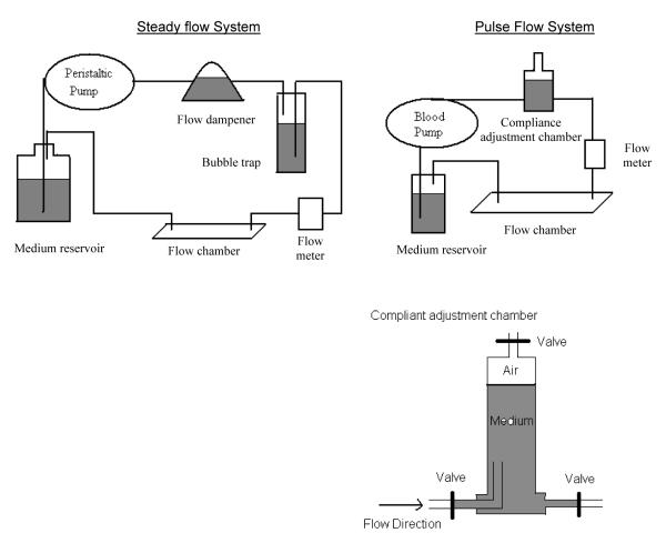
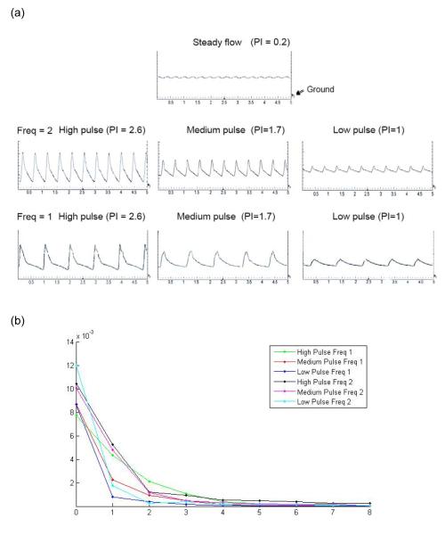
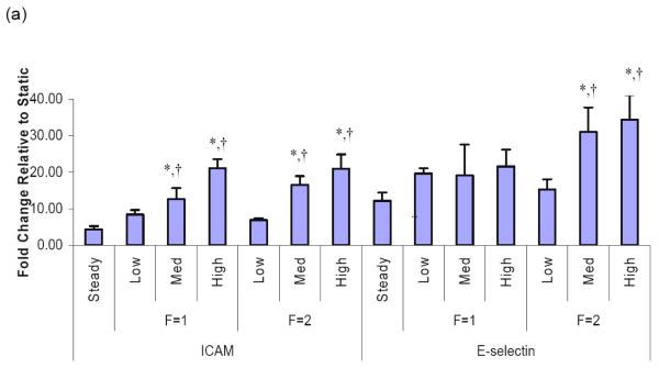
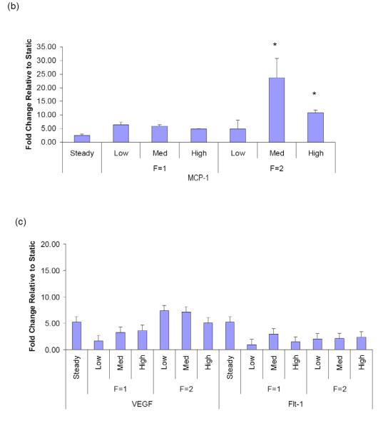
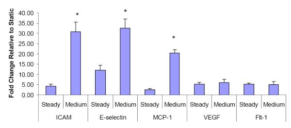
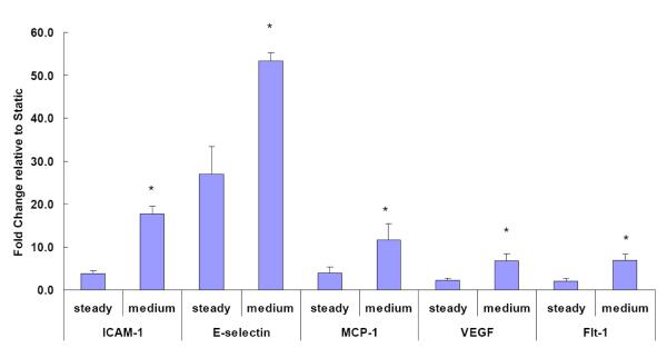
Methods: A flow system was developed to reproduce arterial-like pulse flow waves with the capability of modulating flow pulsatility through regulation of upstream compliance. Pulmonary microvascular endothelial cells (PMVECs) were exposed to steady flow and pulse flow waves of varied pulsatility with varied hemodynamic energy (low: pulsatility index or PI = 1.0; medium: PI = 1.7; high: PI = 2.6) at flow frequency of 1 or 2 Hz for different durations (1 and 6 h). The mean flow rates in all the conditions were kept the same with shear stress at 14 dynes/cm(2). Gene expression was evaluated by analyzing mRNA levels of adhesion molecules (ICAM-1, E-selectin), chemokine (MCP-1) and growth factor/receptor (VEGF, Flt-1) in PMVECs. Functional changes were observed with monocyte adhesion assay.

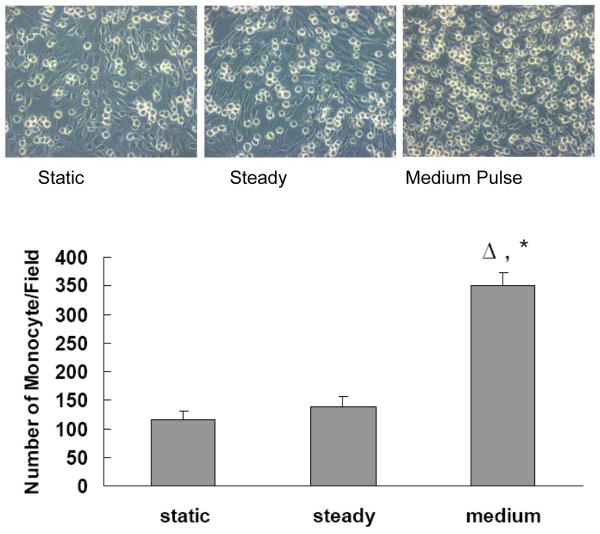
Results: 1) Compared to either steady flow or low pulsatility flow, increased flow pulsatility for 1 h induced significant increases in mRNA levels of ICAM-1, E-selectin and MCP-1. 2) Sustained high pulsatility flow perfusion induced increases in ICAM, E-selectin, MCP-1, VEGF and its receptor Flt-1 expression. 3) Flow pulsatility effects on PMVECs were frequency-dependent with greater responses at 2 Hz and likely associated with the hemodynamic energy level. 4) Pulse flow waves with high flow pulsatility at 2 Hz induced leukocyte adhesion and recruitment to PMVECs.

Conclusion: Increased upstream pulmonary arterial stiffness increases flow pulsatility in distal arteries and induces inflammatory gene expression, leukocyte adhesion and cell proliferation in the downstream PMVECs.


Figures

Figure 1
Figure 1
The system setups for studying effects of steady flow and arterial-like pulse flow conditions
Figure 2
Figure 2
(a) Flow waveforms demonstrated experimental conditions including the steady flow and the arterial-like pulse flow waves with varied pulsatility, i.e. low pulsatility (PI=1), medium pulsatility (PI=1.7) and high pulsatility (PI=2.6), at the frequency of 1Hz or 2Hz. The flow waves were plotted by oscilloscope showing the relationship between voltage representing the flow rate and time. The graphs had time (unit: second) on the x-axis and voltage on the y-axis with minimal division set at 200mV or 2ml/min. Ground shows the point of 0mV. (b) Spectral analysis of the high, medium and low pulse flow waveforms.
Figure 3
Figure 3
Pulsation effects of flows on the mRNA expression in distal PMVECs. Effects of pulsatile flow conditions including peristaltic flow and arterial-like flows with varied pulse magnitudes were compared to those of the steady flow. All the flow conditions had the same mean flow rate with shear stress at 14 dynes/cm2 and the same perfusion time (1 hour). The mRNA expression of (a) adhesion molecules, ICAM-1 and E-selectin, (b) chemokine, monocyte chemoattractant protein (MCP-1), and (c) molecules that regulate cell proliferation in distal PMVECs were examined under various pulsatile flow conditions. The mRNA expression was detected using real-time RT-PCR and expressed as fold change relative to the static condition. Data represent mean ± SEM. “F” means frequency of flow. All the conditions are significantly different from the static condition; ‘*’: significantly different from the steady flow; ‘†’: significantly different from the low pulse flow at the same frequency (P ≤ 0.05).
Figure 3
Figure 3
Pulsation effects of flows on the mRNA expression in distal PMVECs. Effects of pulsatile flow conditions including peristaltic flow and arterial-like flows with varied pulse magnitudes were compared to those of the steady flow. All the flow conditions had the same mean flow rate with shear stress at 14 dynes/cm2 and the same perfusion time (1 hour). The mRNA expression of (a) adhesion molecules, ICAM-1 and E-selectin, (b) chemokine, monocyte chemoattractant protein (MCP-1), and (c) molecules that regulate cell proliferation in distal PMVECs were examined under various pulsatile flow conditions. The mRNA expression was detected using real-time RT-PCR and expressed as fold change relative to the static condition. Data represent mean ± SEM. “F” means frequency of flow. All the conditions are significantly different from the static condition; ‘*’: significantly different from the steady flow; ‘†’: significantly different from the low pulse flow at the same frequency (P ≤ 0.05).
Figure 4
Figure 4
Effects of the medium pulse flow with steady flow preconditioning on gene expressions by distal PMVECs were compared to those of the steady flow. PMVECs were exposed to steady flow before they were exposed to the pulse flow at frequency of 2Hz. Both of the flow conditions had the same mean flow rate with shear stress at 14 dynes/cm2 and the same perfusion time. The mRNA expression was detected using real-time RT-PCR and expressed as fold change relative to the static condition. Data represent mean ± SEM. “F” means frequency of flow. ‘*’: significantly different from the steady flow (P ≤ 0.05).
Figure 5
Figure 5
Effects of the medium pulse flow with extended perfusion at frequency of 2Hz on the gene expression by distal PMVECs were compared to those of the steady flow. The flow perfusion time was extended to 6 hours. Both of the flow conditions had the same mean flow rate with shear stress at 14 dynes/cm2 and the same perfusion time. The mRNA expression was detected using real-time RT-PCR and expressed as fold change relative to the static condition. Data represent mean ± SEM. ‘*’: significantly different from the steady flow (P ≤ 0.05).
Figure 6
Figure 6
Effect of the medium pulse flow on monocyte adhesion: PMVECs were exposed to either the steady flow or the medium pulse flow for 6 hours at frequency of 2Hz with the mean flow shear stress at 14 dynes/cm2. Monocyte adhesion assays were performed by determining the number of monocytes adhered to PMVEC per microscope field. Data represent mean ± SEM, n=6 for each group. ‘Δ’: significantly different from static; ‘*’: significantly different from steady (P ≤ 0.05).

Similar articles

Cited by

References

    1. McVeigh GE, Hamilton PK, Morgan DR. Evaluation of mechanical arterial properties: clinical, experimental and therapeutic aspects. Clin Sci (Lond) 2002;102:51–67. - PubMed
    1. Mattace-Raso F, van der Cammen T, Hofman A. Arterial Stiffness and Risk of Coronary Heart Disease and Stroke: The Rotterdam Study. Circulation. 2006;113:657–663. al. e. - PubMed
    1. Cohn JN. Arterial stiffness, vascular disease, and risk of cardiovascular events. Circulation. 2006;113:601–3. - PubMed
    1. Goldsmith D, MacGinley R, Smith A, Covic A. How important and how treatable is vascular stiffness as a cardiovascular risk factor in renal failure? Nephrol Dial Transplant. 2002;17:965–969. - PubMed
    1. O’Rourke MF. Arterial pressure waveforms in hypertension. Minerva Med. 2003;94:229–50. - PubMed

Publication types