

Finite element simulation of cement-bone interface micromechanics: a comparison to experimental results
- PMID: 19340877
- PMCID: PMC2802538
- DOI: 10.1002/jor.20882
Finite element simulation of cement-bone interface micromechanics: a comparison to experimental results
Abstract
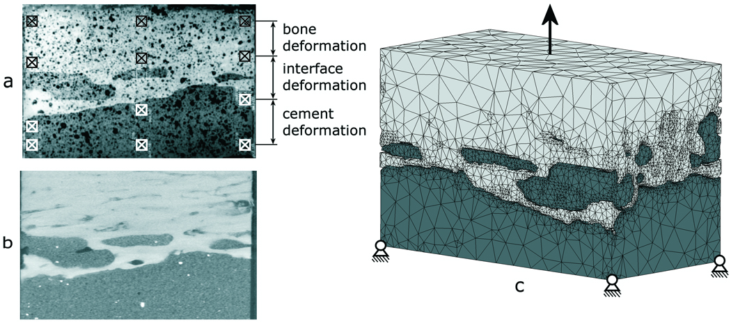
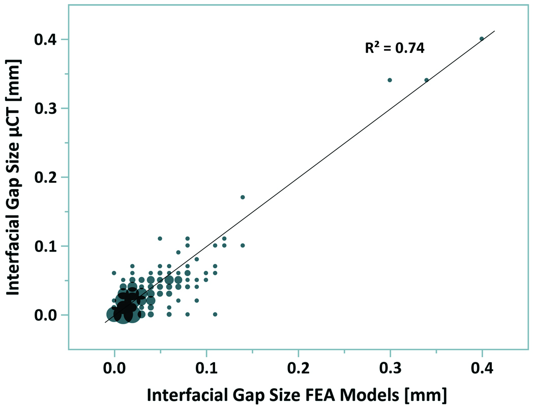
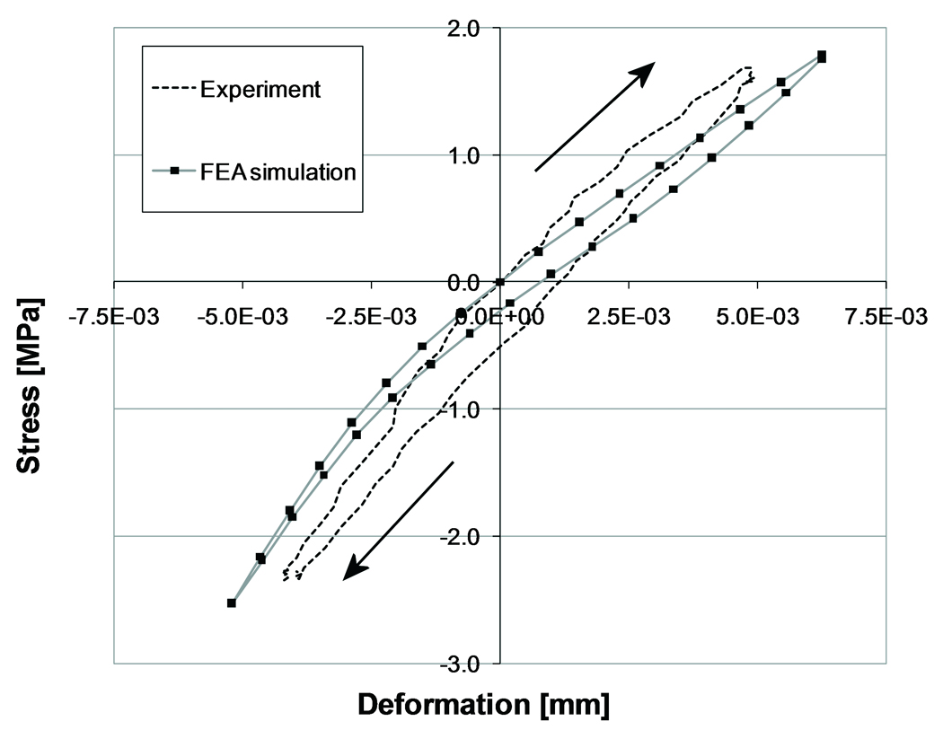
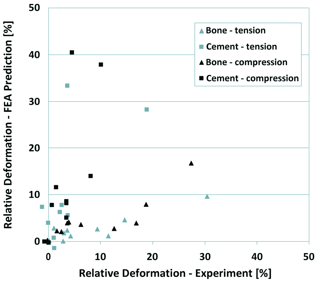
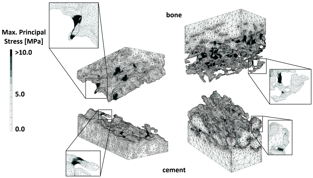
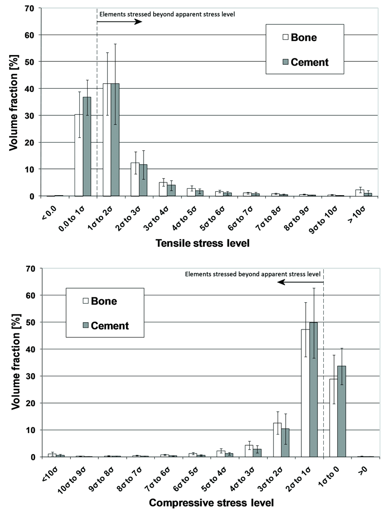
Recently, experiments were performed to determine the micromechanical behavior of the cement-bone interface under tension-compression loading conditions. These experiments were simulated using finite element analysis (FEA) to test whether the micromechanical response of the interface could be captured in micromodels. Models were created of experimental specimens based upon microcomputed tomography data, including the complex interdigitated bone-cement morphology and simulated frictional contact at the interface. The models were subjected to a fully reversed tension-compression load, mimicking the experimental protocol. Similar to what was found experimentally, the simulated interface was stiffer in compression than in tension, and the majority of displacement was localized to the cement-bone interface. A weak correlation was found between the FEA-predicted stiffness and the stiffness found experimentally, with average errors of 8 and 30% in tension and compression, respectively. The hysteresis behavior found experimentally was partially reproduced in the simulation by including friction at the cement-bone interface. Furthermore, stress analysis suggested that cement was more at risk of fatigue failure than bone, concurring with the experimental observation that more cracks were formed in the cement than in the bone. The current study provides information that may help explain the load transfer mechanisms taking place at the cement-bone interface.
(c) 2009 Orthopaedic Research Society. Published by Wiley Periodicals, Inc.
Figures
Similar articles
-
Micro-mechanical modeling of the cement-bone interface: the effect of friction, morphology and material properties on the micromechanical response.J Biomech. 2008 Nov 14;41(15):3158-63. doi: 10.1016/j.jbiomech.2008.08.020. Epub 2008 Oct 10. J Biomech. 2008. PMID: 18848699 Free PMC article.
-
Fatigue creep damage at the cement-bone interface: an experimental and a micro-mechanical finite element study.J Biomech. 2009 Nov 13;42(15):2513-9. doi: 10.1016/j.jbiomech.2009.07.014. Epub 2009 Aug 13. J Biomech. 2009. PMID: 19682690 Free PMC article.
-
The mechanical effects of different levels of cement penetration at the cement-bone interface.J Biomech. 2010 Apr 19;43(6):1167-75. doi: 10.1016/j.jbiomech.2009.11.033. J Biomech. 2010. PMID: 20022010 Free PMC article.
-
3D real-time micromechanical compressive behaviour of bone-cement interface: experimental and finite element studies.J Biomech. 2012 Jan 10;45(2):356-63. doi: 10.1016/j.jbiomech.2011.10.011. Epub 2011 Nov 4. J Biomech. 2012. PMID: 22055427
-
Predicting the failure response of cement-bone constructs using a non-linear fracture mechanics approach.J Biomech Eng. 2002 Aug;124(4):462-70. doi: 10.1115/1.1488167. J Biomech Eng. 2002. PMID: 12188213
Cited by
-
Biomechanical Evaluation of Spinal Column after Percutaneous Cement Discoplasty: A Finite Element Analysis.Orthop Surg. 2022 Aug;14(8):1853-1863. doi: 10.1111/os.13314. Epub 2022 Jul 11. Orthop Surg. 2022. PMID: 35818350 Free PMC article.
-
Can medio-lateral baseplate position and load sharing induce asymptomatic local bone resorption of the proximal tibia? A finite element study.J Orthop Surg Res. 2009 Jul 17;4:26. doi: 10.1186/1749-799X-4-26. J Orthop Surg Res. 2009. PMID: 19615054 Free PMC article.
-
Trabecular level analysis of bone cement augmentation: a comparative experimental and finite element study.Ann Biomed Eng. 2012 Oct;40(10):2168-76. doi: 10.1007/s10439-012-0587-3. Epub 2012 May 31. Ann Biomed Eng. 2012. PMID: 22648574 Free PMC article.
-
The effect of cement creep and cement fatigue damage on the micromechanics of the cement-bone interface.J Biomech. 2010 Nov 16;43(15):3028-34. doi: 10.1016/j.jbiomech.2010.06.031. Epub 2010 Aug 7. J Biomech. 2010. PMID: 20692663 Free PMC article.
-
Evaluation of factors affecting tibial bone strain after unicompartmental knee replacement.J Orthop Res. 2013 May;31(5):821-8. doi: 10.1002/jor.22283. Epub 2012 Nov 28. J Orthop Res. 2013. PMID: 23192787 Free PMC article.
References
-
- Huiskes R, Verdonschot N, Nivbrant B. Migration, stem shape, and surface finish in cemented total hip arthroplasty. Clin Orthop Relat Res. 1998:103–112. - PubMed
-
- Taylor M, Tanner KE, Freeman MA, Yettram AL. Cancellous bone stresses surrounding the femoral component of a hip prosthesis: an elastic-plastic finite element analysis. Med Eng Phys. 1995;17:544–550. - PubMed
-
- Stolk J, Janssen D, Huiskes R, Verdonschot N. Finite element-based preclinical testing of cemented total hip implants. Clin Orthop Relat Res. 2007;456:138–147. - PubMed
-
- Janssen D, Aquarius R, Stolk J, Verdonschot N. Finite-element analysis of failure of the Capital Hip designs. J Bone Joint Surg Br. 2005;87:1561–1567. - PubMed
-
- Lennon AB, Britton JR, MacNiocaill RF, Byrne DP, Kenny PJ, Prendergast PJ. Predicting revision risk for aseptic loosening of femoral components in total hip arthroplasty in individual patients--a finite element study. J Orthop Res. 2007;25:779–788. - PubMed
Publication types
MeSH terms
Substances
Grants and funding
LinkOut - more resources
Full Text Sources
