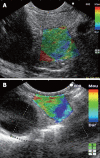
Endoscopic ultrasound elastography for evaluation of lymph nodes and pancreatic masses: a multicenter study
- PMID: 19340900
- PMCID: PMC2669942
- DOI: 10.3748/wjg.15.1587
Endoscopic ultrasound elastography for evaluation of lymph nodes and pancreatic masses: a multicenter study
Abstract
Aim: To evaluate the ability of endoscopic ultrasound (EUS) elastography to distinguish benign from malignant pancreatic masses and lymph nodes.
Methods: A multicenter study was conducted and included 222 patients who underwent EUS examination with assessment of a pancreatic mass (n = 121) or lymph node (n = 101). The classification as benign or malignant, based on the real time elastography pattern, was compared with the classification based on the B-mode EUS images and with the final diagnosis obtained by EUS-guided fine needle aspiration (EUS-FNA) and/or by surgical pathology. An interobserver study was performed.
Results: The sensitivity and specificity of EUS elastography to differentiate benign from malignant pancreatic lesions are 92.3% and 80.0%, respectively, compared to 92.3% and 68.9%, respectively, for the conventional B-mode images. The sensitivity and specificity of EUS elastography to differentiate benign from malignant lymph nodes was 91.8% and 82.5%, respectively, compared to 78.6% and 50.0%, respectively, for the B-mode images. The kappa coefficient was 0.785 for the pancreatic masses and 0.657 for the lymph nodes.
Conclusion: EUS elastography is superior compared to conventional B-mode imaging and appears to be able to distinguish benign from malignant pancreatic masses and lymph nodes with a high sensitivity, specificity and accuracy. It might be reserved as a second line examination to help characterise pancreatic masses after negative EUS-FNA and might increase the yield of EUS-FNA for lymph nodes.
Figures


References
-
- Bhutani MS, Hawes RH, Hoffman BJ. A comparison of the accuracy of echo features during endoscopic ultrasound (EUS) and EUS-guided fine-needle aspiration for diagnosis of malignant lymph node invasion. Gastrointest Endosc. 1997;45:474–479. - PubMed
-
- Tamerisa R, Irisawa A, Bhutani MS. Endoscopic ultrasound in the diagnosis, staging, and management of gastrointestinal and adjacent malignancies. Med Clin North Am. 2005;89:139–158, viii. - PubMed
-
- Vazquez-Sequeiros E, Levy MJ, Clain JE, Schwartz DA, Harewood GC, Salomao D, Wiersema MJ. Routine vs. selective EUS-guided FNA approach for preoperative nodal staging of esophageal carcinoma. Gastrointest Endosc. 2006;63:204–211. - PubMed
-
- Eloubeidi MA, Chen VK, Eltoum IA, Jhala D, Chhieng DC, Jhala N, Vickers SM, Wilcox CM. Endoscopic ultrasound-guided fine needle aspiration biopsy of patients with suspected pancreatic cancer: diagnostic accuracy and acute and 30-day complications. Am J Gastroenterol. 2003;98:2663–2668. - PubMed
-
- Lyshchik A, Higashi T, Asato R, Tanaka S, Ito J, Mai JJ, Pellot-Barakat C, Insana MF, Brill AB, Saga T, et al. Thyroid gland tumor diagnosis at US elastography. Radiology. 2005;237:202–211. - PubMed
Publication types
MeSH terms
LinkOut - more resources
Full Text Sources
Medical

