. 2009 Apr 3;4(4):348-58.
doi: 10.1016/j.stem.2009.01.014.

Small molecules efficiently direct endodermal differentiation of mouse and human embryonic stem cells

Affiliations

Small molecules efficiently direct endodermal differentiation of mouse and human embryonic stem cells

Malgorzata Borowiak et al. Cell Stem Cell. .

Abstract

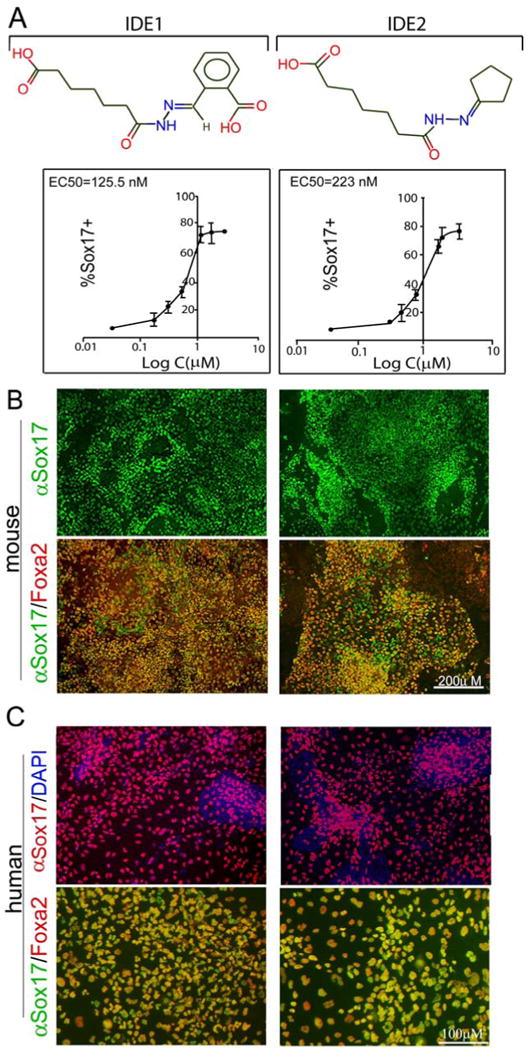
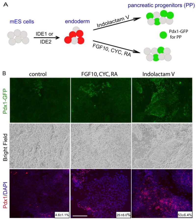
An essential step for therapeutic and research applications of stem cells is the ability to differentiate them into specific cell types. Endodermal cell derivatives, including lung, liver, and pancreas, are of interest for regenerative medicine, but efforts to produce these cells have been met with only modest success. In a screen of 4000 compounds, two cell-permeable small molecules were indentified that direct differentiation of ESCs into the endodermal lineage. These compounds induce nearly 80% of ESCs to form definitive endoderm, a higher efficiency than that achieved by Activin A or Nodal, commonly used protein inducers of endoderm. The chemically induced endoderm expresses multiple endodermal markers, can participate in normal development when injected into developing embryos, and can form pancreatic progenitors. The application of small molecules to differentiate mouse and human ESCs into endoderm represents a step toward achieving a reproducible and efficient production of desired ESC derivatives.


Figures

Figure 1
Figure 1. High throughput screening
(A) Scheme of differentiation into endoderm and evaluation of an endoderm reporter line. Treatment with either Activin A or Nodal induces endoderm in mouse ESC cultures and at day 6 of treatment 45% of total cells are Sox17/dsRed double positive. Every dsRed+ cell stains positively for Sox17 antibody. (B) Overview of the identification of endoderm inducers from small molecule collection. Out of > 4000 screened compounds, 27 primary hits were selected and further evaluated for specificity and toxicity. Markers for definitive endoderm (DE) and extra-embryonic endoderm (EE) were tested by Q-RT-PCR and immunohistochemistry and 2 compounds that induced high levels of DE were indentified. ≥ 3 s.d = more than 3 standard deviations.
Figure 2
Figure 2. Two small molecule inducers of endoderm in mouse and human ESC cultures
(A) Chemical structure of IDE1 and IDE2 that induce endoderm from mouse ESCs. The lower panel shows dose response curves of Sox17 expression (based on immunofluorescence) following treatment with compound for 6 days. The EC50 values and curve fitting were performed with Graph Prism software. Data presented as mean ± s.d. n=4 (B) Representative images of αSox17 immunofluorescence show the highest Sox17 induction in mouse ESCs by compound at day 6 of treatment, quantified as the percent of cells expressing Sox17 (upper panel) out of the total cells. The majority of Sox17+ cells (≥95%) induced by chemical treatment co-express another definitive endoderm marker, FoxA2. (C) HUES cells cultures were treated for 6 days with IDE1, IDE2 and endoderm markers, Sox17 and FoxA2 were analyzed by immunofluorescence. Either of compound treatment leads to Sox17 expression in 55-65% of total cells and ≥ 95% of Sox17+ cells co-expresses also FoxA2.
Figure 3
Figure 3. Time course of endoderm induction and synergy between compound and growth factors
(A) The effect of compounds on the number of Sox17+ and total cells during 14 days of treatment is shown. Endoderm induction by IDE1 and IDE2 peaks at about day 6 and as little as 12hrs treatment with either compound is sufficient to induce Sox17 expression in ˜ 40% of the cells. Activin A treatment induces significant but lower % of Sox17 + cells at all tested time points. Cells were analysed at day 6 (for earlier time points) or 14 of culture. (B) The combined effect of compounds and growth factors on Sox17 expression. Co-treatment of mouse ESCs with compounds and Nodal enables shortening of the treatment time and leads to the induction of Sox17 with a slightly high efficiency (55.6%) expression at day 4. No synergy was observed between IDE1 and IDE2 or for the combination of either compound with Wnt3a. All quantifications were based on the percentage of cells stained by Sox17 antibody out of total cell. Data presented as mean ±s.d. n=4 experiments.
Figure 4
Figure 4. Gene expression analysis of chemically induced endoderm
(A) Expression of definitive endodermal markers in Sox17+ cells induced by compound treatment or isolated form E7.5-8.0 embryos. Sox17/dsRed+ cells were sorted by FACS and expression of endoderm genes was analysed by Illumina microarray. Expression of “endoderm signature” genes normalized to the DMSO treated mouse ESCs is shown. Out of 17 genes, only 2, Spink3 and Tmprss2 (marked by grey circle) were expressed at significantly higher levels (>2 fold change) in Sox17+ cells isolated from E7.75 embryos (endoderm). Each bar represents an average of 3 biological replicates and mean ± s.d. is shown (B) Scatter plots comparing the global gene expression in Sox17/dsRed+ populations sorted out from Sox17/dsRed E7.75 embryos and derived either in vitro by treatment with IDE2 (day 6 of treatment) or with non-treated mouse ESC cultures. Red line in the middle visualizes the equivalent levels in gene expression; two side red lines show two-fold change in gene expression levels between both samples.
Figure 5
Figure 5. Small molecule inducers of endoderm activate TGF-β signaling
(A) Phosphorylation of Smad2 was analysed in lysates of ESCs treated with IDE1, IDE2, DMSO, Activin A or Nodal or in the presence of the ALK4/5/7 inhibitor, SB431542. Treatment with IDE1 or IDE2 leads to activation of the TGF-β pathway after 24 hrs, similar to either Nodal or Activin A treatment. Phosphorylation of Smad2 by either of the two compounds is significantly attenuated in the presence of SB431542. (B) Increase in Nodal expression after treatment with small molecules IDE2, IDE1 and Nodal. Relative expression over DMSO treatment is shown as a mean of triplicate experiments ± s. d.
Figure 6
Figure 6. Functional evaluation of chemically derived endoderm
(A) Scheme of in vivo assay to assess the functional potential of compound induced endoderm. Mouse ESCs treated with chemical inducers incorporate into the developing host gut tube. Cultures of mouse ESC reporter lines expressing constitutive YFP were differentiated into endoderm with IDE1 or IDE2, producing 60-70% Sox17+ cells, then trypsinized and injected into the nascent gut lumen of E8.75 mouse embryos. Dashed line shows an approximate plane of section (B) After 24-30 hours ex vivo culture, mouse embryos were fixed, transversally sectioned and stained with antibodies against FoxA2 and Cldn6 to detect gut epithelial cells and anti-YFP antibodies to visualize injected cells. IDE1 and IDE2 induced endodermal cells incorporate into gut tube and show expression of gut tube markers. In contrast, DMSO treated cells remain clustered in the gut tube lumen 30 hrs after injection and do not incorporate into the gut epithelia nor express gut tube markers.
Figure 7
Figure 7. Developmental potential of chemically derived endoderm
(A) Scheme of mouse ESC differentiation into Pdx1 + pancreatic progenitors. Endoderm was first derived through treatment with either IDE1 or IDE2 and then formation of pancreatic progenitors was monitored using a Pdx1-GFP reporter line. (B) Endoderm enriched cultures were grown for another 6 days in chemically defined media containing: DMSO without any additional growth factors or compounds (control), in presence of growth factors (FGF10, CYC, RA) or in the presence of Indolactam V to induce the expression of Pdx1. At day 12, in cultures treated initially with IDE1 or IDE2 and followed by ILV, 50% of the total cells were Pdx1+, a 10-fold increase above control treatment.

Comment in

References

    1. Agius E, Oelgeschlager M, Wessely O, Kemp C, De Robertis EM. Endodermal Nodal-related signals and mesoderm induction in Xenopus. Development. 2000;127:1173–1183. - PMC - PubMed
    1. Anderson WJ, Zhou Q, Alcalde V, Kaneko OF, Blank LJ, Sherwood RI, Guseh JS, Rajagopal J, Melton DA. Genetic targeting of the endoderm with claudin-6(CreER) Dev Dyn. 2008;237:504–512. - PMC - PubMed
    1. Ang SL, Wierda A, Wong D, Stevens KA, Cascio S, Rossant J, Zaret KS. The formation and maintenance of the definitive endoderm lineage in the mouse: involvement of HNF3/forkhead proteins. Development. 1993;119:1301–1315. - PubMed
    1. Bhushan A, Itoh N, Kato S, Thiery JP, Czernichow P, Bellusci S, Scharfmann R. Fgf10 is essential for maintaining the proliferative capacity of epithelial progenitor cells during early pancreatic organogenesis. Development. 2001;128:5109–5117. - PubMed
    1. Burridge PW, Anderson D, Priddle H, Barbadillo Munoz MD, Chamberlain S, Allegrucci C, Young LE, Denning C. Improved human embryonic stem cell embryoid body homogeneity and cardiomyocyte differentiation from a novel V-96 plate aggregation system highlights interline variability. Stem Cells. 2007;25:929–938. - PubMed

Publication types

MeSH terms