. 2009 Jun 1;330(1):131-41.
doi: 10.1016/j.ydbio.2009.03.021. Epub 2009 Mar 31.

Hand2 is required in the epithelium for palatogenesis in mice

Affiliations

Hand2 is required in the epithelium for palatogenesis in mice

Wei Xiong et al. Dev Biol. .

Abstract

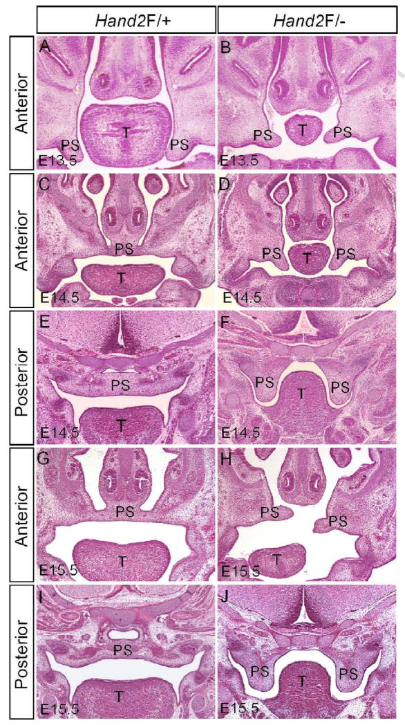
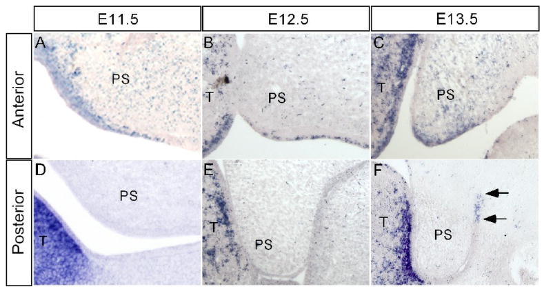
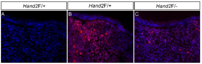
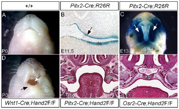
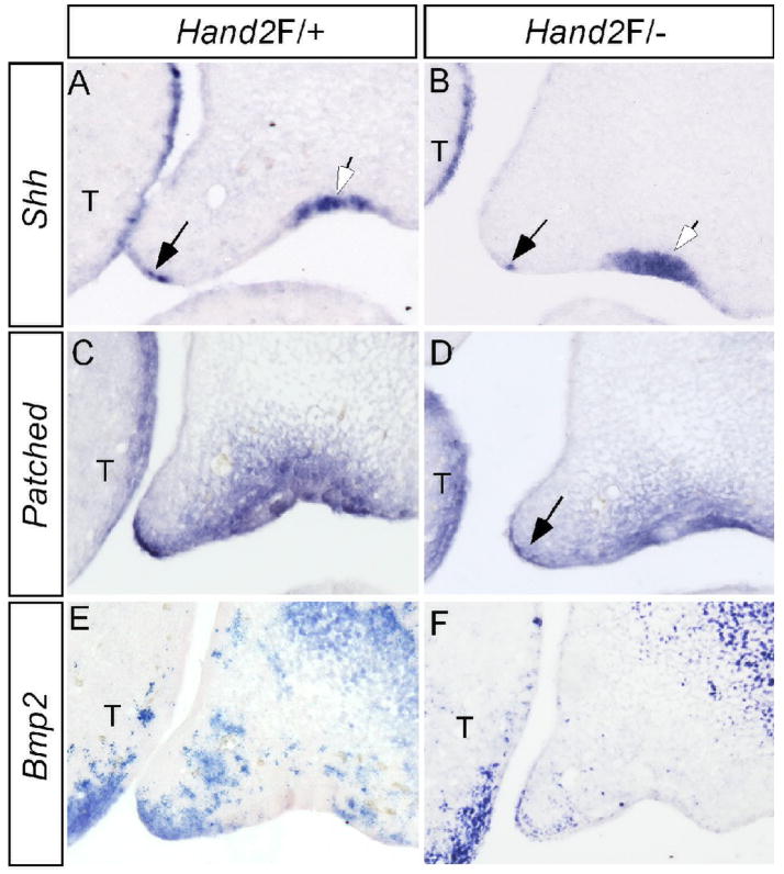
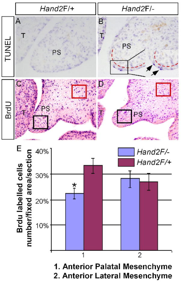
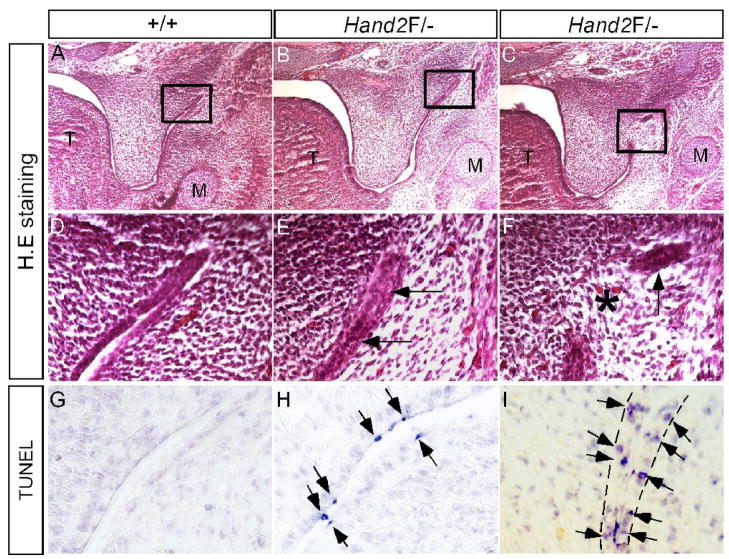
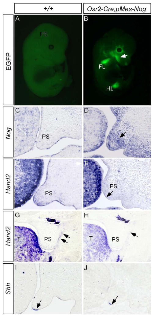
The basic helix-loop-helix (bHLH) transcription factor Hand2 has been implicated in the development of multiple organs, including craniofacial organs. Mice carrying Hand2 hypomorphic alleles (Hand2(LoxP/-)) display a cleft palate phenotype. A specific deletion of the Hand2 branchial arch-specific enhancer also leads to a hypoplastic mandible and cleft palate formation in mice. However, the underlying mechanism of Hand2 regulation of palate development remains unknown. Here we show that Hand2 is expressed in both the epithelium and mesenchyme of the developing palate. While mesenchymal specific inactivation of Hand2 has no impact on palate development, epithelial specific deletion of Hand2 creates a cleft palate phenotype. Hand2 appears to exert distinct roles in the anterior and posterior palate. In the anterior palate of Hand2(LoxP/-) mice, premature death of periderm cells and a down-regulation of Shh are observed in the medial edge epithelium (MEE), accompanied by a decreased level of cell proliferation in the palatal mesenchyme. In the posterior palate, a lower dose of Hand2 causes aberrant periderm cell death on the surface of the epithelium, triggering abnormal fusion between the palatal shelf and mandible and preventing palatal shelf elevation. We further demonstrate that BMP activities are essential for the expression of Hand2 in the palate. We conclude that Hand2 is an intrinsic regulator in the epithelium and is required for palate development.


Figures

Figure 1
Figure 1
Hand2LoxP/− mice exhibit hindrance of palate shelf elevation. (A) A section of an E13.5 wild type embryo shows normal developing palatal shelves. (B) The palatal shelves of an E13.5 Hand2LoxP/− embryo appear relatively small. (C–F) At E14.5, palatal shelves in the wild type control have already elevated to the position above the tongue and fused at the midline (C, E). However, in Hand2LoxP/− mice, the palatal shelves remain in a vertical position (D, F). (G–J) Wild type controls at E15.5 are shown in (G) and (I). In the mutant, the anterior portion of the palatal shelves elevates to the horizontal position above the tongue, but exhibits a misalignment (H), while the posterior portion remains in the vertical position (J). T, tongue; PS, palatal shelf.
Figure 2
Figure 2
Expression of Hand2 in the early developing palatal shelf. (A–C) In the anterior palatal shelf, Hand2 is expressed in the epithelium including MEE and mesenchyme at E11.5 (A), E12.5 (B) and E13.5 (C). (D–F) In the posterior palatal shelf, Hand2 expression is not detectable at E11.5 (D) and E12.5 (E). However, at E13.5, Hand2 transcripts (arrows) are specifically detected in the epithelium of the lateral junction of the palatal shelf and mandible (F). T, tongue; PS, palatal shelf.
Figure 3
Figure 3
Reduction of Hand2 protein in Hand2LoxP/− mandible. (A) A negative control for immunohistochemical staining. (B) Immunohistochemical staining shows abundant Hand2-positve cells in an E13.5 Hand2F/+ mandible. (C) An E13.5 Hand2F/− mandible exhibits significantly reduced immunostaining signals for Hand2 protein in the mesenchymal cells.
Figure 4
Figure 4
Epithelial specific inactivation of Hand2 leads to cleft palate formation. (A) A newborn wild type mouse shows a normal mandible. (B) A section through oral cavity of an E11.5 Pitx2-Cre;R26R embryo shows specific LacZ staining in the palatal epithelium (arrow) and dental epithelium (de). (C) Whole mount LacZ staining of an E13.5 Pitx2-Cre;R26R embryo demonstrates Cre activity in the entire palatal shelf (arrows). (D) A newborn Wnt1-Cre;Hand2LoxP/LoxP mouse (labeled as Wnt1-Cre;Hand2F/F) exhibits a hypoplastic mandible (black arrow) and an intact palate (white arrow). (E) A section through oral cavity of an E16.5 Pitx2-Cre;Hand2LoxP/LoxP embryo (labeled as Pitx2-Cre;Hand2F/F) shows cleft palate defect. Note the palatal shelves remain in a vertical position. (F) A section through oral cavity of an E16.5 Osr2-Cre;Hand2LoxP/LoxP embryo (labeled as Osr2-Cre;Hand2F/F) shows a normal formed palate. T, tongue; de, dental epithelium; PS, palatal shelf.
Figure 5
Figure 5
Down-regulation of Shh and its downstream genes in the palatal shelf of Hand2 hypomorphic mice. (A) Expression of Shh is detected in the MEE (black arrow) and the rugal epithelium (white arrow) of an E13.5 control palate. (B) Shh expression is significantly down-regulated in the MEE (black arrow), but remains unaffected in the rugal epithelium (white arrow) of an E13.5 Hand2LoxP/− embryo. (C, E) Expression of Ptc (C) and Bmp2 (E) in E13.5 control palatal shelves. (D) Ptc expression is down-regulated in the palatal shelf of an E13.5 Hand2LoxP/− embryo. Note specific gene down-regulation in the palatal mesenchyme adjacent to the MEE (arrow). (F) Bmp2 expression is reduced in the palatal shelf of an E13.5 Hand2LoxP/− embryo. Note the expression in the maxillary mesenchyme and the tongue is not affected. T, tongue
Figure 6
Figure 6
Aberrant cell proliferation and apoptosis in the palatal shelf of Hand2 hypomorphic mice. (A, B) TUNEL assay on the anterior portion of palate from E13.5 control (A) and Hand2LoxP/− (B) embryos shows abnormal apoptotic cells (arrows in the insert of panel B) in the MEE of the mutant. Note the apoptotic cells are periderm cells. The red dash lines demarcate the boundary of epithelium and mesenchyme. (C, D) BrdU labeling assays show a reduced level of cell proliferation in the anterior palate of E13.5 Hand2LoxP/− embryo (D), as compared to the control (C). (E) Comparison of BrdU-labeled cells in fixed areas of palate and lateral maxillary mesenchyme in controls and Hand2LoxP/− embryo. Standard deviation values were indicated as the error bars. *: P < 0.01. T, tongue; PS, palatal shelf.
Figure 7
Figure 7
Pathological adhesion/fusion of the palatal shelf and mandible in Hand2 hypomorphic mice. (A, D, G) E13.5 wild type controls show normal histological structure (A, D) and absence of apoptotic cells in the lateral junction of the posterior palate and mandible. (B, E) An E13.5 Hand2LoxP/− embryo shows adherence of epithelia (arrows in E) of the lateral junction of the posterior palate and mandible. (C, F) An E13.5 Hand2LoxP/− embryo shows fusion of the palatal shelf and mandible at the lateral junction. Star denotes a confluence of mesenchyme, and arrow points to a remanent epithelium. (H) An E13.5 Hand2LoxP/− embryo shows apoptotic periderm cells (arrows) in the lateral junction right before epithelial contact/adherence. (I) An E13.5 Hand2LoxP/− embryo shows apoptotic basal layer epithelial cells (arrows) in the lateral junction fusion of the palatal shelf and mandible. The dash lines demarcate the epithelial boundary.
Figure 8
Figure 8
Overexpression of Noggin to the palatal mesenchyme down-regulates the expression of Hand2 and Shh. (A) An E13.5 wild type control embryo shows undetectable fluorescent signals. (B) An E13.5 Osr2-Cre;pMes-Nog embryo shows EGFP signal in the craniofacial region (arrow) and forelimb FL) and hindlimb (HL). (C) An E13.5 wild type control shows barely detectable Noggin transcripts in the developing palatal shelf. Note Noggin expression in the tongue. (D) Strong Noggin expression (arrow) is detected in the palatal mesenchyme of an E13.5 Osr2-Cre;pMes-Nog embryo. (E, G, I) The expression of Hand2 in the anterior palate (E) and the epithelial junction of posterior palate (G) and Shh expression (I) in the MEE are detected in E13.5 controlled palatal shelves. (F, H, J) The expression of Hand2 is down-regulated in the anterior palate (F) and the epithelial junction of posterior palate (H) of E13.5 Osr2-Cre;pMes-Nog embryo. Shh expression is also down-regulated in the MEE of E13.5 Osr2-Cre;pMes-Nog palate (J). Note unaltered strong Hand2 expression in the tongue in (F). Arrows in (F, G, H) point to the MEE, and in (G, H) point to the epithelial junction of posterior palate and mandible.

Similar articles

Cited by

References

    1. Alappat SR, Zhang Z, Suzuki K, Zhang X, Liu H, Jiang R, Yamada G, Chen YP. The cellular and molecular etiology of the cleft secondary palate in Fgf10 mutant mice. Dev Biol. 2005;277:102–113. - PubMed
    1. Barbosa AC, Funato N, Chapman S, McKee MD, Richardson JA, Olson EN, Yanagisawa H. Hand transcription factors cooperatively regulate development of the distal midline mesenchyme. Dev Biol. 2007;310:154–168. - PMC - PubMed
    1. Casey LM, Yu L, Eui-Sic C, Maltby KM, Thomas G, Jiang R. Jag2-Notch1 signaling regulates oral epithelial differentiation and palate development. Dev Dyn. 2006;235:1830–1844. - PMC - PubMed
    1. Chai Y, Jiang X, Ito Y, Bringas P, Han J, Rowitch DH, Soriano P, McMahon AP, Sucov HM. Fate of the mammalian cranial neural crest during tooth and mandibular morphogenesis. Development. 2000;127:1671–1679. - PubMed
    1. Charite J, McFadden DG, Olson EN. The bHLH transcription factor dHAND controls Sonic hedgehog expression and establishment of the zone of polarizing activity during limb development. Development. 2000;127:2461–2470. - PubMed

Publication types

Substances