. 2009 Oct 1;47(4):1701-10.
doi: 10.1016/j.neuroimage.2009.03.050. Epub 2009 Mar 31.

On a basal ganglia role in learning and rehearsing visual-motor associations

Affiliations

On a basal ganglia role in learning and rehearsing visual-motor associations

Patrick Bédard et al. Neuroimage. .

Abstract

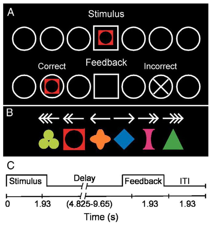
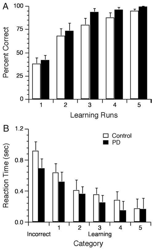
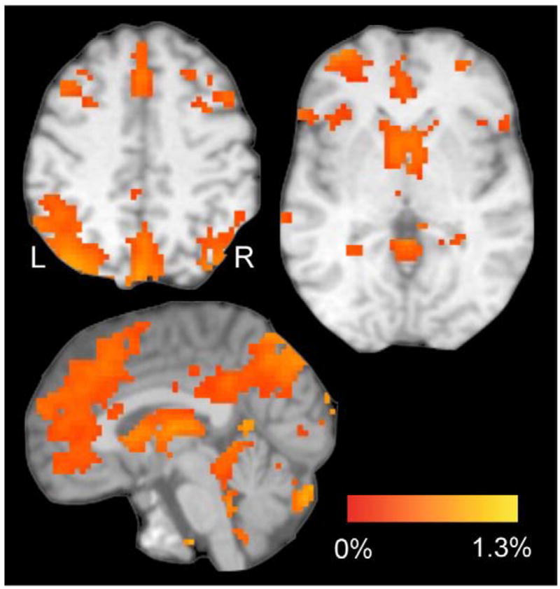
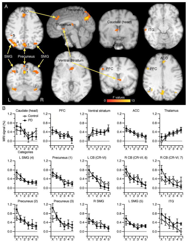
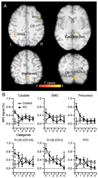
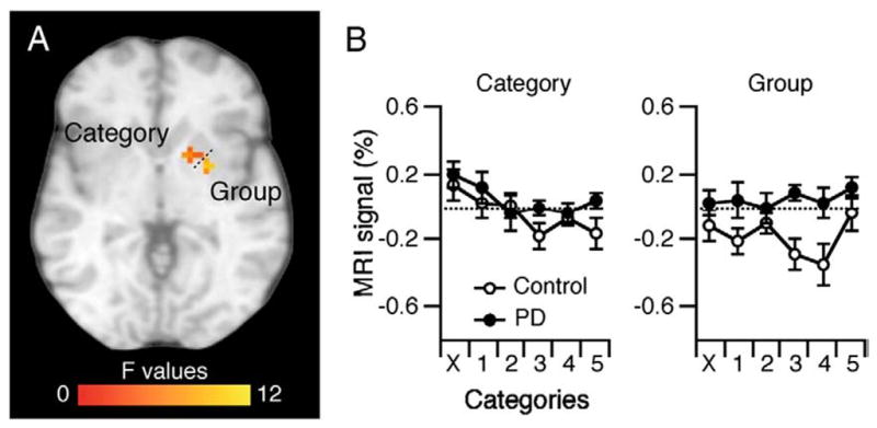
Fronto-striatal circuitry interacts with the midbrain dopaminergic system to mediate the learning of stimulus-response associations, and these associations often guide everyday actions, but the precise role of these circuits in forming and consolidating rules remains uncertain. A means to examine basal ganglia circuit contributions to associative motor learning is to examine these process in a lesion model system, such as Parkinson's disease (PD), a basal ganglia disorder characterized by the loss of dopamine neurons. We used functional magnetic resonance imaging (MRI) to compare brain activation of PD patients with a group of healthy aged-match participants during a visual-motor associative learning task that entailed discovering and learning arbitrary associations between a set of six visual stimuli and corresponding spatial locations by moving a joystick-controlled cursor. We tested the hypothesis that PD would recruit more areas than age-matched controls during learning and also show increased activation in commonly activated regions, probably in the parietal and premotor cortices, and the cerebellum, perhaps as compensatory mechanisms for their disrupted fronto-striatal networks. PD had no effect in acquiring the associative relationships and learning-related activation in several key frontal cortical and subcortical structures. However, we found that PD modified activation in other areas, including those in the cerebellum and frontal, and parietal cortex, particularly during initial learning. These results may suggest that the basal ganglia circuits become active more so during the initial formation of rule-based behavior.


Figures

Fig. 1
Fig. 1
Schematic of experiment. (A) Visual stimuli used in the experiment: arrows (Movement task) and symbols (associative learning task). (B) Task schematic. Participants viewed a stimulus (arrow or symbol) and move the joystick that displaced a cursor on the screen from the center home position (square) to one of the six targets (circles) and then received feedback for correctness. (C) Trial Events. A trial started with the presentation of a stimulus during 1.93 s. Then, a variable delay of 4.825–9.65 s (randomly presented) allowed time for participants to move to a target. After the delay, feedback appeared for 1.93 s. An inter-stimulus interval (ITI) of 1.93 s allowed time for participants to replace the cursor in the home position before the next trial started. See text for additional details.
Fig. 2
Fig. 2
Behavioral results. (A) Percent of correct responses increased for the control and PD groups across runs as revealed by a two-way 2 Group×5 Runs ANOVA that yielded a main effect of Runs, F(4, 72)=46.21, p=0.02e–14, no interaction F(4, 72)<1, and no main effect of Group F(1, 18)=2.14, p>0.16. (B) RT decreased across the 6 categories of trials for the control and PD groups as revealed by a two-way 2 Group×6 Category ANOVA that yielded a main effect of Category, F(5, 90)=30.92, p<0.01e–13, indicating a decrease of RT going from the Incorrect to Correct-1 and Correct-2 with no further significant decrease. The ANOVA yielded no significant main effect of Group, F(1, 18)<1, or interaction F(5, 90)<1.
Fig. 3
Fig. 3
Task-related brain activation (% MRI signal) for both groups. These regions represent a contrast (t-test) between the Associative task (averaged across the 6 trials categories) vs. the Movement task on both groups (voxel threshold: p≤0.001, t (19)≥3.863; corrected for multiple comparisons at p≤0.05 for 21 contiguous voxels as implemented in AFNI). Activation occurred in frontal and parietal cortices, thalamus, basal ganglia, and cerebellum. The color bar represents the % MRI signal change of the contrast aforementioned. L, left hemisphere.
Fig. 4
Fig. 4
Regions depicting a main effect of Category. (A) These included the ACC (BA32), PFC (BA9), precuneus, SMG, cerebellum (CR-I), head of the caudate nucleus, ventral striatum, ITG, and thalamus. The color bar represents the F statistics of the Category main effect. See Table 2 for more details. L, left hemisphere. (B) MRI signal (%) for each group across all 6 learning categories. Most clusters showed decreased activation across the learning categories but two showed an increase (ventral striatum and thalamus). CB, cerebellum (Cr: Crus); ACC, anterior cingulate cortex; PFC, prefrontal cortex; SMG, supramarginal gyrus; ITG, inferior temporal gyrus.
Fig. 5
Fig. 5
Regions depicting an interaction effect of Group×Category. (A) These included the SMG (BA40), precuneus (BA7), PFC (BA9), cerebellum, and tail of the caudate nucleus. The color bar represents the F statistics of the interaction. See Table 3 for more details. L, left hemisphere. (B) MRI signal (%) for each group across all 6 learning categories. The interaction indicates that PD had high level of activation in the early learning phase but decreased it across learning whereas controls had sustained activation throughout (see text). CS, central sulcus.
Fig. 6
Fig. 6
Putamen ROI analysis. We found clusters within the right putamen with a main effect of Group and a main effect of Category (p≤0.05).

Similar articles

Cited by

References

    1. Alexander GE, DeLong MR, Strick PL. Parallel organization of functionally segregated circuits linking basal ganglia and cortex. Annu Rev Neurosci. 1986;9:357–381. - PubMed
    1. Berardelli A, Rothwell JC, Thompson PD, Hallett M. Pathophysiology of bradykinesia in Parkinson’s disease. Brain. 2001;124:2131–2146. - PubMed
    1. Bezard E, Gross CE, Brotchie JM. Presymptomatic compensation in Parkinson’s disease is not dopamine-mediated. Trends Neurosci. 2003;26:215–221. - PubMed
    1. Boettiger CA, D’Esposito M. Frontal networks for learning and executing arbitrary stimulus–response associations. J Neurosci. 2005;25:2723–2732. - PMC - PubMed
    1. Botvinick MM, Cohen JD, Carter CS. Conflict monitoring and anterior cingulate cortex: an update. Trends Cogn Sci. 2004;8:539–546. - PubMed

Publication types