. 2009 May-Jun;32(3-4):195-200.
doi: 10.1016/j.jaut.2009.02.021. Epub 2009 Apr 1.

B cells contribute to ischemia/reperfusion-mediated tissue injury

Affiliations

B cells contribute to ischemia/reperfusion-mediated tissue injury

Jie Chen et al. J Autoimmun. 2009 May-Jun.

Abstract

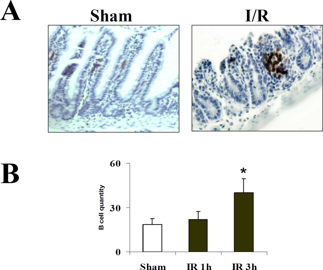
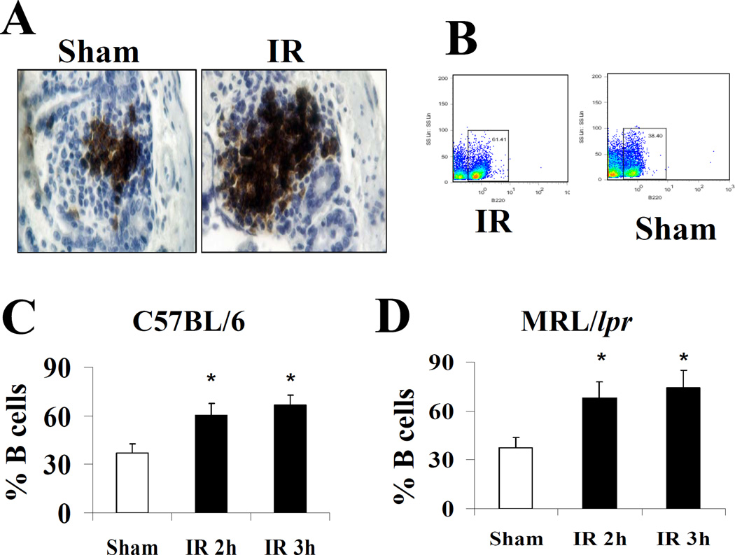
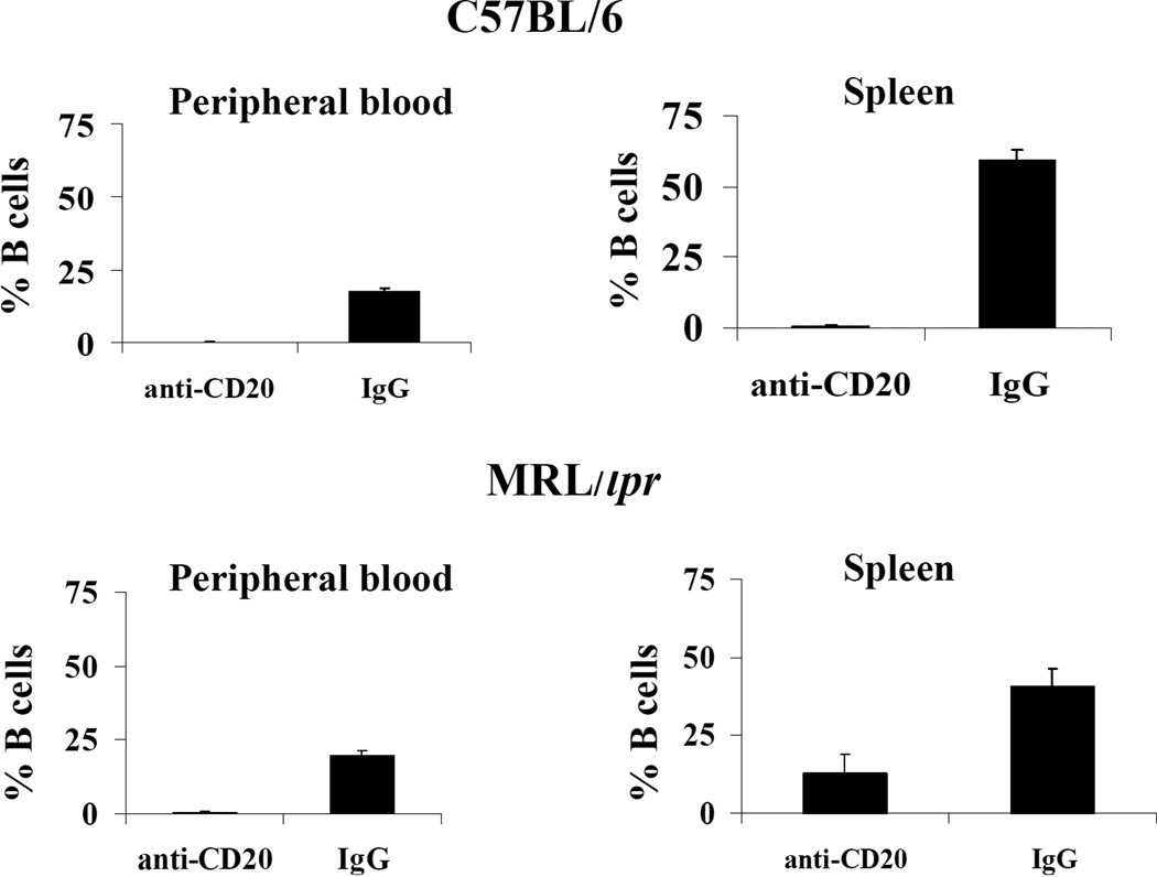
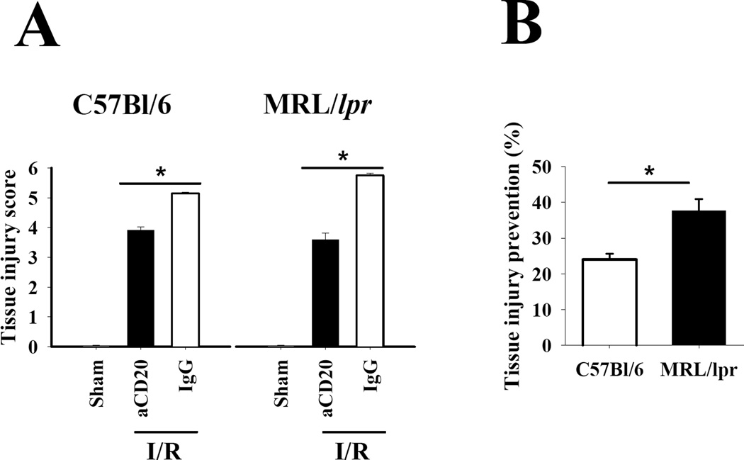
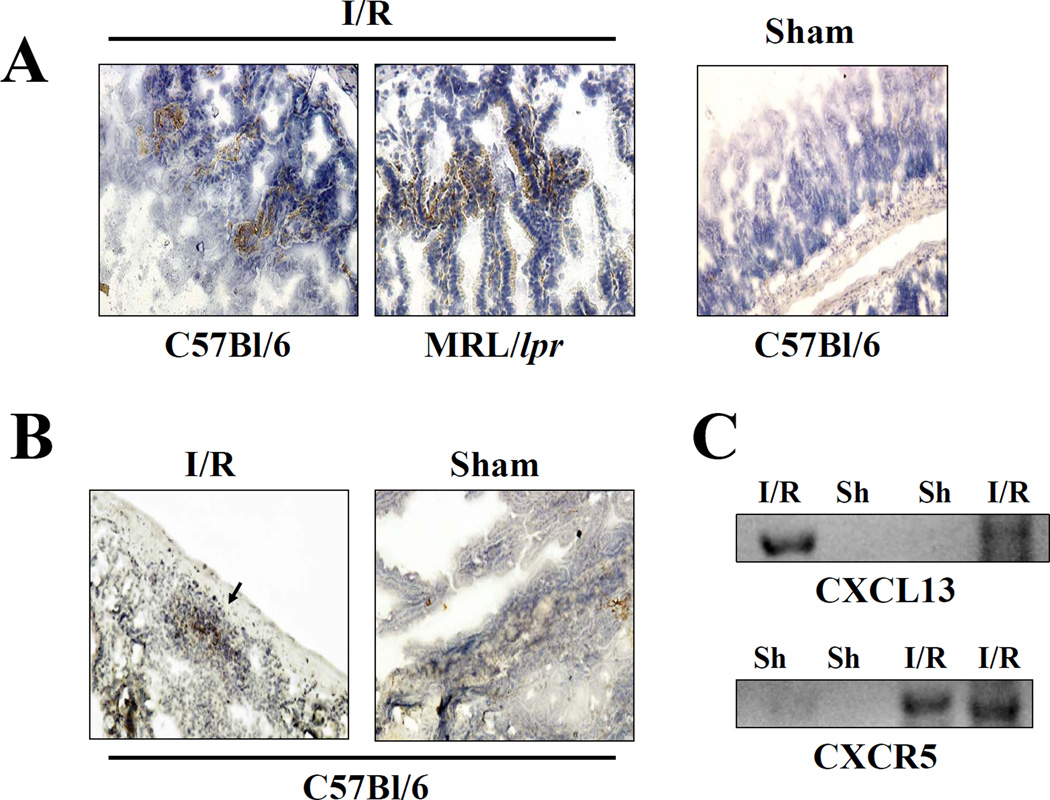
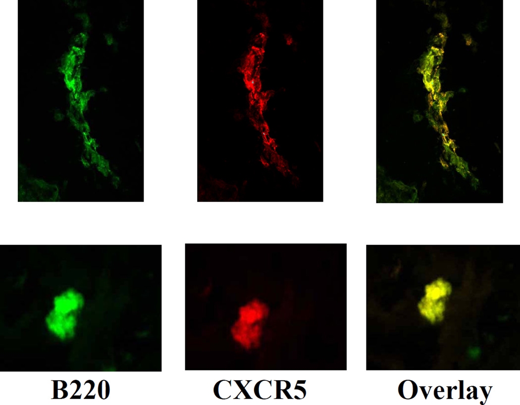
Multiple elements are known to participate in ischemia/reperfusion (I/R)-mediated tissue injury. Amongst them, B cells have been shown to contribute by the production of antibodies that bind to ischemic cells and fix complement. It is currently unknown whether B cells participate through antibody-independent mechanisms in the pathogenesis of I/R. In a mesenteric I/R model we found that B cells infiltrate the injured intestine of normal and autoimmune mice 2h after reperfusion is established. B cell depletion protected mice from the development of I/R-mediated intestinal damage. The protection conferred by B cell depletion was significantly greater in MRL/lpr mice. Finally, we show that ischemic tissue expressed the B cell-attractant CXCL13 and infiltrating B cells expressed the corresponding receptor CXCR5. Our data grant B cells an antibody-independent role in the pathogenesis of intestinal I/R and suggest that B cells accumulate in the injured tissue in response to the chemokine CXCL13.


Figures

Figure 1
Figure 1. B cells infiltrate ischemic intestine during reperfusion
A. C57Bl/6 mice were subjected to sham procedure or I/R. Immunohistochemistry was performed with an anti-B220 antibody. Magnification 200×. B. B cell infiltration was quantified with image analysis software. *p<0.001 when compared to sham mice.
Figure 2
Figure 2. I/R leads to an increase in the number of B cells within Payer’s patches
A. The number of B220+ cells in Payer’s patches increases following I/R (magnification 200×). B. Payer’s patches were isolated and cells were stained with anti-B220 antibody. B cells were quantified in a flow cytometer. C. Cumulative data of flow cytometry experiments (n=7 per group). *p<0.001 when compared to Sham.
Figure 3
Figure 3. MRL/lpr mice are resistant to complete B cell depletion
A depleting anti-mouse CD20 antibody was administered in a single dose to mice 9 days before I/R was induced. C57Bl/6 mice received 20 µg per mice; MRL/lpr received 250 µg per mice. Cellular suspensions from blood and spleen were stained with anti-B220 and analyzed by flow cytometry.
Figure 4
Figure 4. B cell depletion reduces I/R-induced intestinal tissue injury
A. Tissue injury score was significantly reduced in C57Bl/6 (left panel) and MRL/lpr (right panel) mice depleted of B cells (black bars) when compared to B cell-sufficient mice (white bars). * p<0.01. B. The reduction in tissue injury scores was significantly greater in MRL/lpr mice than in control mice. * p<0.05.
Figure 5
Figure 5. CXCL13 expressed by injured tissue recruits CXCR5+ cells
Anti-CXCL13 (A) and anti-CXCR5 (B) antibodies were used to stain frozen sections from C57Bl/6 and MRL/lpr mice subjected to I/R (left panels) or sham (right panel) procedures (n=5). Magnification 200×. C. Total RNA was extracted from intestinal tissue of mice that had undergone I/R or sham (Sh) procedures. CXCL13 and CXCR5 transcripts were amplified by reverse transcriptase-PCR.
Figure 6
Figure 6. CXCR5 expression is limited to B cells
Intestinal sections C57Bl/6 mice subjected to I/R were stained with FITC-labeled anti-B220 and PE-labeled anti-CXCR5 antibody and scanned in a confocal microscope.

Similar articles

Cited by

References

    1. Fleming SD, Tsokos GC. Complement, natural antibodies, autoantibodies and tissue injury. Autoimmun Rev. 2006;5:89–92. - PubMed
    1. Huang Y, Rabb H, Womer KL. Ischemia-reperfusion and immediate T cell responses. Cell Immunol. 2007;248:4–11. - PMC - PubMed
    1. Edgerton C, et al. IL-17 producing CD4+ T cells mediate accelerated ischemia/reperfusion-induced injury in autoimmunity-prone mice. Clin Immunol. 2008 in press. - PMC - PubMed
    1. Tsokos GC. B cells, be gone--B-cell depletion in the treatment of rheumatoid arthritis. N Engl J Med. 2004;350:2546–2548. - PubMed
    1. Burne MJ, et al. Identification of the CD4(+) T cell as a major pathogenic factor in ischemic acute renal failure. J Clin Invest. 2001;108:1283–1290. - PMC - PubMed

Publication types

MeSH terms