. 2009 Apr;174(4):1291-308.
doi: 10.2353/ajpath.2009.080295.

Inhibition of autoregulated TGFbeta signaling simultaneously enhances proliferation and differentiation of kidney epithelium and promotes repair following renal ischemia

Affiliations

Inhibition of autoregulated TGFbeta signaling simultaneously enhances proliferation and differentiation of kidney epithelium and promotes repair following renal ischemia

Hui Geng et al. Am J Pathol. 2009 Apr.

Abstract

We studied autocrine transforming growth factor (TGF)beta signaling in kidney epithelium. Cultured proximal tubule cells showed regulated signaling that was high during log-phase growth, low during contact-inhibited differentiation, and rapidly increased during regeneration of wounded epithelium. Autoregulation of signaling correlated with TGFbeta receptor and Smad7 levels, but not with active TGFbeta, which was barely measurable in the growth medium. Confluent differentiated cells with low receptor and high Smad7 levels exhibited blunted responses to saturating concentrations of exogenously provided active TGFbeta, suggesting that TGFbeta signaling homeostasis was achieved by cell density-dependent modulation of signaling intermediates. Antagonism of Alk5 kinase, the TGFbeta type I receptor, dramatically accelerated the induction of differentiation in sparse, proliferating cultures and permitted better retention of differentiated features in regenerating cells of wounded, confluent cultures. Alk5 antagonism accelerated the differentiation of cells in proximal tubule primary cultures while simultaneously increasing their proliferation. Consequently, Alk5-inhibited primary cultures formed confluent, differentiated monolayers faster than untreated cultures. Furthermore, treatment with an Alk5 antagonist promoted kidney repair reflected by increased tubule differentiation and decreased tubulo-interstitial pathology during the recovery phase following ischemic injury in vivo. Our results show that autocrine TGFbeta signaling in proliferating proximal tubule cells exceeds the levels that are necessary for physiological regeneration. To that end, TGFbeta signaling is redundant and maladaptive during tubule repair by epithelial regeneration.


Figures

Figure 1
Figure 1
BUMPT cells undergo density-dependent growth arrest and differentiation. Cells were plated at 13,000/cm2 and examined after 1, 2, 3, or 4 days. A: Cells were counted (n = 4). B: SDS extracts were immunoblotted for cyclin D, c-Myc, and p27kip1. C: SDS extracts were immunoblotted for differentiation markers NDRG1, Na+/K+ ATPase, DPP IV, NEP, and E-cadherin. D: Fixed cells were examined by phase contrast (top panels) and immunofluorescence for E-cadherin and ZO-1 (middle and bottom panels). Magnification: phase contrast, ×200; immunofluorescence, ×400. SDS extracts from the same experiment were used for Figures 1 (B) and (C); glyceraldehyde-3-phosphate dehydrogenase loading controls from the same extracts are shown in (C) only.
Figure 2
Figure 2
TGFβ signaling is suppressed in BUMPT cells undergoing density-dependent growth arrest and differentiation. Cell-autonomous TGFβ signaling requires extracellular TGFβ ligand, but autoregulated signaling does not depend on variations in the composition of growth medium. The signaling pathway becomes refractory in dense, growth-arrested cultures. BUMPT or BM-Lux cells were plated at 13,000/cm2 and examined after 1, 2, 3, or 4 days. Cells were growth arrested and differentiated by days 3 to 4 as shown in Figure 1. A: SDS extracts of cells were immunoblotted for TGFβ receptors (TβRI and TβRII), cell-associated TGFβ, Smad7, phospho-Smad2 (S465/467), and Smad2. B: Luciferase activity was measured after 1, 2, 3, and 4 days of seeding in each of five clones of BUMPT cells carrying stably incorporated p3TP-Lux TGFβ reporter (BM-Lux cells). C: Neutralizing TGFβ antibodies or non-immune IgG were added to the culture medium of subconfluent (Day 1 density) cells in two divided doses of 15 μg/ml each over 36 hours, after which SDS extracts were immunoblotted for Smad2 and phospho-Smad2 (S465/467). D: BM-Lux cells (Clone 5) were grown to subconfluent (day 1) or contact-inhibited (day 4) density and luciferase activity was measured in cells that had been in medium with serum (FCS+) or without serum (FCS−) for 16 hours. The middle pair of bars shows luciferase activity following cross-transfer of conditioned medium from subconfluent cells to confluent cells and vice versa for 6 hours (FCS+/FCS− media switch; mean ± SE, n = 3). E: Subconfluent (day 1) and contact-inhibited (day 4) BM-Lux cells were incubated without or with 0.05 to 2.0 ng/ml TGFβ1 in growth medium, and luciferase activity was measured 6 hours later; mean ± SE, n = 3. F: Subconfluent (day 1) and growth-arrested (day 4) cells were exposed to 2 ng/ml TGFβ1 in growth medium for 6 hours. SDS extracts were immunoblotted for Smad2/3, phospho-Smad2 (S465/467), and phospho-Smad3 (S423/425). (Phospho-Smad2 signals in cells without TGFβ are not evident in panel 2F for technical reasons; the times of exposure to the electrochemiluminescence reagent needed to visualize them would have resulted in unacceptably large signals in the TGFβ-stimulated lanes).
Figure 3
Figure 3
TGFβ Receptor I kinase antagonism by mutant receptor Alk5KR or inhibitory Smad7 induces epithelial clustering and accelerated differentiation of subconfluent cells. Subconfluent BUMPT cells were infected with adenoviral vectors: 10 MOI of AdAlk5KR or empty AdCMV; 5 MOI of AdSmad7 or empty AdCMV. A: After 48 hours, cultures were photographed (original magnification ×200) and then extracted with SDS for electrophoresis. B, C: SDS extracts were immunoblotted with antibodies against HA (for Alk5KR), FLAG (for Smad7), phospho-Smad2 (S465/467), and Smad2, NEP, and E-cadherin.
Figure 4
Figure 4
TGFβ receptor I kinase antagonism by a chemical inhibitor accelerates the development of epithelial phenotype in subconfluent BUMPT cells. To ensure that the cells continued to be subconfluent and in log phase growth over the 4-day duration of the experiment, they were plated at 830/cm2, or 16-fold lower than in Figure 1 (except that the cells shown in Figure 5C which were plated at 13,000/cm2). After 2 hours to allow attachment, SB431542 or DMSO vehicle was added and cells were examined after 48, 72, or 96 hours. A: Cells were examined by immunofluorescence for E-cadherin (top panel) and ZO-1 (bottom panel). B: Cells were examined by immunofluorescence for actin (top panel) and β-catenin (bottom panel); Original magnification ×400. C: After 48 hours of treatment with SB431542 or DMSO, subconfluent cells (day 2 density – Figure 1) were fixed and examined by electron microscopy; (original magnification ×5000 in left and right panels). D: Cells were counted after 48 and 96 hours of treatment with SB431542 or vehicle (mean ± SE, n = 3).
Figure 5
Figure 5
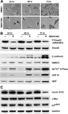
TGFβ receptor I kinase antagonism by a chemical inhibitor promotes differentiation of subconfluent BUMPT cells. To ensure that the cells continued to be subconfluent and in log-phase growth over the duration of the experiment, they were plated at 830/cm2, or 16-fold lower than in Figure 1. Cells were treated with 1 μmol/L SB431542 or vehicle and examined after 24, 48, and 72 hours. A: Formaldehyde-fixed cells were stained with toluidine blue/basic fuchsin. The cell densities correspond to cultures extracted for protein analysis in (B) and (C); magnification = original ×200. B: SDS extracts were immunoblotted for phospho-Smad2 (S465/467) and Smad2, E-cadherin, NDRG1, Na+/K+ ATPase, DPP IV, and NEP. C: SDS extracts were immunoblotted for cyclin D, c-Myc, and p27kip1.
Figure 6
Figure 6
TGFβ receptor I kinase antagonism by a chemical inhibitor increases DNA synthesis, cell proliferation, and Rb phosphorylation but concurrently accelerates the development of epithelial phenotype in subconfluent primary cultures of PT cells. Primary cultures of PT cells were seeded at low density. Following cell attachment, 2 μmol/L SB431542 or vehicle were added and the cells were examined after durations of up to 6 days. A: Cells were incubated with BrdU for 1 hour before fixation and the percentage of BrdU-labeled nuclei determined by immunofluorescence (n = 5, mean ± SE P < 0.05 for differences at 2 and 4 days). B: Cells were counted using a hemacytometer (n = 4, mean ± SE P < 0.05 for differences at 4 and 6 days). C: Phase contrast micrographs after 4 days of treatment (original magnification ×200). D: Cells were fixed after 3 days of treatment and examined by immunofluorescence for Ksp-cadherin (magnification = original ×400). E: After 12 and 24 hours of treatment, SDS extracts were immunoblotted for phospho-Smad2 (S465/467), Smad2, phospho-Rb (S608 - S601 of mouse Rb), phospho-Rb (S807/811 - S800/804 of mouse Rb), and Rb.
Figure 7
Figure 7
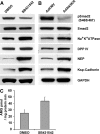
TGFβ receptor I kinase antagonism by chemical inhibitor or mutant receptor AlK5KR promotes differentiation in primary cultures of PTs. A and B: Primary cultures of PTs were seeded at low density. After attachment, 2 μmol/L SB431542 or vehicle only was added (A) or, after 24 hours of seeding, cells were infected with 5 MOI of either empty adenovirus (AdCMV) or adenovirus carrying Alk5KR (B). After 4 days (A) or 2 days (B) SDS extracts were immunoblotted for phospho-Smad2 (S465/467), Smad2, Na+K+ATPase, DPP IV, NEP, and Ksp-cadherin. C: Primary cultures of PTs were seeded at subconfluent density. After attachment, 2 μmol/L SB431542 or vehicle was added and the cells were allowed to reach confluent growth arrest. Phloridzin-sensitive, sodium-dependent glucose transport was then measured (n = 5, mean ± SE, P < 0.05 for difference between SB431542 and DMSO).
Figure 8
Figure 8
A: Contact-inhibited (day 4) cells were wounded, after which ∼200-μm wide concentric strips of epithelium remained, alternating with ∼800-μm wide wounds (left panel). Migration of “activated” and polarized surviving cells into the wounds is shown in the right panels. Formaldehyde-fixed cells were stained with toluidine blue/basic fuchsin. (B) After 6 and 12 hours of wounding, SDS extracts of wounded cells, “0 time” controls and non-wounded “time controls” were immunoblotted for phospho-Smad2 (S465/467), Smad2, TβRII, TβRI, and Smad7. C: Contact-inhibited (day 4) BM-Lux cells were wounded and incubated with 2 μmol/L SB431542 or vehicle for 6 hours before measurement of luciferase activity; mean ± SE, n = 3. P < 0.005 for all differences. D: Contact-inhibited (day 4) BM-Lux cells were wounded and 15 μg/ml of neutralizing TGFβ antibodies or non-immune IgG were added to the incubation medium, 6 hours before measurement of luciferase activity; mean ± SE, n = 3. P < 0.05. E: After 6 and 12 hours of wounding, SDS extracts of wounded cells and unwounded controls were immunoblotted for the differentiation marker NEP and E-cadherin. F: After 12 hours of wounding and treatment with SB431542 or vehicle, SDS extracts of wounded cells and unwounded controls were immunoblotted for phospho-Smad2 (S465/467), and Smad2, or the differentiation marker NEP and E-cadherin. G: Unwounded controls and wounded cells incubated for 12 hours with SB431542 or vehicle only were fixed with formaldehyde and assessed by immunofluorescence for E-cadherin.
Figure 9
Figure 9
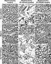
Treatment with Alk5 inhibitor SD-208 does not modify azotemia or its remission after ischemic kidney injury but ameliorates tubulo-interstitial pathology. A: Serum creatinine in control rats (vehicle and SD-208 treated rats pooled), and rats with ischemia-reperfusion (IR) treated with vehicle alone or with SD-208. B: SDS extracts of the outer stripe of outer medulla from normal rats without surgery (NK), nephrectomized controls (C), rats with ischemia-reperfusion (IR) (3, 5, and 7 days after surgery) and rats with IR treated with SD-208 (SD) 3 days after surgery were immunoblotted for phospho-Smad2 (S465/467), Smad2, TβRI, and TβRII. C: Kidney sections stained with H&E from control rat, and rats with ischemia-reperfusion treated with vehicle only or with SD-208, 14 days after surgery. Magnification = original ×100. D: Kidney sections stained with Periodic Acid Schiff stain from rats with ischemia-reperfusion treated with vehicle only or with SD-208, 14 days after surgery. The areas shown are from the outer stripe of outer medulla and were selected to highlight the type of tubulo-interstitial pathology and tubule morphology that was analyzed by morphometry and grading. Magnification = original ×200. E, F: Tubulo-interstitial index and tubule differentiation index calculated from representative photographs of the outer stripe of outer medulla. All relevant difference were significant (n = 7 for controls and vehicle treated IR; n = 6 for SD-208 treated IR); vehicle treatment versus nephrectomized control, *P < 0.001; SD-208 treatment versus vehicle treatment, **P < 0.001).
Figure 10
Figure 10
Immunohistochemistry for Ksp-cadherin, meprin, and Na+K+ATPase in kidney sections from control rats and rats with ischemia-reperfusion of the kidney treated with vehicle only or with SD-208, 14 days after surgery. Photographs are from outer stripe of outer medulla. Magnification = original ×200.
Figure 11
Figure 11
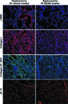
Immunofluorescence for α-SMA, collagen I, collagen IV, and endothelial marker aminopeptidase P (JG12) in cryosections from rats with ischemia-reperfusion of the kidney treated with vehicle only or with SD-208, 14 days after surgery. In the third panel from top, sections were doubly stained for type IV collagen (Green) and monocyte marker, EDI (Red). Photographs are from outer stripe of outer medulla. Magnification = original ×200.

Similar articles

Cited by

References

    1. Nony PA, Schnellmann RG. Mechanisms of renal cell repair and regeneration after acute renal failure. J Pharmacol Exp Ther. 2003;304:905–912. - PubMed
    1. Bonventre JV. Dedifferentiation and proliferation of surviving epithelial cells in acute renal failure, J Am Soc Nephrol. 2003;14 Suppl 1:S55–S61. - PubMed
    1. Devarajan P. Update on mechanisms of ischemic acute kidney injury. J Am Soc Nephrol. 2006;17:1503–1520. - PubMed
    1. Siegel PM, Massague J. Cytostatic and apoptotic actions of TGF-beta in homeostasis and cancer. Nat Rev Cancer. 2003;3:807–821. - PubMed
    1. Derynck R, Zhang YE. Smad-dependent and Smad-independent pathways in TGF-beta family signalling. Nature. 2003;425:577–584. - PubMed

Publication types

MeSH terms

Substances