. 2009 Jun 5;284(23):15676-84.
doi: 10.1074/jbc.M809787200. Epub 2009 Apr 2.

Biological functions of miR-29b contribute to positive regulation of osteoblast differentiation

Affiliations

Biological functions of miR-29b contribute to positive regulation of osteoblast differentiation

Zhaoyong Li et al. J Biol Chem. .

Erratum in

Abstract

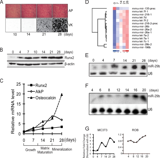
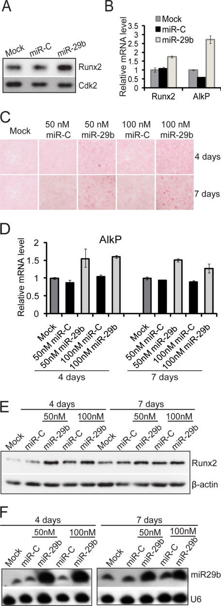
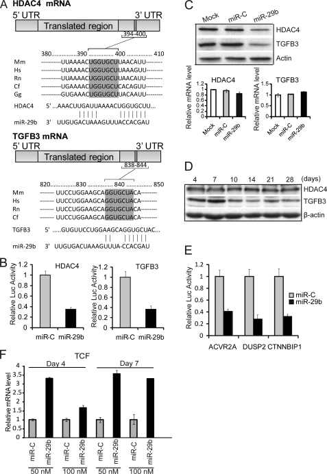
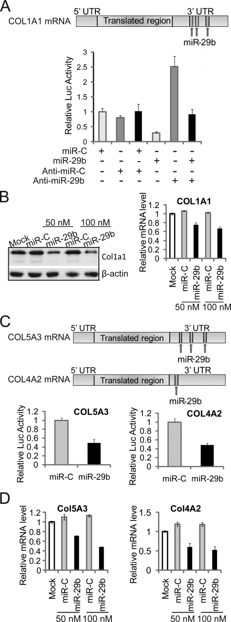
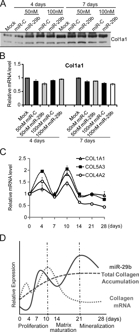
Bone tissue arises from mesenchymal cells induced into the osteoblast lineage by essential transcription factors and signaling cascades. MicroRNAs regulate biological processes by binding to mRNA 3'-untranslated region (UTR) sequences to attenuate protein synthesis. Here we performed microRNA profiling and identified miRs that are up-regulated through stages of osteoblast differentiation. Among these are the miR-29, miR-let-7, and miR-26 families that target many collagens and extracellular matrix proteins. We find that miR-29b supports osteoblast differentiation through several mechanisms. miR-29b decreased and anti-miR-29b increased activity of COL1A1, COL5A3, and COL4A2 3'-UTR sequences in reporter assays, as well as endogenous gene expression. These results support a mechanism for regulating collagen protein accumulation during the mineralization stage when miR-29b reaches peak levels. We propose that this mechanism prevents fibrosis and facilitates mineral deposition. Our studies further demonstrate that miR-29b promotes osteogenesis by directly down-regulating known inhibitors of osteoblast differentiation, HDAC4, TGFbeta3, ACVR2A, CTNNBIP1, and DUSP2 proteins through binding to target 3'-UTR sequences in their mRNAs. Thus, miR-29b is a key regulator of development of the osteoblast phenotype by targeting anti-osteogenic factors and modulating bone extracellular matrix proteins.


Figures

FIGURE 1.
FIGURE 1.
miR-29b expression profile during MC3T3 osteoblast differentiation. A, MC3T3 preosteoblasts cells cultured in differentiation medium for 28 days. Histochemical staining of alkaline phosphatase (AP) activity and von Kossa (VK) for mineral deposition at 10, 14, 21, and 28 days is shown. B, Western blot for Runx2 protein which increases during osteoblast differentiation. β-Actin protein was used as control. C, quantitative mRNA normalized by GAPDH for osteoblastic markers Runx2, AlkP, and osteocalcin on selected days during MC3T3 osteoblast differentiation used in miR profiling studies. D, significantly changed microRNAs that putatively target extracellular cellular matrix genes. Total RNA of MC3T3 cells during differentiation time points (0, 7, 14, 21, and 28 days) was used for miRNA microarray analysis. Relative fold changes of the microRNAs were hierarchically clustered by using dChip software. E and F, representative Northern blot analysis of miR-29b using total RNA isolated from mouse (MC3T3) (E) and rat primary osteoblasts (F), which were induced to differentiate. U6 RNA was used as a loading control. G, densitometric quantitation of miR-29b in indicated osteoblasts normalized to U6. The average volumes from two different MC3T3 studies are shown, and one time course from primary osteoblasts is shown.
FIGURE 2.
FIGURE 2.
miR-29b activates osteogenic differentiation in osteoblasts. A and B, MC3T3 osteoblast cells were transfected with miR-29b mature miRNA oligonucleotides (50 nm), miRNA negative Control 1 (miR-C) (50 nm), or transfection reagent only (Mock) for 48 h for protein and RNA analysis. Both expression of Runx2 protein (A) and Runx2 and AlkP mRNA were up-regulated in miR-29b overexpressing cells (B). C–E, miR-29b promotes osteoblast differentiation. After transfection with 50 or 100 nm miR-29b or miRNA Control (miR-C) for 72 h, confluent MC3T3 cells were treated with α-minimum essential medium containing 10 mm β-glycerophosphate and 50 μg/ml ascorbic acid for 4 or 7 days. The cells were fixed in 2% paraformaldehyde for histochemical detection of alkaline phosphatase (C). The mRNA level of AlkP was detected by quantitative PCR normalized by GAPDH (D). Runx2 and β-actin protein (as control) were detected by Western blot (E). F, Northern blot analysis shows endogenous (miR-C lanes) and overexpressed miR-29b levels in the samples shown in E. U6RNA is the loading control.
FIGURE 3.
FIGURE 3.
miR-29b targets negative regulators of osteoblast differentiation. A, one putative target site of miR-29b predicted by the TargetScan program was contained in the HDAC4 or TGFβ3 mRNA 3′-UTR, and both were highly conserved in vertebrate species. The numbers represent the position of the “seed region” match to miR-29b within the UTR sequences. Mm, mouse; Hs, human; Rn, rat; Cf, dog; Gg, chicken. B, MC3T3 cells were co-transfected with 100 nm RNA of miR-29b or miRNA control (miR-C), phRL-null (Renilla plasmid), and the luciferase constructs (firefly) carrying HDAC4 3′-UTR or TGFβ3 3′-UTR. Luciferase assays were performed 36 h after transfection. The ratio of reporter Firefly to control Renilla luciferase in relative luminescence units was plotted. The error bars represent the standard error for n = 3. C, 30–50% confluent osteoblast cell line MC3T3 was transfected with 100 nm miR-29b RNA, miRNA negative control, or transfection reagent only (Mock). Protein and RNA analysis was performed at 48 h after transfection. HDAC4, TGFβ3, and β-actin (as control) protein were monitored by Western blot. HDAC4 and TGFβ3 mRNA level were detected by quantitative RT-PCR normalized by GAPDH. D, HDAC4 and TGFβ3 expression during osteoblast differentiation. Differentiating MC3T3 time points were analyzed by Western for HDAC4, TGFβ3, and β-actin (as control) protein. E, MC3T3 cells were co-transfected with reagents in the panel, and the luciferase constructs carrying 3′-UTR of activin A receptor type IIA, DUSP2, or CTNNBIP1. Functional activity of the luciferase reporter plasmid was assessed as described above for B. The values represent the means ± S.E. for n = 3. F, shown is the effect of miR-29b overexpression on endogenous mRNA levels of TCF1, a marker of Wnt/β-catenin signaling that is inhibited by CTNNBIP1 (see supplemental Fig. S2, B). TCF1 is increased during osteoblast differentiation (days 4 and 7). The experimental design is described for Fig. 2C.
FIGURE 4.
FIGURE 4.
miR-29b functions as an attenuator for collagen in differentiated osteoblasts. A, Col1a1 is a direct target of miR-29b. Three putative target sites of miR-29b are predicted to be in Col1a1 mRNA 3′-UTR by TargetScan, PicTar, and miRanda program (upper panel). MC3T3 cells were co-transfected with the luciferase constructs, phRL-null (Renilla plasmid) and 100 nm RNA oligonucleotides of miR-29b, miRNA negative control (miR-C), Anti-miR-29b or Anti-miR negative control (Anti-miR-C), as shown in the lower panel. Luciferase assays were performed 36 h after transfection. The ratio of reporter (firefly) to control phRL-null plasmid (Renilla) in relative luminescence units was plotted. The error bars represent the standard error for n = 3. B, miR-29b overexpression inhibits Col1a1 expression in osteoblasts. 30–50% confluent MC3T3 osteoblast cells were transfected with 50 or 100 nm miR-29b RNA oligonucleotides or miRNA negative control (miR-C). The cells were harvested for protein and RNA analysis at 48 h after transfection. Col1A1 and β-actin (as control) protein were monitored by Western blot (left panel) and Col1a1 mRNA level was detected by quantitative RT-PCR normalized by GAPDH (right panel). C, miR-29b target Col5a3 and Col4a2 directly by mRNA 3′-UTR. Three and one putative target site for miR-29b is contained in the Col5a3 and Col4a2 mRNA 3′-UTR, respectively. MC3T3 cells were co-transfected with luciferase constructs carrying Col5a3 or Col4a2 3′-UTR, phRL-null, miR-29b, or miRNA negative control (miR-C). Luciferase activity assays and analysis were described as in A. D, mRNA of Col5a3 and Col4a2 were detected in MC3T3 overexpressed with RNA oligonucleotides as described for B.
FIGURE 5.
FIGURE 5.
miR-29b regulates collagen expression in differentiated osteoblasts. A and B, miR-29b has no effect on collagen gene expression in early osteoblast differentiation. MC3T3 cells were transfected with 50 or 100 nm miR-29b RNA mimics or miRNA Control (miR-C) and cultured with differentiation-inducing medium described as in Fig. 2C. Col1a1 protein was monitored by Western blot (A), and mRNA levels detected by quantitative PCR normalized by GAPDH (B). C, mRNA expression levels of Col1a1, Col5a3, and Col4a2 during MC3T3 osteogenic differentiation. Differentiation was initiated 3 days after plating cells (day 0). The cells were harvested at indicated time points for quantitative RT-PCR normalized to GAPDH. D, reciprocal expression of miR-29b and COL Type I (days 21–28); miR-29b quantitated from Northern blot analyses; total collagen measured from densitometry of Western blots; COL Type I mRNA determined by quantitative PCR.

References

    1. Lian J. B., Stein G. S., Javed A., van Wijnen A. J., Stein J. L., Montecino M., Hassan M. Q., Gaur T., Lengner C. J., Young D. W. ( 2006) Rev. Endocr. Metab. Disord. 7, 1– 16 - PubMed
    1. Stein G. S., Zaidi S. K., Stein J. L., Lian J. B., van Wijnen A. J., Montecino M., Young D. W., Javed A., Pratap J., Choi J. Y., Ali S. A., Pande S., Hassan M. Q. ( 2008) J. Cell. Biochem. 104, 2016– 2026 - PMC - PubMed
    1. Komori T. ( 2006) J. Cell. Biochem. 99, 1233– 1239 - PubMed
    1. Nakashima K., Zhou X., Kunkel G., Zhang Z., Deng J. M., Behringer R. R., de Crombrugghe B. ( 2002) Cell 108, 17– 29 - PubMed
    1. Bodine P. V., Komm B. S. ( 2006) Rev. Endocr. Metab. Disord. 7, 33– 39 - PubMed

Publication types