Review
. 2009 Jun;296(6):G1151-66.
doi: 10.1152/ajpgi.90649.2008. Epub 2009 Apr 2.

A guide to Ussing chamber studies of mouse intestine

Affiliations
Review

A guide to Ussing chamber studies of mouse intestine

Lane L Clarke. Am J Physiol Gastrointest Liver Physiol. 2009 Jun.

Abstract

The Ussing chamber provides a physiological system to measure the transport of ions, nutrients, and drugs across various epithelial tissues. One of the most studied epithelia is the intestine, which has provided several landmark discoveries regarding the mechanisms of ion transport processes. Adaptation of this method to mouse intestine adds the dimension of investigating genetic loss or gain of function as a means to identify proteins or processes affecting transepithelial transport. In this review, the principles underlying the use of Ussing chambers are outlined including limitations and advantages of the technique. With an emphasis on mouse intestinal preparations, the review covers chamber design, commercial equipment sources, tissue preparation, step-by-step instruction for operation, troubleshooting, and examples of interpretation difficulties. Specialized uses of the Ussing chamber such as the pH stat technique to measure transepithelial bicarbonate secretion and isotopic flux methods to measure net secretion or absorption of substrates are discussed in detail, and examples are given for the adaptation of Ussing chamber principles to other measurement systems. The purpose of the review is to provide a practical guide for investigators who are new to the Ussing chamber method.


Figures

Fig. 1.
Fig. 1.
Classic Ussing chamber design. A: assembled apparatus with water-jacketed reservoirs, Ussing chamber (intestinal preparation is mounted vertically; red arrow) secured by thumb wheel screws (white arrow), and electrodes attached to voltage clamp head stage. B: close-up view of voltage (Vt)-measuring and short-circuit current (Isc)-passing pathways. Calomel half-cell electrodes used for Vt measurements are connected by 3 M KCl salt bridges at each side of intestinal preparation (red arrow). Ag-AgCl electrodes used for Isc passing across the intestinal preparation are connected to the chamber by Krebs bicarbonate Ringer (KBR) salt bridges at each end of the chamber. C: schematic cut-away diagram of Vt and Isc circuits of the Ussing chamber. Short circuiting (Isc) is provided by an automatic voltage clamp (symbol). Note superfusion circulation of KBR is driven by gas lift using 95%O2-5% CO2. Intestinal preparation (red disc) separates the mucosal and serosal baths.
Fig. 2.
Fig. 2.
Murine small intestinal preparation (black arrow) mounted on pins of the serosal half of an acrylic Ussing chamber. Note corresponding holes for pins in opposing chamber half and guideposts for assembly of chamber.
Fig. 3.
Fig. 3.
Modular Ussing chamber systems. Commercially available systems include acrylic blocks where the chamber is made integral with reservoirs and gas lift. A: modular system available from Navicyte (Harvard Apparatus). B: modular system available from Physiologic Instruments. Inset: “slider” with pins and aperture for mounting murine intestinal preparations.
Fig. 4.
Fig. 4.
Seromuscular stripping method for murine intestine. A: intestinal preparation (duodenum; note pylorus at upper end) is pinned mucosal side down to a cooled Sylgard-cast plate under a dissection stereomicroscope with bottom illumination (magnification = ×7) B: scalpel blade is used to score the seromuscular layer (white dashed line; note occasional full thickness cuts). The seromuscular layer is reflected using fine microforceps by blunt dissection (white arrow). C: higher magnification of reflected seromuscular layer (magnification = ×32).
Fig. 5.
Fig. 5.
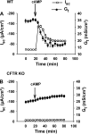
Effect of cystic fibrosis transmembrane conductance regulator (CFTR) activation on transepithelial conductance (Gt). A: time course of short-circuit current (Isc) and transepithelial conductance responses of wild-type (WT) murine jejunum (not seromuscular stripped) to treatment with a cocktail of 10 μM forskolin and 100 μM IBMX (cAMP). Note that the decline in Gt after cAMP treatment slightly precedes the decline in the Isc from its maximal value (n = 6). B: time course of Isc and Gt responses of CFTR knockout (KO) murine jejuna to cAMP treatment (n = 6). [Modified from Gawenis et al. (15).]
Fig. 6.
Fig. 6.
Common problems encountered in Ussing chamber studies of murine intestine. A: representation of forskolin (Forsk) contamination in Ussing chamber apparatus. Short-circuit (Isc) measurements during sequential additions of forskolin (10 μM, mucosal and serosal addition) and glucose (10 mM, mucosal addition). Black trace shows normal Isc response to forskolin and glucose additions. *Red trace shows elevated baseline Isc and reduced response (ΔIsc) to forskolin addition attributable to forskolin contamination from previous experiments. Although the system was thoroughly rinsed and washed, soft plastic tubing was not replaced and served as the source of forskolin contamination. B: time period required for 22Na added to the mucosal bath to attain a steady-state flux into the serosal bath. Samples taken from the serosal (sink) bath after addition of 3 μCi addition of 22Na into the mucosal (source) bath attained a steady-state rate after 20–30 min. C: representation showing diffusion barrier to bumetanide (50 μM, serosal addition) attributable to the seromuscular layer. Black trace shows sequential Isc responses to forskolin (10 μM, mucosal and serosal addition), bumetanide (50 μM, serosal addition), and glucose (10 mM, mucosal addition) in murine jejunal preparation stripped of the seromuscular layers. Bumetanide effect (inhibition of electrogenic Cl secretion) occurs within 5 min. Red trace shows identical experiment using a whole-thickness murine jejunal preparation. *Note that the bumetanide inhibition is only 40% complete at 5 min.
Fig. 7.
Fig. 7.
Difficulties with interpretation of Ussing chamber experiments performed using murine intestine. A: effect of nonphysiological buffer (HEPES) on changes of intracellular pH (pHi) induced by application of the Pept1 substrate (glycine-sarcosine, gly-sar). Gly-sar (25 mM) induces an Isc response in the Ussing chamber attributable to H+-dipeptide transport via Pept1. Measurement of pHi in the upper villous epithelium was performed on murine duodenum situated in a horizontal Ussing chamber. In the presence of physiological buffer, i.e., CO2/HCO3 in KBR (•), pHi is unaffected by gly-sar transport. In contrast, in the presence of HEPES-buffered Ringers solution (○), pHi becomes markedly acidic during gly-sar exposure. B: effect of sequential additions of cadmium (100 μM, mucosal addition) and glucose (10 mM, mucosal addition) on the Isc and Gt of murine jejunum. Cd2+ treatment was predicted to reduce the Isc as a means to evaluate the contribution of CLC-2 Cl channels to the residual Isc in CFTR knockout mouse jejunum. Although the experiment could be interpreted positively on the basis of the abrupt decrease in Isc, the large increases in Gt (as indicated by the upward deflections of the Isc in response to repeating 5 mV command pulses) and the lack of response to glucose indicated metabolic insult and loss of tissue viability.
Fig. 8.
Fig. 8.
Effects of unbalanced physical forces on the Gt of murine intestine in Ussing chamber experiments. A: effect of hypertonic medium (+300 mOsm/l mannitol) in either the mucosal (•) or serosal (○) bath on the Gt of murine jejunum. Note that mucosal hypertonicity results in an abrupt decrease in Gt attributable to the collapse of the lateral intercellular spaces, whereas serosal hypertonicity moderately increases Gt. B: effect of small increases in serosal hydrostatic pressure on the Gt of murine jejunum. Either 0 (no gradient), +2.9, or +3.7 mmHg was applied by increasing the fluid height of the serosal bath for 10 min on mouse jejunum in the Ussing chamber. Changes in Gt under all conditions were adjusted for the mean decrease in Gt (−9.9 mS/cm2) that normally occurs when no hydrostatic pressure gradient is applied. a,bMeans with different letters are significantly different (P < 0.05). [Modified from Gawenis et al. (15).]
Fig. 9.
Fig. 9.
Flow chart for performing 22Na36Cl flux studies of murine intestine.
Fig. 10.
Fig. 10.
A: changes in pH of an unbuffered luminal solution during pH stat measurements of fast and slow rates of HCO3 secretion. Traces showing changes in pH of the luminal solution during pH stat studies. Note the pH excursions about the target pH 7.4 (solid line) for an epithelium with high HCO3 secretory flux (Jsm) (pink trace and arrows) and low HCO3 secretory flux (blue trace and arrow). [Modified from Krouse et al. (24).] B: pH stat measurements of HCO3 secretion (JsmHCO3) and Isc across WT murine duodenum during sequential treatments with forskolin (cAMP; 10 μM, mucosal and serosal addition) and bumetanide (Bumet; 50 μM, serosal addition). Compare the rapid increase in Isc (blue line and symbols) with the slower changes in JsmHCO3 (green line and symbols) after cAMP treatment. The different time courses for Isc and JsmHCO3 demonstrate the lag in pH measurement attributable to mixing of the superfusate. Bumetanide treatment inhibits Cl secretion and reduces the Isc without affecting the JsmHCO3. Vertical dashed lines indicate 3 successive steady-state flux periods (30 min each). n = 5–7 duodenal preparations.
Fig. 11.
Fig. 11.
Adaptation of microfluorimetry to measurement of pHi in intact villous epithelium of murine jejunum using a horizontal Ussing chamber system. A: duodenal villi loaded with BCECF, a pH-sensitive ratiometric dye, and observed using a water-immersion objective. B: higher magnification of a single villus immobilized for pHi measurements using a nylon mesh overlay (not seen). C: higher magnification of upper villous epithelium with regions of interest for capture of fluorescence emission. [Reprinted by permission from Simpson et al. (44).]

Comment in

References

    1. Akiba Y, Furukawa O, Guth PH, Engel E, Nastaskin I, Kaunitz JD. Acute adaptive cellular base uptake in rat duodenal epithelium. Am J Physiol Gastrointest Liver Physiol 280: G1083–G1092, 2001. - PubMed
    1. Argenzio RA, Whipp SC. Effect of theophylline and heat-stable enterotoxin of Escherichia coli on transcellular and paracellular ion movement across isolated porcine colon. Can J Physiol Pharmacol 61: 1138–1148, 1983. - PubMed
    1. Binder HJ, Rawlins CL. Electrolyte transport across isolated large intestinal mucosa. Am J Physiol 225: 1232–1239, 1973. - PubMed
    1. Binder HJ, Sandle GI. Electrolyte absorption and secretion in the mammalian colon. In: Physiology of the Gastrointestinal Tract, edited by Johnson LR. New York: Raven, 1987, pp. 1389–1418.
    1. Bookstein C, Depaoli AM, Xie Y, Niu P, Musch MW, Rao MC, Chang EB. Na+/H+ exchanger, NHE-1 and NHE-3, of rat intestine. J Clin Invest 93: 106–113, 1994. - PMC - PubMed

Publication types

LinkOut - more resources