. 2009 Jun;296(6):G1277-86.
doi: 10.1152/ajpgi.90257.2008. Epub 2009 Apr 2.

Role of iron in the pathogenesis of cysteamine-induced duodenal ulceration in rats

Affiliations

Role of iron in the pathogenesis of cysteamine-induced duodenal ulceration in rats

Tetyana Khomenko et al. Am J Physiol Gastrointest Liver Physiol. 2009 Jun.

Abstract

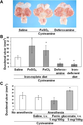
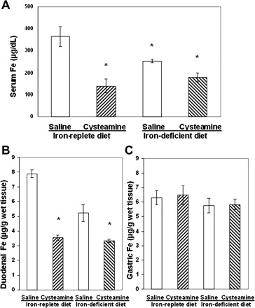
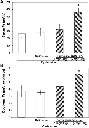
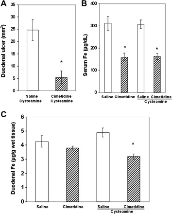
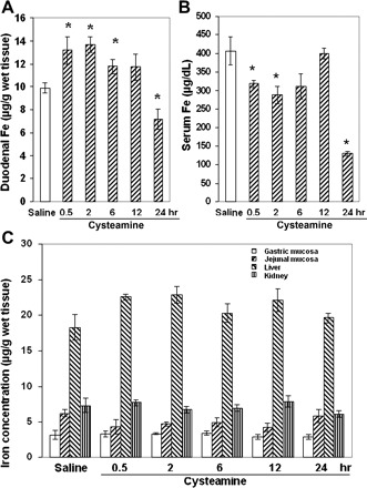
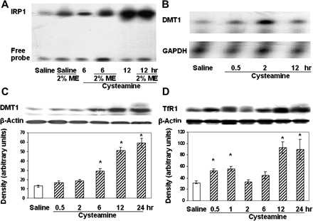
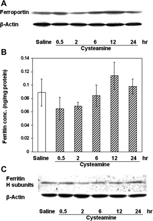
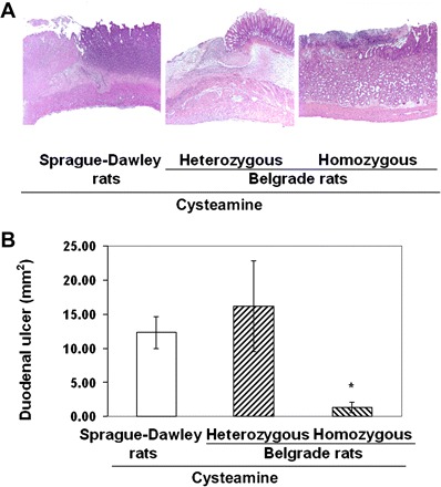
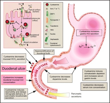
Cysteamine induces perforating duodenal ulcers in rats within 24-48 h. This reducing aminothiol generates hydrogen peroxide in the presence of transition metals (e.g., ferric iron), producing oxidative stress, which may contribute to organ-specific tissue damage. Since most intestinal iron absorption takes place in the proximal duodenum, we hypothesized that cysteamine may disrupt regulation of mucosal iron transport, and iron may facilitate cysteamine-induced duodenal ulceration. We show here that cysteamine-induced ulceration was aggravated by pretreatment of rats with Fe(3+) or Fe(2+) compounds, which elevated iron concentration in the duodenal mucosa. In contrast, feeding rats an iron-deficient diet was associated with a 4.6-fold decrease in ulcer formation, accompanied by a 34% decrease (P < 0.05) in the duodenal mucosal iron concentration. Administration of deferoxamine inhibited ulceration by 65%. We also observed that the antiulcer effect of H2 receptor antagonist cimetidine included a 35% decrease in iron concentration in the duodenal mucosa. Cysteamine-induced duodenal ulcers were also decreased in iron-deficient Belgrade rats (P < 0.05). In normal rats, cysteamine administration increased the iron concentration in the proximal duodenal mucosa by 33% in the preulcerogenic stage but at the same time decreased serum iron (P < 0.05). Cysteamine also enhanced activation of mucosal iron regulatory protein 1 and increased the expression of divalent metal transporter 1 mRNA and protein. Transferrin receptor 1 protein expression was also increased, although mucosal ferroportin and ferritin remained almost unchanged. These results indicate an expansion of the intracellular labile iron pool in the duodenal mucosa, increasing its susceptibility to oxidative stress, and suggest a role for iron in the pathogenesis of organ-specific tissue injury such as duodenal ulcers.


Figures

Fig. 1.
Fig. 1.
Stimulation or inhibition of cysteamine-induced duodenal ulcerogenesis in rats with increased (Fe+3, Fe+2) or decreased iron levels. A: gross appearance of duodenal ulcers 48 h after cysteamine administration. Animals were pretreated with saline (n = 7), FeSO4 (n = 10), or deferoxamine (n = 10) 15 min before each dose of cysteamine. B: duodenal ulcer size was scored 48 h after cysteamine in animals pretreated with saline, FeCl3, FeSO4, or deferoxamine or fed an iron-deficient diet (chronic experiments, n = 5). The duodenal ulcer crater dimensions were measured in millimeters, and the ulcer areas were calculated using the ellipsoid formula. C: aggravation of duodenal ulcers by ferric gluconate. Duodenal ulcers were compared without and with anesthesia by isoflurane inhalation. Saline and ferric gluconate injections were given intravenously (i.v.) 30 min before cysteamine administration. Results are expressed as means ± SE (*P < 0.01, n = 5).
Fig. 2.
Fig. 2.
Comparison of the iron concentrations in serum and duodenal mucosa of rats fed with either an iron-replete diet or a low-iron diet for 4 wk before cysteamine treatment. A: serum iron concentration 48 h after saline or cysteamine administration in rats fed with iron-replete or iron-deficient diets. B: decreased iron concentration in 2.5 cm of ulcerated duodenal mucosa 48 h after cysteamine in rats fed iron-replete or iron-deficient diets compared with saline treated animals (*P < 0.05, n = 5 for each group). C: iron concentration in gastric mucosa did not change in animals fed a low-iron diet or an iron-replete diet.
Fig. 3.
Fig. 3.
Treatment of anesthetized rats with ferric gluconate (5 mg/100 g) 0.5 h before cysteamine increased serum iron concentration (A) and iron concentration in the proximal duodenum (B) vs. animals treated with cysteamine only, without anesthesia (first column) and anesthetized rats pretreated with saline (i.v.) or ferric gluconate (1 mg/100 g); *P < 0.05, n = 5 for each group.
Fig. 4.
Fig. 4.
Effect of the histamine H2 receptor antagonist cimetidine on cysteamine-induced duodenal ulcers. A: cimetidine inhibited cysteamine-induced duodenal ulcers. The rats were pretreated with cimetidine 3 and 0.5 h before cysteamine and euthanized 3 h or 48 h after the first cysteamine dose. B: cimetidine decreased serum iron concentration alone or with cysteamine- vs. only saline-treated animals (3 h after cysteamine administration). C: cimetidine given to rats before cysteamine decreased iron concentration in the proximal duodenum vs. saline- or cysteamine-treated rats (3 h after cysteamine administration). Results are expressed as means ± SE (*P < 0.01, n = 5 for each group).
Fig. 5.
Fig. 5.
Effect of cysteamine on iron concentration in different organs of rats within the preulcerogenic stage of duodenal ulcer development. A: temporal increase of iron concentration in the proximal duodenum within the first 12 h after cysteamine compared with saline administration. B: iron concentration in rat serum within the first 24 h after cysteamine administration. C: iron concentrations in gastric, jejunal, liver, and kidney tissue of rats treated with cysteamine or saline (*P < 0.05, n = 5 for each group).
Fig. 6.
Fig. 6.
Effect of the duodenal ulcerogen cysteamine on iron regulatory protein 1 (IRP1) activity and divalent metal transporter 1 (DMT1) and transferrin receptor 1 (TfR1) expression in rat duodenal mucosa during the early stages of duodenal ulcer development. A: IRP1-iron-responsive element (IRE) binding activity in the rat duodenal mucosa 6 and 12 h after treatment with cysteamine or saline. 2-Mercaptoethanol (ME, 2%) was added to protein samples to maximize IRE binding activity. The experiments were performed at least 5 times, and representative results are shown. B: RNase protection assay showing DMT1 and GAPDH mRNA expression in the duodenal mucosa 0.5, 2, and 12 h after treatment with cysteamine. C: DMT1 protein expression in rat duodenal mucosa was detected by Western blot at various times after cysteamine treatment. Samples from saline-treated rats were used as controls. D: TfR1 was detected by Western blot in rats treated with saline or cysteamine for various periods of time. The experiments were performed at least 4 times, and representative results are shown.
Fig. 7.
Fig. 7.
Ferroportin and ferritin protein expression in the duodenal mucosa of rats treated with cysteamine. A: ferroportin was detected by Western blot. B: cysteamine did not induce significant changes in ferritin concentration. C: ferritin (heavy subunit) was also detected by Western blot in rat duodenal mucosa after cysteamine treatment in the same group of rats as used in B. The experiments were performed at least 4 times, and representative results are shown.
Fig. 8.
Fig. 8.
Cysteamine-induced duodenal ulcers in Sprague-Dawley rats and in heterozygous (+/b) and homozygous (b/b) Belgrade rats. A: light microscopy of duodenal ulcers in rats treated with cysteamine (25 mg/100 g) in Sprague-Dawley rats (deep ulcer, left), heterozygous (deep ulcer, middle), and homozygous Belgrade rats (small ulcer, right) (hematoxylin and eosin staining). B: duodenal ulcer crater dimensions in the same groups of rats were measured in millimeters, and the ulcer areas were calculated using the ellipsoid formula. Size of duodenal ulcers is shown as mm2 (means ± SE, *P < 0.01).
Fig. 9.
Fig. 9.
Proposed role of iron in the pathogenesis of cysteamine-induced ulceration in the proximal duodenum of rats. Cysteamine may promote dysregulation of iron absorption by simultaneously increasing iron uptake by duodenal mucosal enterocytes and inhibiting transfer of mucosal iron to the systemic circulation. Cysteamine may enhance iron availability for uptake by duodenal enterocytes through 1) direct reduction of Fe3+ to Fe2+, 2) increased gastric acid secretion following elevation of gastrin levels and inhibition of somatostatin, 3) decreased bicarbonate secretion, and 4) delayed gastric emptying. Within duodenal enterocytes (illustrated here by a composite drawing representing both villus enterocytes and crypt cells expressing TfR1), the reducing agent cysteamine activates IRP1, which normally would be inactivated by increased intracellular iron. Activation of IRP1 in turn would increase DMT1 (IRE) expression, which otherwise would be decreased in the presence of increased intracellular iron, and the increased DMT1 would lead to increased iron transport across the apical membrane, thus further expanding the labile iron pool. IRP1 activation also leads to translational inhibition of ferritin mRNA, which typically would otherwise be increased, and stabilization of TfR1 mRNA, which normally should be decreased, in the presence of increased intracellular iron. TfR1 may take up circulating transferrin-bound iron across the basolateral membrane of crypt cells, thereby also expanding the labile iron pool. Under the conditions of these experiments, however, ferritin protein levels were not affected, suggesting possible competing effects of increased IRP1 activation by cysteamine and increased labile iron pool levels. Although ferroportin 1 mRNA contains IRE or IRE-like moieties, suggesting the possibility of posttranscriptional regulation, the functionality of ferroportin IREs is unclear, and cysteamine administration did not change ferroportin levels. However, iron must be oxidized to Fe3+ for transfer from the enterocyte to the systemic circulation, and the reducing effect of cysteamine may compete with this step, thereby decreasing iron transport across the basolateral membrane and favoring increased intracellular iron levels. Taken together, these processes may expand the labile iron pool and thereby potentiate oxidative stress, which in turn may trigger duodenal ulceration.

Similar articles

Cited by

References

    1. Adamama-Moraitou K, Rallis T, Papasteriadis A, Roubies N, Kaldrimidou H. Iron, zinc, and copper concentration in serum, various organs, and hair of dogs with experimentally induced exocrine pancreatic insufficiency. Dig Dis Sci 46: 1444–1457, 2001 - PubMed
    1. Adler RS, Gallagher GT, Szabo S. Duodenal ulcerogens cysteamine and propionitrile decrease duodenal neutralization of acid in the rat. Dig Dis Sci 288: 716–723, 1983 - PubMed
    1. Anderson GJ, Vulpe CD. Regulation of intestinal iron transport. In: Molecular and Cellular Iron Transport, edited by D. Templeton New York: Marcel Dekker, 2001, pp. 559–596.
    1. Aziz N, Munro HN. Iron regulates ferritin mRNA translation through a segment of its 5′ untranslated region. Proc Natl Acad Sci USA 84: 8478–8482, 1987 - PMC - PubMed
    1. Berruyer C, Martin FM, Castellano R, Macone A, Malergue F, Garrido-Urbani S, Millet V, Imbert J, Dupre S, Pitari G, Naquet P, Galland F. Vanin-1−/− mice exhibit a glutathione-mediated tissue resistance to oxidative stress. Mol Cell Biol 24: 7214–7224, 2004 - PMC - PubMed

Publication types

MeSH terms