. 2009 May 8;324(5928):794-7.
doi: 10.1126/science.1171085. Epub 2009 Apr 2.

Apicomplexan parasites co-opt host calpains to facilitate their escape from infected cells

Affiliations

Apicomplexan parasites co-opt host calpains to facilitate their escape from infected cells

Rajesh Chandramohanadas et al. Science. .

Abstract

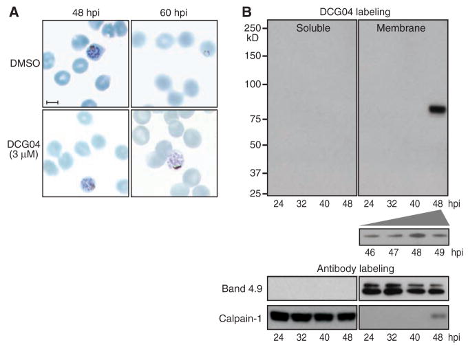
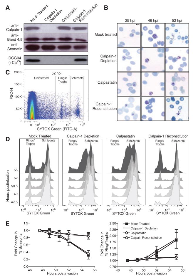
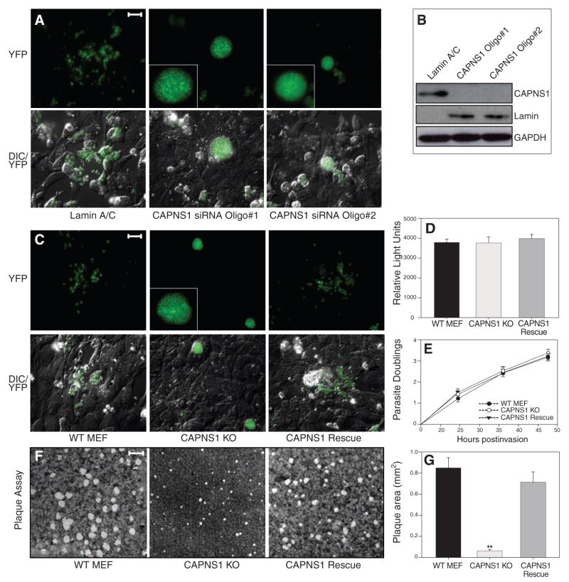
Apicomplexan parasites, including Plasmodium falciparum and Toxoplasma gondii (the causative agents of malaria and toxoplasmosis, respectively), are responsible for considerable morbidity and mortality worldwide. These pathogenic protozoa replicate within an intracellular vacuole inside of infected host cells, from which they must escape to initiate a new lytic cycle. By integrating cell biological, pharmacological, and genetic approaches, we provide evidence that both Plasmodium and Toxoplasma hijack host cell calpain proteases to facilitate parasite egress. Immunodepletion or inhibition of calpain-1 in hypotonically lysed and resealed erythrocytes prevented the escape of P. falciparum parasites, which was restored by adding purified calpain-1. Similarly, efficient egress of T. gondii from mammalian fibroblasts was blocked by either small interfering RNA-mediated suppression or genetic deletion of calpain activity and could be restored by genetic complementation.


Figures

Fig. 1
Fig. 1
Host calpain-1 is associated with P. falciparum egress. (A) Human RBCs were infected with 3D7 strain parasites, synchronized in sorbitol, treated at 42 hours postinfection (hpi) with either dimethyl sulfoxide (control) or 3 μM DCG04, and fixed and stained 6 to 18 hours later. Normal schizonts were evident in both samples at 48 hours, but whereas controls emerged to establish new ring stage infections by 60 hours, parasites treated with DCG04 appeared unable to egress, remaining arrested as schizonts. Scale bar, 5 μm. (B) Synchronized infected cultures were treated with 3 μM DCG04 for 2 hours beginning at various times throughout the intraerythrocytic life cycle, followed by incubation with 0.02% saponin to permeabilize the RBC and parasitophorous vacuole membrane (but not parasites). Parasites were removed by centrifugation, and the remaining material fractionated to yield a host + parasitophorous vacuole membrane pellet and a soluble fraction. Blotting identified proteins biotinylated by DCG04 (labeling any active cysteine protease), as well as the RBC membrane marker band 4.9, and calpain-1 (a cytoplasmic RBC protein when inactive). The single prominent ~80-kD band observed in membranes isolated from RBCs harboring schizont-stage parasites was identified as host cell calpain-1 (table S1).
Fig. 2
Fig. 2
Host calpain-1 is required for P. falciparum egress. Uninfected RBCs were hypotonically lysed, immunodepleted with monoclonal anti–calpain-1 conjugated to Sepharose (or mock-treated with Sepharose beads only), and resealed under hypotonic conditions in the presence of 1 μM calpastatin, purified calpain-1, or buffer alone. (A) Immunoblots showed specific removal of calpain-1 in immunodepleted RBCs and reconstitution with purified enzyme; equal loading was confirmed with the use of anti-band 4.9 and anti-stomatin. Labeling of resealed RBCs with 5 μM DCG04 in the presence of calcium demonstrated that calpain activity was eliminated by either immunodepletion or calpastatin loading; activity was restored by reconstitution with exogenous calpain-1. (B) Purified schizont-stage parasites were mixed with resealed RBCs and incubated for various times before fixation. Giemsa-stained smears showed incomplete egress from calpain-depleted or calpastatin-loaded cells, but proper egress from RBCs reconstituted with purified calpain-1. Scale bar = 5 μm. (C) During late schizogony, infected resealed RBCs were harvested at 90-min intervals, fixed, and stained for flow cytometry based on forward scatter (FSC-H) and parasite DNA content (SYTOX-Green). Boxes indicate gates defining uninfected cells, rings/trophozoites, and schizont-stage parasites (sample plot; see fig. S2B). (D) Flow cytometric data was gated to exclude uninfected erythrocytes and converted to a two-dimensional plot highlighting the progress of calpain-reconstituted cultures from schizonts to ring-stage infection as efficiently as mock treated controls, in contrast to the arrest of calpain-depleted and calpastatin-treated cells in schizogony, indicating an egress defect. (E) Quantitation of flow cytometric data (percentage of rings/trophozoites or schizonts relative to time t = 0, ± SE, from four independent experiments).
Fig. 3
Fig. 3
Host calpain facilitates the egress of T. gondii. (A) U2OS cells were transfected with siRNAs targeting either the common calpain regulatory subunit CAPNS1 (two different siRNAs) or lamin A/C, infected 24 hours post-transfection with T. gondii parasites expressing yellow fluorescent protein (YFP), and examined by fluorescence microscopy 56 hours later. Parasites ruptured out of most infected cells in controls, but not the knockdown cultures, where infected cells were grossly swollen with intracellular parasites. Insets show enlarged images of parasite-containing vacuoles. Scale bar, 30 μm. (B) Suppression of CAPNS1 protein levels was assessed in uninfected cells 72 hours post transfection using antibodies to CAPNS1, lamin A/C, and glyceraldehyde phosphate dehydrogenase (GAPDH). (C) YFP parasites were used to follow replication and egress in CAPNS1 KO host cells. By 56 hours postinfection, parasites had ruptured out of most cells in the WT and CAPNS1 rescue cultures, whereas CAPNS1 KO cells were heavily infected but unlysed. Scale bar, 30 μm. (D) To follow infection, cells were inoculated with luciferase-expressing transgenic parasites, producing comparable numbers of intracellular tachyzoites 4 hours postinfection. (E) To assay intracellular replication, host cell monolayers were infected with parasites, and parasitophorous vacuoles were scored every 12 hours for the number of parasites per vacuole, demonstrating comparable growth rates throughout the intracellular growth cycle (represented as log2 parasite count). (F) Confluent fibroblast monolayers were infected with T. gondii tachyzoites, and plaques were stained with crystal violet at 7 days, demonstrating comparable rates of infection but very small plaques in the CAPNS1 KO cultures, as a consequence of the egress defect in this mutant host cell line. Scale bar, 2 mm. (G) Quantitation of plaque size revealed a substantial decrease in the CAPNS1 KOs versus WT and CAPNS1 rescued cells (average radius of 20 randomly selected plaques per sample ± SE). Asterisks indicate the statistical significance using a Student’s two-tailed t test comparing CAPNS1 KO to WT, which yielded a P value of 1.91 × 10⁻⁷.

References

    1. Nishi M, Hu K, Murray JM, Roos DS. J Cell Sci. 2008;121:1559. - PMC - PubMed
    1. Hu K, et al. Mol Biol Cell. 2002;13:593. - PMC - PubMed
    1. Glushakova S, Yin D, Li T, Zimmerberg J. Curr Biol. 2005;15:1645. - PubMed
    1. Salmon BL, Oksman A, Goldberg DE. Proc Natl Acad Sci USA. 2001;98:271. - PMC - PubMed
    1. Black MW, Arrizabalaga G, Boothroyd JC. Mol Cell Biol. 2000;20:9399. - PMC - PubMed

Publication types

MeSH terms