. 2009 Jun;29(6):802-8.
doi: 10.1161/ATVBAHA.109.186189. Epub 2009 Apr 2.

Amplification of coronary arteriogenic capacity of multipotent stromal cells by epidermal growth factor

Affiliations

Amplification of coronary arteriogenic capacity of multipotent stromal cells by epidermal growth factor

Souad Belmadani et al. Arterioscler Thromb Vasc Biol. 2009 Jun.

Abstract

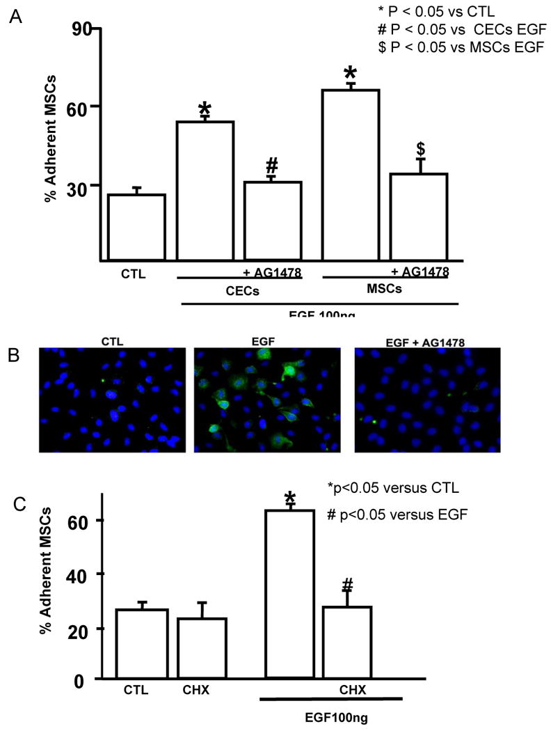
Objective: We determined whether increasing adherence of multipotent stromal cells (MSCs) would amplify their effects on coronary collateral growth (CCG).

Methods and results: Adhesion was established in cultured coronary endothelials cells (CECs) or MSCs treated with epidermal growth factor (EGF). EGF increased MSCs adhesion to CECs, and increased intercellular adhesion molecule (ICAM-1) or vascular cell adhesion molecule (VCAM-1) expression. Increased adherence was blocked by EGF receptor antagonism or antibodies to the adhesion molecules. To determine whether adherent MSCs, treated with EGF, would augment CCG, repetitive episodes of myocardial ischemia (RI) were introduced and CCG was measured from the ratio of collateral-dependent (CZ) and normal zone (NZ) flows. CZ/NZ was increased by MSCs without treatment versus RI-control and was further increased by EGF-treated MSCs. EGF-treated MSCs significantly improved myocardial function versus RI or RI+MSCs demonstrating that the increase in collateral flow was functionally significant. Engraftment of MSCs into myocardium was also increased by EGF treatment.

Conclusions: These results reveal the importance of EGF in MSCs adhesion to endothelium and suggest that MSCs may be effective therapies for the stimulation of coronary collateral growth when interventions are used to increase their adhesion and homing (in vitro EGF treatment) to the jeopardized myocardium.


Figures

Figure 1
Figure 1
A. Adhesion of MSCs to endothelial cells. % of MSCs adherent to CECs after 1, 2, 4 and 24 hours of incubation of both MSCs and CECs. Data are expressed as mean ± SEM. * p<0.05 versus 1 or 2 hr, n = 4. B. Effect of EGF on MSCs adhesion to CECs. % of MSCs adherent to CECs after pretreatment (16 hours) of CECs, MSCs or both with different concentrations of EGF. Data are expressed as mean ± SEM. * p<0.05 versus untreated control (CTL), n = 4.
Figure 2
Figure 2
Effect of AG1478 on EGF induced adhesion of MSCs to CECs. A. % of MSCs adherents to CECs after pretreatment of MSCs or CECs with AG1478 prior to the stimulation with EGF, n=4. B. Representative pictures of CECs in CTL, with EGF +/- AG1478 showing the effects of EGF and AG1478 on EGF-R phosphorylation. The pictures are representative of 4 experiments. C. Effect of Cyclohexamide on EGF induced adhesion of MSCs to CECs. % of MSCs adherents to CECs after pretreatment of CECs with Cyclohexamide prior to the stimulation with EGF, n = 4. Data are expressed as mean ± SEM. * p<0.05 versus CTL. # p<0.05 versus EGF.
Figure 3
Figure 3
Importance of VCAM-1 and ICAM-1 on EGF induced adhesion of MSCs to CECs. A. Effect of blocking ICAM-1 and VCAM-1 antibodies on EGF induced MSCs adhesion, % of MSCs adherents to CECs after pretreatment of CECs and MSCs with ICAM-1 and VCAM-1 antibodies. Data are expressed as mean ± SEM. * p<0.05 versus CTL. # p<0.05 versus CECs EGF. $ p<0.05 versus MSCs EGF. B. Representative western blots for ICAM-1 and VCAM-1 expression in MSCs or CECs (CTL, EGF and EGF+AG1478). Equal amounts of protein extracted from CECs and MSCs were separated by SDS-PAGE (10% gel), transferred to PVDF membranes, and immunoblotted with antibodies to ICAM-1 and VCAM-1 and developed by ECL. Each figure is representative of 4 experiments.
Figure 4
Figure 4
Effects of MSCs EGF stimulation on LV remodeling and function. A. Representative M-mode echocardiograms obtained from CTL with no occlusion, Repetitive occlusion RI with MSCs or MSCs+EGF. Arrows show left ventricular chamber. B. Evaluation of LV function: %FS = % fractional shortening, LVEDD= left ventricular end-diastolic dimension, LVESD = left ventricular end-systolic dimension and LVPWD = Posterior wall thickness. Data are expressed as mean ± SEM. * p<0.05 versus CTL with no occlusion. # p<0.05 versus IR. $ p<0.05 versus IR MSC. (CTL n = 5, IR n = 7, IR+ MSC, n = 5, IR+MSCs+EGF n = 6).
Figure 5
Figure 5
A. Effects of MSCs EGF stimulation on coronary collateral growth. CZ/NZ flow ratio in IR, IR MSCs and IR EGF MSCs treated. Data are expressed as mean ± SEM. * p<0.05 versus IR. # p<0.05 versus IR+MSCs. (IR n=7, IR+ MSC, n = 5, IR+MSCs+EGF n = 6). B-E. Presence of MSCs (red color, labeled with DiI, some MSCs noted by arrows) in the heart as shown by fluorescence microscopy. All images were obtained from tissues after 10 days of repetitive ischemia. B. Untreated MSCs in normal myocardial tissue. C. EGF-treated MSCs in normal myocardial tissue. D. Untreated MSCs in the collateral-dependent zone. E. EGF-treated MSCs in the collateral-dependent zone. Note that in the collateral-dependent region the numbers of MSCs were greater than in the normal myocardium, and note that EGF-treatment increases the numbers of MSCs in the collateral zone.
Figure 6
Figure 6
A,D: Green, smooth muscle cells labeled with anti-α-actin. G,J: Green, endothelial cells labeled with anti-Von Willebrand factor. B,E,H,K: Red, MSCs labeled with DiI. C,F,I,L: Overlay of the images. Yellow indicates Red labeled MSC co-localizing with α-actin (C,F), or vWF (I,L). A-C and G-I are low power (20×) images, and D-F and J-L are higher power views, respectively. In A-F the oval shows MSCs that co-localize with α-actin. The circles in G-L shows MSCs that co-localize with vWF. Note in G-L that not all the MSCs co-localize with vWF.

References

    1. Lopez AD, Mathers CD, Ezzati M, Jamison DT, Murray CJ. Global and regional burden of disease and risk factors, 2001: systematic analysis of population health data. Lancet. 2006;367:1747–1757. - PubMed
    1. Vismara LA, Miller RR, Price JE, Karem R, DeMaria AN, Mason DT. Improved longevity due to reduction of sudden death by aortocoronary bypass in coronary atherosclerosis. Am J Cardiol. 1977;39:919–924. - PubMed
    1. Holmes DR, Jr, Kim LJ, Brooks MM, Kip KE, Schaff HV, Detre KM, Frye RL. The effect of coronary artery bypass grafting on specific causes of long-term mortality in the Bypass Angioplasty Revascularization Investigation. J Thorac Cardiovasc Surg. 2007;134:38–46. 46 e31. - PubMed
    1. Minguell JJ, Erices A. Mesenchymal stem cells and the treatment of cardiac disease. Exp Biol Med (Maywood) 2006;231:39–49. - PubMed
    1. Mangi AA, Noiseux N, Kong D, He H, Rezvani M, Ingwall JS, Dzau VJ. Mesenchymal stem cells modified with Akt prevent remodeling and restore performance of infarcted hearts. Nat Med. 2003;9:1195–1201. - PubMed

Publication types

MeSH terms