

Human CD133+ progenitor cells promote the healing of diabetic ischemic ulcers by paracrine stimulation of angiogenesis and activation of Wnt signaling
- PMID: 19342601
- PMCID: PMC2821014
- DOI: 10.1161/CIRCRESAHA.108.192138
Human CD133+ progenitor cells promote the healing of diabetic ischemic ulcers by paracrine stimulation of angiogenesis and activation of Wnt signaling
Abstract
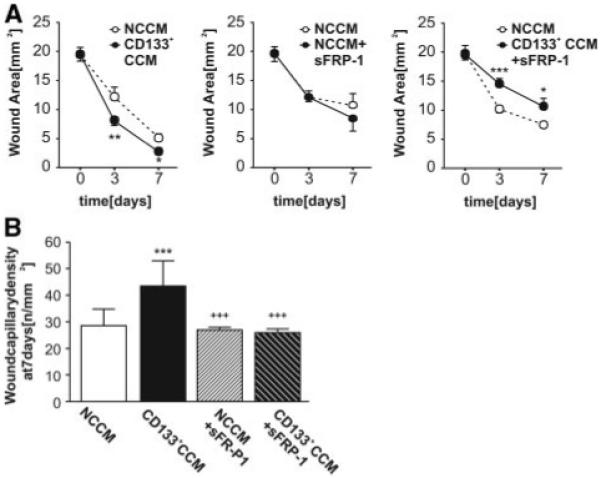
We evaluated the healing potential of human fetal aorta-derived CD133(+) progenitor cells and their conditioned medium (CD133(+) CCM) in a new model of ischemic diabetic ulcer. Streptozotocin-induced diabetic mice underwent bilateral limb ischemia and wounding. One wound was covered with collagen containing 2x10(4) CD133(+) or CD133(-) cells or vehicle. The contralateral wound, covered with only collagen, served as control. Fetal CD133(+) cells expressed high levels of wingless (Wnt) genes, which were downregulated following differentiation into CD133(-) cells along with upregulation of Wnt antagonists secreted frizzled-related protein (sFRP)-1, -3, and -4. CD133(+) cells accelerated wound closure as compared with CD133(-) or vehicle and promoted angiogenesis through stimulation of endothelial cell proliferation, migration, and survival by paracrine effects. CD133(+) cells secreted high levels of vascular endothelial growth factor (VEGF)-A and interleukin (IL)-8. Consistently, CD133(+) CCM accelerated wound closure and reparative angiogenesis, with this action abrogated by co-administering the Wnt antagonist sFRP-1 or neutralizing antibodies against VEGF-A or IL-8. In vitro, these effects were recapitulated following exposure of high-glucose-primed human umbilical vein endothelial cells to CD133(+) CCM, resulting in stimulation of migration, angiogenesis-like network formation and induction of Wnt expression. The promigratory and proangiogenic effect of CD133(+) CCM was blunted by sFRP-1, as well as antibodies against VEGF-A or IL-8. CD133(+) cells stimulate wound healing by paracrine mechanisms that activate Wnt signaling pathway in recipients. These preclinical findings open new perspectives for the cure of diabetic ulcers.
Figures


Comment in
-
Vascular progenitor cells in diabetes mellitus: roles of Wnt signaling and negatively charged low-density lipoprotein.Circ Res. 2009 May 8;104(9):1038-40. doi: 10.1161/CIRCRESAHA.109.198051. Circ Res. 2009. PMID: 19423862 No abstract available.
-
Risk and benefit of CD133+ progenitors.Circ Res. 2009 Jul 17;105(2):e2. doi: 10.1161/CIRCRESAHA.109.201814. Circ Res. 2009. PMID: 19608984 No abstract available.
References
-
- Posnett J, Franks PJ. The burden of chronic wounds in the UK. Nurs Times. 2008;104:44–45. - PubMed
-
- Moulik PK, Mtonga R, Gill GV. Amputation and mortality in new-onset diabetic foot ulcers stratified by etiology. Diabetes Care. 2003;26:491–494. - PubMed
-
- Campbell WB, Ponette D, Sugiono M. Long-term results following operation for diabetic foot problems: arterial disease confers a poor prognosis. Eur J Vasc Endovasc Surg. 2000;19:174–177. - PubMed
-
- Wieman TJ, Becaplermin Gel Studies Group Clinical efficacy of becaplermin (rhPDGF-BB) gel. Am J Surg. 1998;176:74S–79S. - PubMed
-
- Bennett SP, Griffiths GD, Schor AM, Leese GP, Schor SL. Growth factors in the treatment of diabetic foot ulcers. Br J Surg. 2003;90:133–146. - PubMed
Publication types
MeSH terms
Substances
Grants and funding
LinkOut - more resources
Full Text Sources
Other Literature Sources
Medical
Research Materials
