Comparative Study
. 2009 May 8;104(9):1085-94.
doi: 10.1161/CIRCRESAHA.108.189316. Epub 2009 Apr 2.

Contribution of insulin and Akt1 signaling to endothelial nitric oxide synthase in the regulation of endothelial function and blood pressure

Affiliations
Comparative Study

Contribution of insulin and Akt1 signaling to endothelial nitric oxide synthase in the regulation of endothelial function and blood pressure

J David Symons et al. Circ Res. .

Abstract

Impaired insulin signaling via phosphatidylinositol 3-kinase/Akt to endothelial nitric oxide synthase (eNOS) in the vasculature has been postulated to lead to arterial dysfunction and hypertension in obesity and other insulin resistant states. To investigate this, we compared insulin signaling in the vasculature, endothelial function, and systemic blood pressure in mice fed a high-fat (HF) diet to mice with genetic ablation of insulin receptors in all vascular tissues (TTr-IR(-/-)) or mice with genetic ablation of Akt1 (Akt1-/-). HF mice developed obesity, impaired glucose tolerance, and elevated free fatty acids that was associated with endothelial dysfunction and hypertension. Basal and insulin-mediated phosphorylation of extracellular signal-regulated kinase 1/2 and Akt in the vasculature was preserved, but basal and insulin-stimulated eNOS phosphorylation was abolished in vessels from HF versus lean mice. In contrast, basal vascular eNOS phosphorylation, endothelial function, and blood pressure were normal despite absent insulin-mediated eNOS phosphorylation in TTr-IR(-/-) mice and absent insulin-mediated eNOS phosphorylation via Akt1 in Akt1-/- mice. In cultured endothelial cells, 6 hours of incubation with palmitate attenuated basal and insulin-stimulated eNOS phosphorylation and NO production despite normal activation of extracellular signal-regulated kinase 1/2 and Akt. Moreover, incubation of isolated arteries with palmitate impaired endothelium-dependent but not vascular smooth muscle function. Collectively, these results indicate that lower arterial eNOS phosphorylation, hypertension, and vascular dysfunction following HF feeding do not result from defective upstream signaling via Akt, but from free fatty acid-mediated impairment of eNOS phosphorylation.


Figures

Figure 1
Figure 1
Metabolic characteristics HF and CON mice. Body weights (A), blood glucose during a GTT (B), insulin at minute 0 of the GTT but not at minute 30 (C), and fasting FFAs (D) are elevated in HF vs CON mice. Data are means±SEM from 10 to 12 mice per group. *P<0.05 HF vs CON.
Figure 2
Figure 2
Metabolic characteristics of TTr-IR−/− and WT mice. mRNA for the insulin receptor (IR) was lower in aortae from TTr-IR−/− vs WT mice, whereas insulin-like growth factor 1 receptor (IGF1R) mRNA was similar between groups (A). Body weight gain over time (B) and blood glucose during the GTT (C) were similar between TTr-IR−/− and WT mice. Insulin concentrations at minute 0 and minute 30 during the GTT were greater in TTr-IR−/− vs WT mice (D). Data are means±SEM from 4 (A) and 11 to 13 mice per group (B through D). *P<0.05 TTr-IR−/− vs WT.
Figure 3
Figure 3
Vascular signal transduction in HF and CON mice. Compared to vehicle (−), insulin (+) increased p-ERK1/2 (A), p-Akt S473/T308 (B), and p-eNOS S1177 (C) in aortae from CON mice. Insulin-stimulated p-ERK was intact in aorta from HF mice, but insulin-stimulated p-Akt S473/T308 was blunted, and insulin-stimulated p-eNOS S1177 was abolished. Basal p-eNOS S1177 was minimal in aorta from HF vs CON mice. Immunoblots (top) and densitometric measurements (bottom) of p-protein to total protein (arbitrary units) from aortae of 7 to 9 mice per group are shown. *P<0.05 (+) vs (−); **P<0.05 HF vs CON.
Figure 4
Figure 4
Vascular signal transduction in TTr-IR−/− and WT mice. Compared to vehicle (−), insulin (+) increased p-ERK1/2 (A), p-Akt S473/T308 (B), and p-eNOS S1177 (C) in WT but not TTr-IR−/− mice (n=7 to 9 per group). *P<0.05 (+) vs (−).
Figure 5
Figure 5
Vascular signal transduction in Akt1−/− and WT mice. Compared to vehicle (−), insulin (+) increased p-ERK1/2 (A), p-Akt S473/T308 (B), and p-eNOS S1177 (C) equivalently in aortae from Akt1−/− and WT mice (n=7 to 9 per group). *P<0.05 (+) vs (−). To explore whether insulin-mediated Akt phosphorylation in vessels from Akt1−/− mice was mediated via the Akt2 isoform, Akt2 was immunoprecipitated from vascular homogenates obtained from Akt1−/− and WT mice after stimulation with insulin or vehicle, and immunoblotted (IB) with phospho-specific antibodies (D). Compared to vehicle (−), insulin (+) increased the ratio of p-Akt S473 and p-Akt T308 to total Akt2 in both groups (n=4 mice per group).
Figure 6
Figure 6
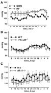
Blood pressure. Blood pressure was higher in HF vs CON animals (n=8 to 10 mice per group) (A) but similar between TTr-IR−/− and WT mice (n=8 to 10 per group) (B) and Akt1−/− and WT mice (n=5 per group) (C). *P<0.05 CON vs HF for all time points.
Figure 7
Figure 7
Vascular function. Percentage (%) relaxation to acetylcholine (ACh), L-NMMA–evoked tension development, and percentage relaxation to sodium nitroprusside (SNP), respectively, in HF (A through C), TTr-IR−/− (D through F), and Akt1−/− mice (G through I). Data are means±SEM from 10 to 14 (TTr-IR−/− and HF) or 4 to 8 (Akt1−/−) mice and their respective controls. *P<0.05 CON vs HF.
Figure 8
Figure 8
Treatment of BAECs and arteries with palmitate. Basal (−) and insulin stimulated (+) p-eNOS S1177 is blunted and abolished, respectively, in BAECs incubated for 6 hours with 500 μmol/L palmitate (Pal) vs vehicle (Veh) (A), whereas basal and insulin-stimulated p-Akt (B) and p-ERK (C) are intact. Insulin-stimulated NO production is abolished in BAECs treated for 6 hours with Pal (D). *P<0.05 (+) vs (−); **P<0.05 vs respective Veh treatment (n=6 to 14 experiments per treatment for A through D). ACh-evoked percentage vasorelaxation is impaired in femoral arteries after 3 hours of incubation with 500 μmol/L Pal (n=10 vessels) (E). *P<0.05 pre- vs post-Pal. Basal p-eNOS is impaired in BAECs after 3 hours of incubation with 500 μmol/L Pal (F). *P<0.05 Pal vs Veh (n=12 experiments per treatment).

Comment in

References

    1. Kim JA, Montagnani M, Koh KK, Quon MJ. Reciprocal relationships between insulin resistance and endothelial dysfunction: molecular and pathophysiological mechanisms. Circulation. 2006;113:1888–1904. - PubMed
    1. Jiang ZY, Lin YW, Clemont A, Feener EP, Hein KD, Igarashi M, Yamauchi T, White MF, King GL. Characterization of selective resistance to insulin signaling in the vasculature of obese Zucker (fa/fa) rats. J Clin Invest. 1999;104:447–457. - PMC - PubMed
    1. Vicent D, Ilany J, Kondo T, Naruse K, Fisher SJ, Kisanuki YY, Bursell S, Yanagisawa M, King GL, Kim JA. The role of endothelial insulin signaling in the regulation of vascular tone and insulin resistance. J Clin Invest. 2003;111:1373–1380. - PMC - PubMed
    1. Muniyappa R, Montagnani M, Koh KK, Quon MJ. Cardiovascular actions of insulin. Endocr Rev. 2007;28:463–491. - PubMed
    1. Fulton D, Gratton JP, McCabe TJ, Fontana J, Fujio Y, Walsh K, Franke TF, Papapetropoulos A, Sessa WC. Regulation of endothelium-derived nitric oxide production by the protein kinase Akt. Nature. 1999;399:597–601. - PMC - PubMed

Publication types

MeSH terms