Review
. 2009 Apr;89(2):411-52.
doi: 10.1152/physrev.00029.2007.

Supramolecular assemblies and localized regulation of voltage-gated ion channels

Affiliations
Review

Supramolecular assemblies and localized regulation of voltage-gated ion channels

Shuiping Dai et al. Physiol Rev. 2009 Apr.

Abstract

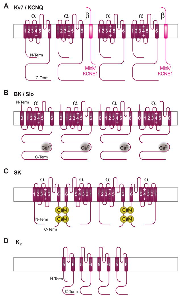
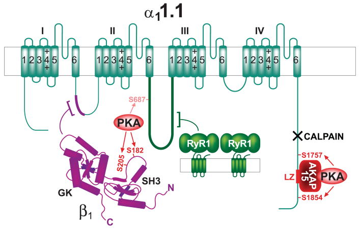
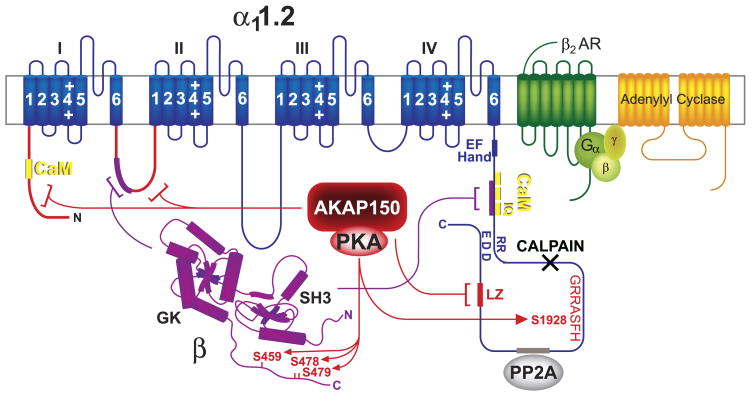
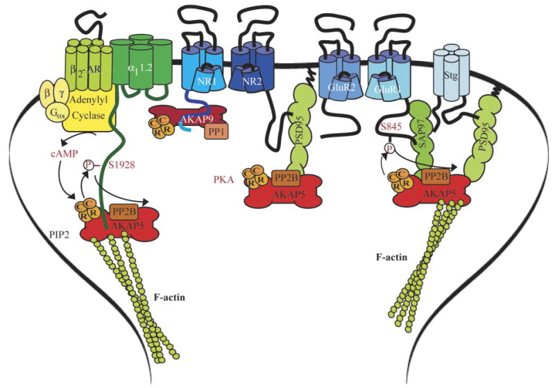
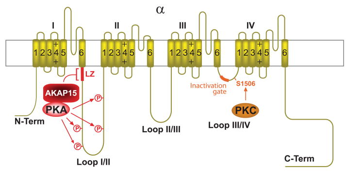
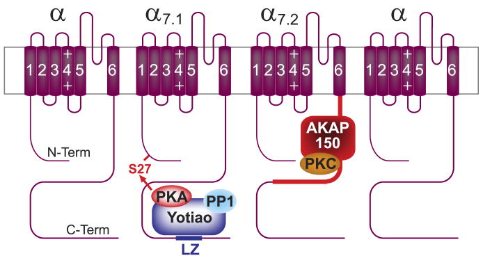
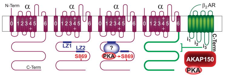
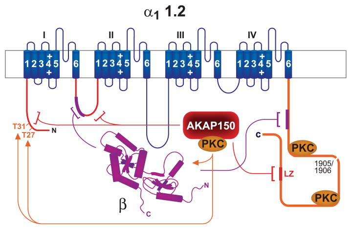
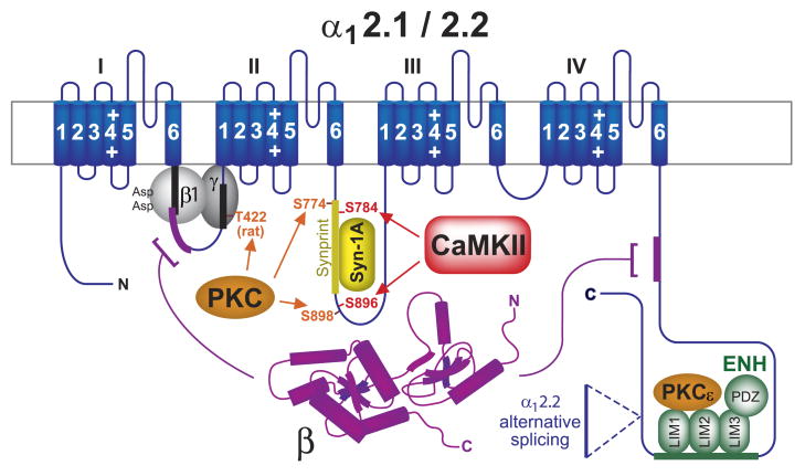
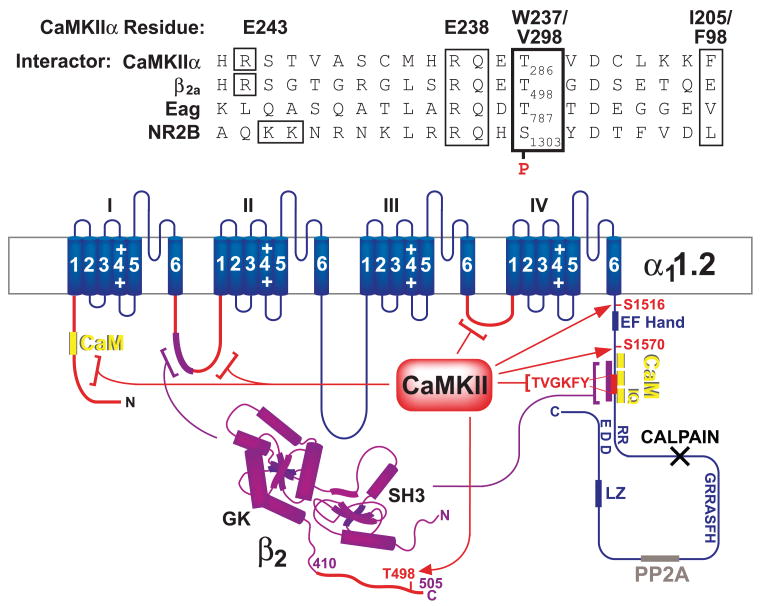
This review addresses the localized regulation of voltage-gated ion channels by phosphorylation. Comprehensive data on channel regulation by associated protein kinases, phosphatases, and related regulatory proteins are mainly available for voltage-gated Ca2+ channels, which form the main focus of this review. Other voltage-gated ion channels and especially Kv7.1-3 (KCNQ1-3), the large- and small-conductance Ca2+-activated K+ channels BK and SK2, and the inward-rectifying K+ channels Kir3 have also been studied to quite some extent and will be included. Regulation of the L-type Ca2+ channel Cav1.2 by PKA has been studied most thoroughly as it underlies the cardiac fight-or-flight response. A prototypical Cav1.2 signaling complex containing the beta2 adrenergic receptor, the heterotrimeric G protein Gs, adenylyl cyclase, and PKA has been identified that supports highly localized via cAMP. The type 2 ryanodine receptor as well as AMPA- and NMDA-type glutamate receptors are in close proximity to Cav1.2 in cardiomyocytes and neurons, respectively, yet independently anchor PKA, CaMKII, and the serine/threonine phosphatases PP1, PP2A, and PP2B, as is discussed in detail. Descriptions of the structural and functional aspects of the interactions of PKA, PKC, CaMKII, Src, and various phosphatases with Cav1.2 will include comparisons with analogous interactions with other channels such as the ryanodine receptor or ionotropic glutamate receptors. Regulation of Na+ and K+ channel phosphorylation complexes will be discussed in separate papers. This review is thus intended for readers interested in ion channel regulation or in localization of kinases, phosphatases, and their upstream regulators.


Figures

FIG. 1
FIG. 1
Membrane topology of Ca2+ channels. The central pore-forming subunit α1 (dark blue) consists of the four homologous domains I–IV that are linked to each other by the intracellular loops I/II, II/III, and III/IV, each containing six transmembrane segments and a P-loop between segments 5 and 6. The auxiliary subunits α2 (light blue) and β (magenta; Refs. 62, 308, 402) directly interact with α1 [the precise interaction sites of α1 with α2 and γ (medium blue) have not been defined]. Magenta: β subunits generally bind with their GK domains to loop I/II connecting domains I and II (AID); black X: calpain cleavage region.
FIG. 2
FIG. 2
Membrane topology of Na+ channels. The central pore-forming subunit α (yellow) consists of the four homologous domains I–IV that are linked to each other by the intracellular loops I/II, II/III, and III/IV, each containing six transmembrane segments and a P-loop between segments 5 and 6. Some complexes contain the auxiliary subunits β1 and β2, which span the plasma membrane once (green). The extracellular interaction of β1 with the exctracellular segment preceding IVS6 is indicated (bracket).
FIG. 3
FIG. 3
Membrane topology of K+ channels. The pore is formed by four homologous α subunits (purple), which interact with each other via their NH2 termini. α Subunits of Kv (A), BK (B), and SK (C) are formed by six transmembrane segments and a P-loop between segments 5 and 6, whereas Kir (D) lacks S1–S4. BK channel subunits contain an additional transmembrane segment (S0) NH2 terminal to the conserved S1 segment. Kv7/KCNQ channels (A) are typically associated with the auxiliary single transmembrane MinK/KCNE subunit (magenta), BK channels bind Ca2+ with their COOH termini (B), and SK channel α subunits dimerize through binding two CaM molecules (C).
FIG. 4
FIG. 4
α-Actinin coimmunoprecipitates with SK2 from rat brain. Rat forebrains were homogenized in 1% Triton X-100, and nonsolubilized proteins were removed by ultracentrifugation before immunoprecipitation with anti-α-actinin or control IgG and immunoblotting with two different antibodies against SK2 (53–5 and 73–2) (see Refs. 153, for more technical details). Both SK2 antibodies detected a single band of the expected molecular mass in anti-α-actinin but not control precipitates and in total lysate.
FIG. 5
FIG. 5
The Cav1.1-AKAP15-PKA complex. Shown are the subunits α11.1 (dark green) and β1 (magenta). Dark red, AKAP15 and AKAP15 leucine zipper binding site on α11.1 (LZ); light red, PKA and identified phosphorylation sites for PKA on α1 and β1 (arrows). The main PKA sites in full-length α11.1 (serines 1757 and 1854) are removed by calpain (black X, calpain cleavage region). Magenta, β2 and its interaction with α11.1. The main in vitro PKA site of the truncated α11.1 is serine 687. Green, RyR1 (for simplicity only 2 of the 4 subunits that form one pore complex in the sarcoplasmic reticulum are shown). The large cytosolic foot structure of RyR1 directly interacts with α11.1.
FIG. 6
FIG. 6
The Cav1.2-AKAP150-PKA complex. Blue, α11.2; magenta, β2 and its interactions with α11.2; yellow, CaM binding sites on α11.2 [there is one binding site in the NH2 terminus and three binding sites in tandem in the COOH terminus; the latter region also interacts with β subunits (magenta bracket and segment)]; red, AKAP79/150 binding sites (LZ, brackets, and segments), PKA, and PKA phosphorylation sites on α1 and β2 (arrows); gray, PP2A binding site; black X, calpain cleavage region; green, β2 AR; yellow-green, heterotrimeric G protein complex; yellow-orange, adenylyl cyclase.
FIG. 7
FIG. 7
Cardiac α11.2 and its association with AKAP150. Cav1.2 was solubilized from rat heart extracts with 1% Triton X-100 before ultracentrifugation to remove nonsolublized material, immunoprecipitation (IP) with anti-α11.2 or control antibody (IgG), and immunoblotting (IB) with antibodies against α11.2 and AKAP150. Top: α11.2 long and short forms are present in a ratio of ∼1:1. Note the rather diffuse appearance of the two α11.2 bands, which suggests heterogeneity likely due to minor variations by differential splicing and other factors. Bottom: AKAP150 is prominent in total rat heart extract (data not shown) and coprecipitates with Cav1.2.
FIG. 8
FIG. 8
Exemplary Cav1.2 currents and their regulation by cAMP/PKA in HEK293 cells. HEK293 cells were grown on poly-d-lysine-coated coverslips in Dulbecco's modified Eagle's medium plus 10% fetal bovine serum at 37°C under 5% CO2 and transfected with full-length cardiac α11.2, β2a with FuGENE 6. Whole cell patch Ca2+ currents were elicited by depolarization from a holding potential of −70 to 0 mV for 200 ms at 24°C (leak-subtracted using p/4; extracellular: 125 mM NaCl, 10 mM tetraethylammonium chloride, 5 mM CaCl2, 5.4 mM CsCl, 1 mM 4-aminopyridine, 1 mM MgCl2, 10 mM HEPES-NaOH, 10 mM glucose, pH 7.4; intracellular: 120 mM CsCl, 10 mM tetraethylammonium chloride, 10 mM EGTA, 1 mM MgCl2, 3 mM MgATP, 0.5 mM Na3GTP, 10 mM HEPES-CsOH, pH 7.3). DClcBIMPS (closed circles) but not vehicle (open) induced in 7 of 14 experiments (see box blots) an increase in current of >20% (compare a and b), which readily reversed upon wash-out (c).
FIG. 9
FIG. 9
Direct interactions between the NH2-terminal half of AKAP150 and the β2 AR. Fusion proteins were expressed in E. coli and extracted using sarcosyl as described (81, 153, 236). GST fusion proteins of the first, second, and third intracellular loops of the β2 AR (β2-i1, -i2, -i3, respectively) or its cytosolic COOH terminus (β2-C), or GST alone (GST) were immobilized on glutathione Sepharose. Resins were incubated with E. coli lysates of 6xHis- and V5-tagged AKAP79 fragments encoding residues 1–120, 100–220, 200–320, and 300–428 (these constructs cover the full length of AKAP75). Top: immunoblotting with antibodies against V5 showed specific binding of the first two AKAP75 fragments to the COOH terminus of the β2 AR. Weak binding of the second fragment to the third intracellular loop of the β2 AR (β2-i3) was also detectable to variable degrees. Bottom: reprobing with anti-GST demonstrated that comparable amounts of the full-length GST fusion proteins were present, although loop fragments were also substantially degraded.
FIG. 10
FIG. 10
Postsynaptic A kinase anchor protein (AKAP)/PKA complexes. Left: the Cav1.2 signaling complex containing the β2-AR, adenylyl cyclase (AC), Gs, and the PKA holoenzyme (2C plus 2R), which is linked to the complex via AKAP150 (AKAP5). The PKA phosphorylation site serine-1928 is indicated. Middle: the NMDAR complex with Yotiao (AKAP9; binds to the C1 segment in the NR1 COOH terminus) and AKAP150. Yotiao functionally links not only PKA but also the counteractive phosphatase PP1 to the NMDAR. AKAP150 interacts with PSD-95 (or its homologs), which in turn bind to the very COOH-terminal ESDL-COO motif of NR2A and 2B. A potential function of AKAP150 in NMDAR regulation is not known. Right: anchoring of PKA and PP2B by AKAP150 via SAP97 and via PSD-95/stargazin (stg) to the AMPAR GluR1 subunit. SAP97 and PSD-95 bind AKAP150 with their COOH-terminal portion containing SH3 and GK (the exact interaction sites have proven difficult to dissect). PKA and PP2B phosphorylation and dephosphorylation are indicated at serine-845 on GluR1.
FIG. 11
FIG. 11
The Nav1.2-AKAP15-PKA complex. Yellow, sodium channel α1.2 subunit; dark red, AKAP15 binding site (LZ, bracket and segment); light red, PKA, four phosphorylation sites for PKA within loop I/II (arrows); orange, PKC, PKC phosphorylation site serine-1506, and the inactivation gate sequence within loop III/IV.
FIG. 12
FIG. 12
The Kv7.1-Yotiao-PKA/PP2A and Kv7.2-AKAP150-PKC complexes. Purple, α7.1/7.2; blue, Yotiao binding site on α7.1 (LZ); magenta, AKAP150 binding site on α7.2; bright red, PKA, identified phosphorylation site for PKA in NH2 terminus of α7.1; dark red, AKAP150 and AKAP150 binding sites on α7.1; orange, PKC binding to AKAP150.
FIG. 13
FIG. 13
The BK-PKA complex. Purple, α subunit of BK; red, PKA, identified PKA phosphorylation sites; blue, leucine zipper motifs (LZ) within BK, unidentified AKAP that recruits PKA for serine-869 phosphorylation; green, β2 AR and its interactions with the BK α subunit; dark red, AKAP150.
FIG. 14
FIG. 14
The Cav1.2(-AKAP150-)PKC complex. Blue, α11.2; red, AKAP79/150 binding sites (brackets and segments); orange, identified direct binding sites independent of AKAP150 (segments) and phosphorylation sites (arrows, residues followed by number) for PKC on α1. Two separate fragments of the α1 COOH terminus (1509–1905, 1906–2170; Ref. 445) bind PKC. Magenta, β2 and its interactions with α11.2.
FIG. 15
FIG. 15
Cav2.2 PKC complexes. PKC increases channel activity by antagonizing the donwregulation by binding of Gβγ to loop I/II and of syntaxin to the synprint region in loop II/III. Blue, α12.2; orange, PKC phosphorylation site threonine-422 in loop I/II; this site is present in rat but not rabbit α12.2 and specifically antagonizes inhibition by Gβ1, which carries two unique aspartates, but not other trimeric Gβ subunits in rat. Other unidentified phosphorylation sites are responsible for the antagonistic action of PKC with respect to other Gβγ interactions. Grey, β1 and γ subunits of G proteins; black, segments within loop I/II that bind G protein β1 and γ subunits. Also shown in orange are PKC phosphorylation sites in and near the synprint region in loop II/III. Yellow, synprint region within loop II/III and inhibitory interacting protein Syntaxin 1A (Syn-1A); red, CaMKII, identified CaMKII phosphorylation sites in and near the synprint region in loop II/III; green, ENH binds with its LIM domains to both the COOH terminus of Cav2.2 (but not Cav2.1) and to PKC-ε (orange), thereby recruiting PKC-ε to the channel complex.
FIG. 16
FIG. 16
Interaction of CaMKII with Cav1.2 and related targets. Blue, α11.2; magenta, β2 and its interactions with α11.2; red, CaMKII binding sites (brackets and segments) and defined phosphorylation sites (arrows and residues followed by number). Evidence for functional importance is available for CaMKII binding inside the CaM binding region (TVGKFY) and the COOH terminus of β2. Yellow, one CaM binding site has been identified in the NH2 terminus and a cluster of three sites in the COOH terminus; the cluster also interacts with β subunits (magenta arrow). Gray, PP2A binding site; black X, calpain cleavage region. Top: sequence alignment of the autoinhibitory domain of CaMKII with CaMKII binding sites on ion channel subunits. Residues at the top refer to interactions between residues in the segment of the CaMKII autoinhibitory domain that interacts with the P site under unstimulated conditions and residues in the large lobe of CaMKII. I205 is also important for binding of CaMKII to the NMDA receptor NR2B subunit (18). Boxes indicate residues that interact (or are homologous to other proteins) with CaMKII residues above.

References

    1. Allen D, Fakler B, Maylie J, Adelman JP. Organization and regulation of small conductance Ca2+-activated K+ channel multi-protein complexes. J Neurosci. 2007;27:2369–2376. - PMC - PubMed
    1. Allen PB, Ouimet CC, Greengard P. Spinophilin, a novel protein phosphatase 1 binding protein localized to dendritic spines. Proc Natl Acad Sci USA. 1997;94:9956–9961. - PMC - PubMed
    1. Altier C, Dubel SJ, Barrere C, Jarvis SE, Stotz SC, Spaetgens RL, Scott JD, Cornet V, De Waard M, Zamponi GW, Nargeot J, Bourinet E. Trafficking of L-type calcium channels mediated by the postsynaptic scaffolding protein AKAP79. J Biol Chem. 2002;277:33598–33603. - PubMed
    1. Altier C, Khosravani H, Evans RM, Hameed S, Peloquin JB, Vartian BA, Chen L, Beedle AM, Ferguson SS, Mezghrani A, Dubel SJ, Bourinet E, McRory JE, Zamponi GW. ORL1 receptor-mediated internalization of N-type calcium channels. Nat Neurosci. 2006;9:31–40. - PubMed
    1. An R, Heath BM, Higgins JP, Koch WJ, Lefkowitz RJ, Kass RS. Beta2-adrenergic receptor overexpression in the developing mouse heart: evidence for targeted modulation of ion channels. J Physiol. 1999;516:19–30. - PMC - PubMed

Publication types

MeSH terms

Substances

LinkOut - more resources