. 2009 Apr 15;182(8):4891-8.
doi: 10.4049/jimmunol.0803776.

Group V secretory phospholipase A2 modulates phagosome maturation and regulates the innate immune response against Candida albicans

Affiliations

Group V secretory phospholipase A2 modulates phagosome maturation and regulates the innate immune response against Candida albicans

Barbara Balestrieri et al. J Immunol. .

Abstract

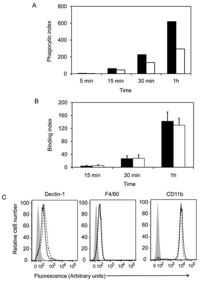
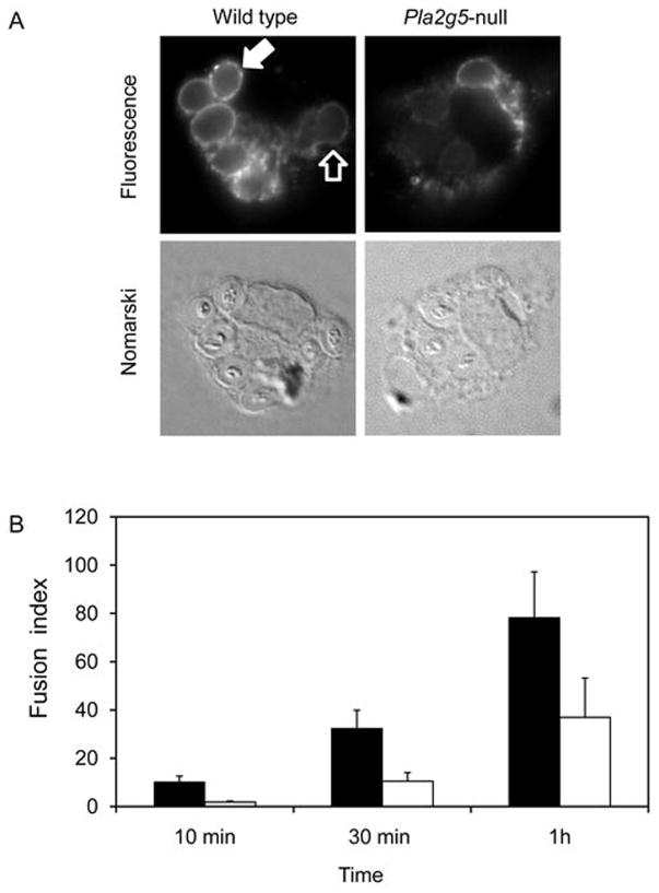
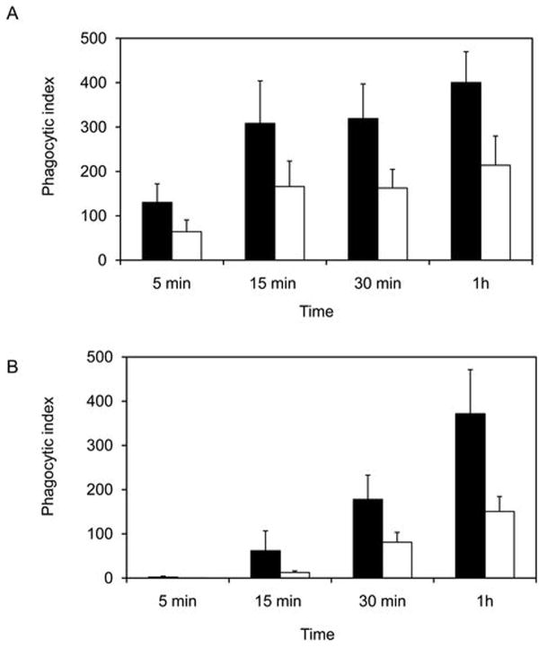
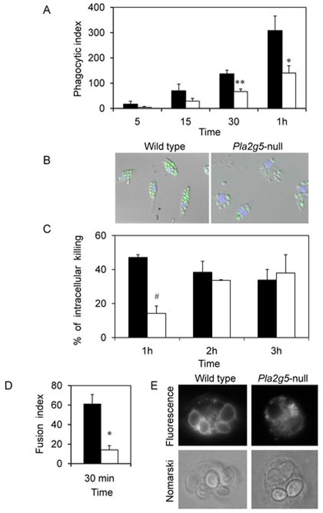
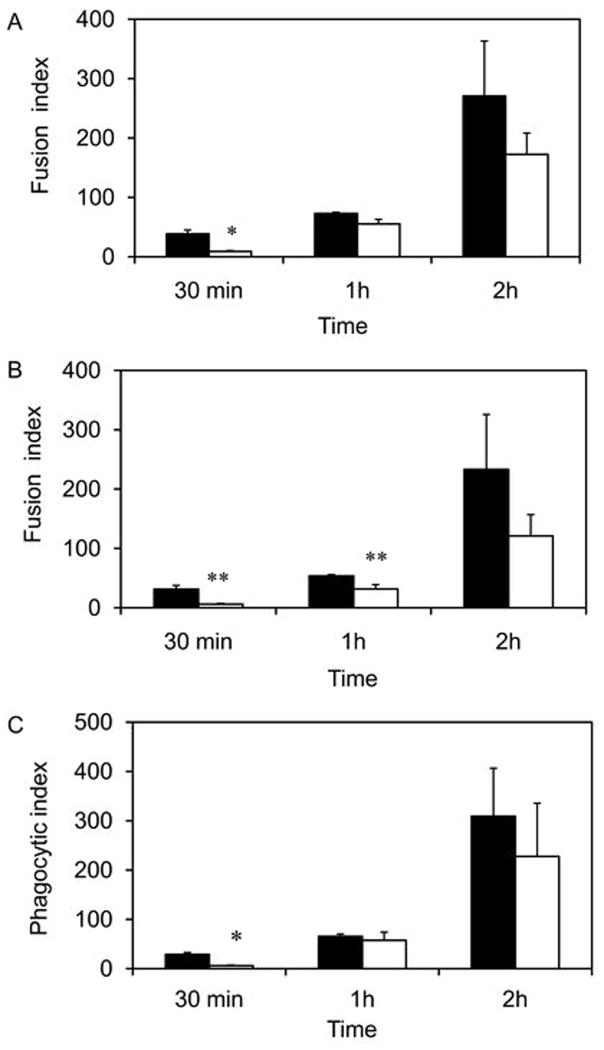
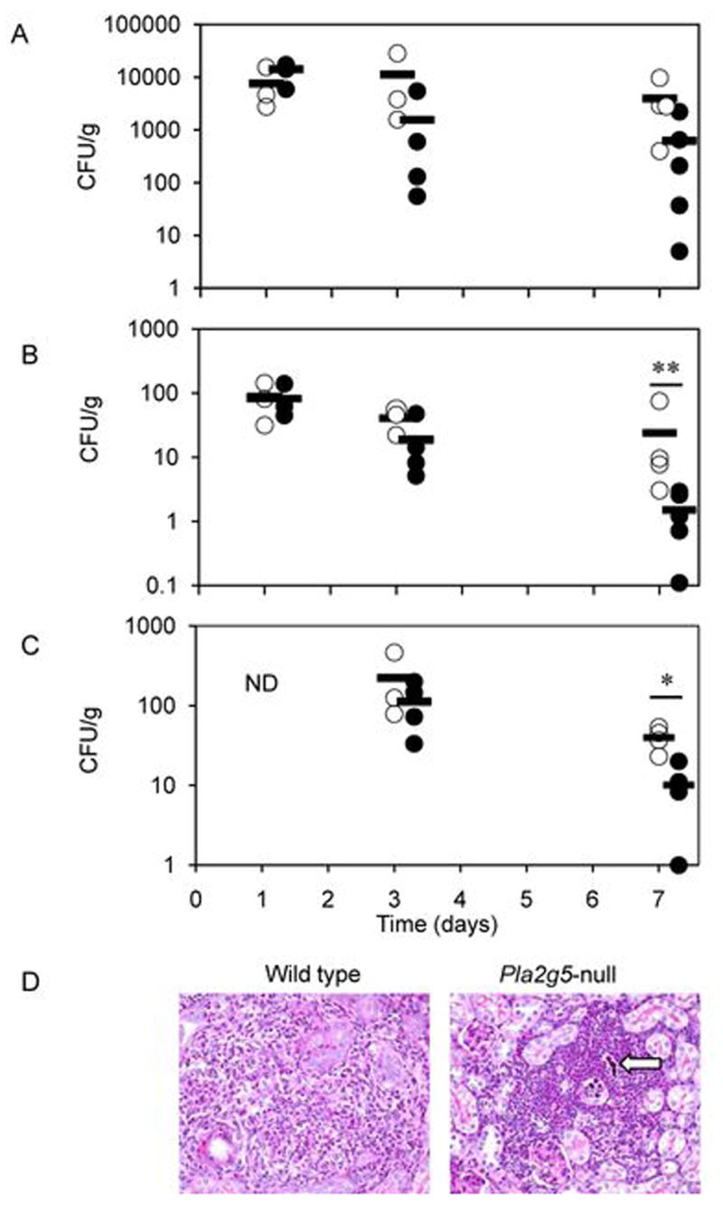
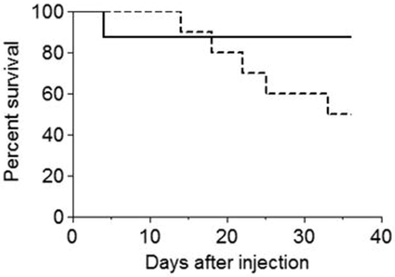
Phospholipase A(2) (PLA(2)) hydrolyzes the sn-2 position of cell membrane phospholipids to release fatty acids and lysophospholipids. We have previously reported that group V secretory PLA(2) (sPLA(2)) translocates from the Golgi and recycling endosomes of mouse peritoneal macrophages to newly formed phagosomes and regulates the phagocytosis of zymosan, suggesting a role in innate immunity. Here we report that in macrophages lacking group V sPLA(2), phagosome maturation was reduced 50-60% at early time points while the binding of zymosan was unimpaired. The ability of group V sPLA(2) to regulate phagocytosis extended to phagocytosis of IgG- and complement-opsonized sheep RBC. Moreover, macrophages lacking group V sPLA(2) had delays in phagocytosis, phagosome maturation, and killing of Candida albicans. Cytokine production and eicosanoid generation were not impaired by the lack of group V sPLA(2). Furthermore, in a model of systemic candidiasis, mice lacking group V sPLA(2) had an increased fungal burden in the kidney, liver, and spleen at day 7 postinfection and increased mortality. Thus, group V sPLA(2) regulates phagocytosis through major phagocytic receptors and contributes to the innate immune response against C. albicans by regulating phagocytosis and killing through a mechanism that is likely dependent on phagolysosome fusion.


Conflict of interest statement

Disclosures

The authors have no financial conflict of interest.

Figures

Figure 1
Figure 1
Binding of zymosan and expression of Dectin-1. (A) Peritoneal macrophages isolated from wild type (black bars) and Pla2g5-null (open bars) mice were stimulated with zymosan (10 ppc) for 5 min to 1 h. Cells were fixed and stained with Diff-Quick. The phagocytic index was obtained as previously described (18). (B) Unopsonized zymosan was added to macrophages pre-treated with cytochalasin D (4μM) for 30 min and incubated at 37°C for 15 min to 1 h. After two washes to remove unbound particles, cells were stained with Diff-Quick and bound particles were counted using an optical microscope. The binding index was obtained as described in “Material and Methods”. (C) Dectin-1 expression was analyzed by immunofluoresence staining and flow cytometry on mouse peritoneal macrophages isolated from wild type (solid line) and Pla2g5-null macrophages (dashed line). Shaded histogram represents isotype control Ab. Data are mean ± SEM of three experiments (B), or representative of two experiments with duplicate samples (A, C).
Figure 2
Figure 2
Pla2g5-null macrophages have a delay in phagosome maturation. Indirect immunofluorescence microscopy was used to visualize the distribution of Lamp-1 in macrophages phagocytosing FITC-zymosan (2.5 ppc) for 10 min to 1 h as described under “Material and methods”. (A) Representative images at 1h. Closed arrow: Lamp-1-positive phagosomes. Open arrow: Lamp-1-negative phagosomes (B) The fusion index was calculated for wild type (closed bars) and Pla2g5-null (open bars) macrophages, as described under “Material and Methods”. Data are mean ± SEM of at least three experiments.
Figure 3
Figure 3
Phagocytosis of IgG- and iC3b-opsonized sheep red blood cells is impaired in Pla2g5-null peritoneal macrophages. Peritoneal macrophages isolated from Pla2g5-null (open bars) and wild type (black bars) mice were stimulated with IgG-sRBC (A) or iC3b-sRBC (B) (10 ppc) for 5 min to 1 h. Cells were fixed and stained with Diff-Quick. The phagocytic index was obtained as described in “Material and Methods”. Data are mean ± SEM of three experiments.
Figure 4
Figure 4
Impaired phagocytosis and killing of C. albicans by Pla2g5-null peritoneal macrophages. (A) Peritoneal macrophages isolated from Pla2g5-null (open bars) and wild type (black bars) mice were incubated with C. albicans from 5 min to 1 h (yeast to cell ratio 5:1). The phagocytic index was obtained as described under “Material and Methods”. (B) C. albicans viability was distinguished by fluorescence staining with FUN-1. Nuclei were stained in blue by Hoechst dye. (C) Viability of ingested C. albicans was assessed by a limiting dilution technique and is expressed as a percentage of viable yeast initially ingested, as described under “Material and Methods”. (D) Indirect immunofluorescence microscopy was used to visualize the distribution of Lamp-1 in macrophages phagocytosing C. albicans (m.o.i. 2) for 30 min. The fusion index was calculated for wild type (closed bars) and Pla2g5-null (open bars) macrophages, as described under “Material and Methods”. (E) Representative images at 30 min. Data are mean ± SEM of three experiments (A), of at least four independent experiments, each with quadruplicate measurement (C), or of two experiments (D).
Figure 5
Figure 5
Delayed phagosome-lysosome fusion in Pla2g5-null peritoneal macrophages. Peritoneal macrophages isolated from wild type (black bars) and Pla2g5-null (open bars) mice were incubated with C. albicans from 30 min to 2 h (m.o.i. 2). To synchronize phagocytosis, particles were first allowed to bind at 4°C, excess unbound particles were washed away then cell transferred at 37°C. Indirect immunofluorescence microscopy was used to visualize the distribution of Lamp-1 (A) or cathepsin D (B). The fusion index (A, B) and phagocytic index (C) were calculated as described in “Materials and Methods”. Data are mean ± SEM of two experiments.
Figure 6
Figure 6
Pla2g5-null mice are more susceptible than wild type mice to live C. albicans infection. Mice were infected with C. albicans by the intravenous injection of 200 μl of 0.9% NaCl containing 5x10⁵ CFU. To assess outgrowth of the microorganism and its ability to invade organs, subgroups of 3 to 5 Pla2g5-null (open circles) and wild type (closed circles) mice were sacrificed 1, 3, and 7 days after injection. The outgrowth of C. albicans was measured by serial dilution and expressed as CFU per gram of tissue. Individual data points are shown for kidney (A), liver (B) and spleen (C); means are indicated by horizontal bars. ND, not determined. (D) Histopathology of the kidney of C. albicans-infected mice. Histological sections of representative wild type and Pla2g5-null kidneys 7 days after infection are stained with Periodic-Acid Schiff. Arrow points to pseudohyphae. Magnification 40X for both panels.
Figure 7
Figure 7
Survival of mice infected with C. albicans. Wild type (filled line) and Pla2g5-null (dotted line) mice were infected i.v. with 1x10⁶ CFU/mouse and followed for 33 days. Data are from one experiment using 11 Pla2g5-null and 9 wild type mice and are expressed as a Kaplan Meier Plot. Similar data were reproduced in a second experiment.

References

    1. Aderem A, Underhill DM. Mechanisms of phagocytosis in macrophages. Annu Rev Immunol. 1999;17:593–623. - PubMed
    1. Underhill DM, Ozinsky A. Phagocytosis of microbes: complexity in action. Annu Rev Immunol. 2002;20:825–852. - PubMed
    1. Brown GD, Taylor PR, Reid DM, Willment JA, Williams DL, Martinez-Pomares L, Wong SY, Gordon S. Dectin-1 is a major beta-glucan receptor on macrophages. J Exp Med. 2002;196:407–412. - PMC - PubMed
    1. Taylor PR, Brown GD, Herre J, Williams DL, Willment JA, Gordon S. The role of SIGNR1 and the beta-glucan receptor (dectin-1) in the nonopsonic recognition of yeast by specific macrophages. J Immunol. 2004;172:1157–1162. - PubMed
    1. Brown GD, Gordon S. Immune recognition. A new receptor for beta-glucans. Nature. 2001;413:36–37. - PubMed

Publication types

MeSH terms