. 2009 Apr 15;182(8):5024-31.
doi: 10.4049/jimmunol.0803603.

The pattern recognition receptor CD36 is a chondrocyte hypertrophy marker associated with suppression of catabolic responses and promotion of repair responses to inflammatory stimuli

Affiliations

The pattern recognition receptor CD36 is a chondrocyte hypertrophy marker associated with suppression of catabolic responses and promotion of repair responses to inflammatory stimuli

Denise L Cecil et al. J Immunol. .

Abstract

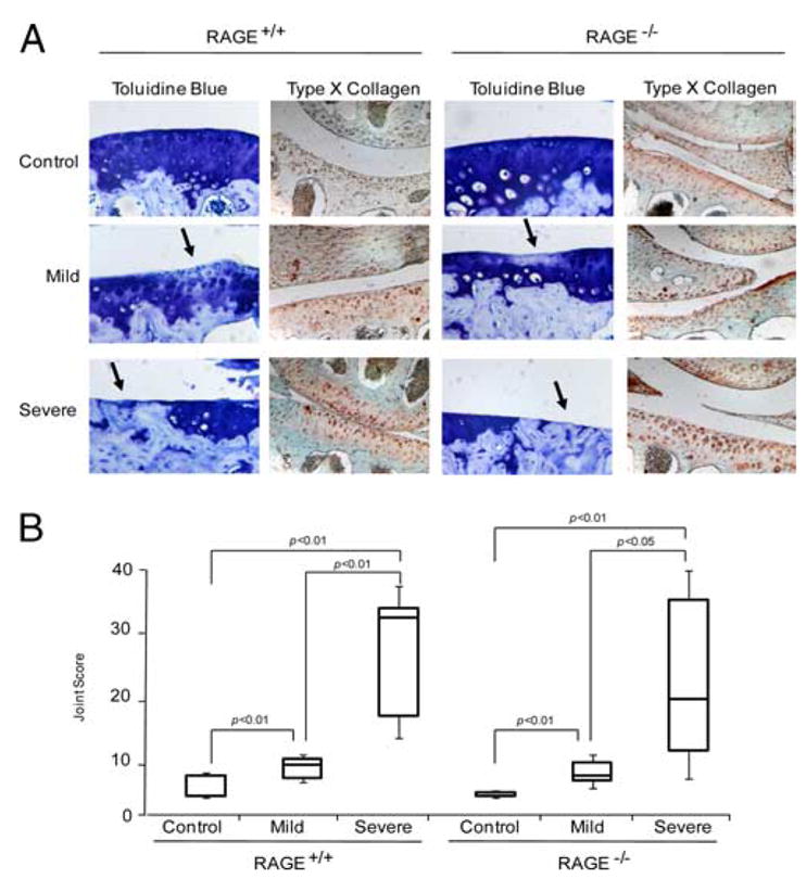
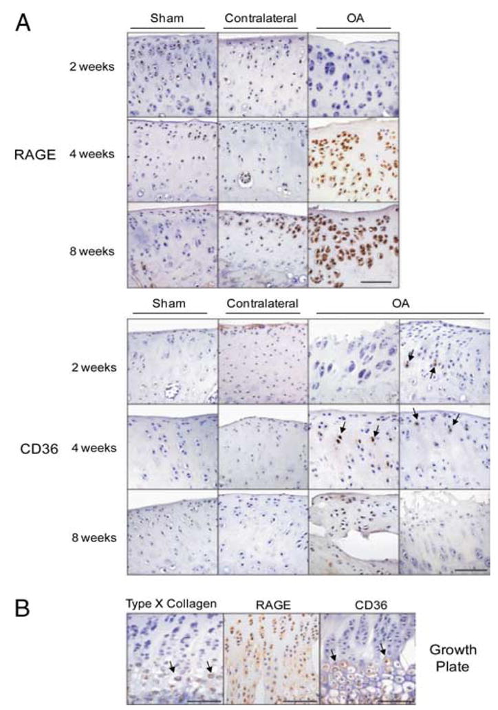
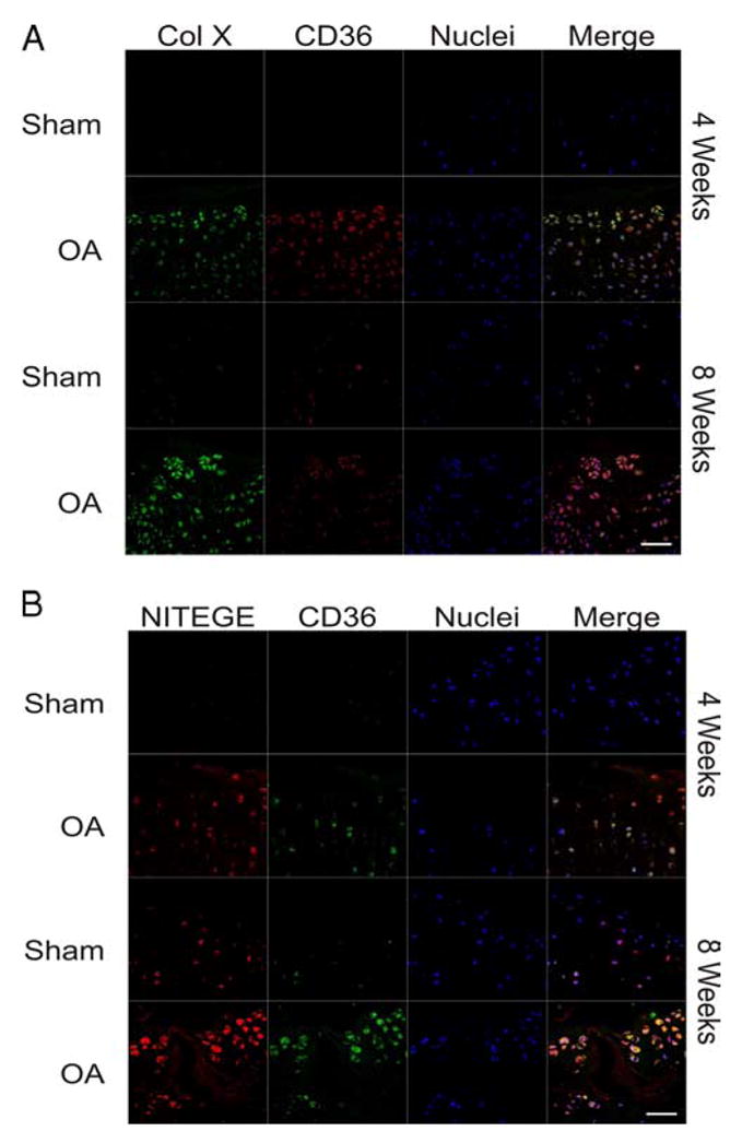
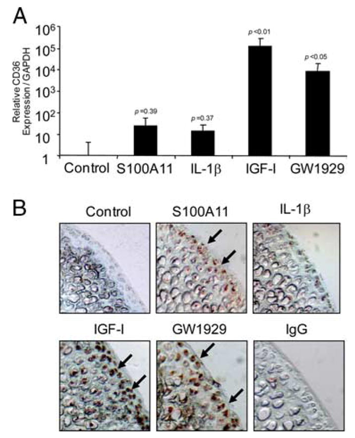
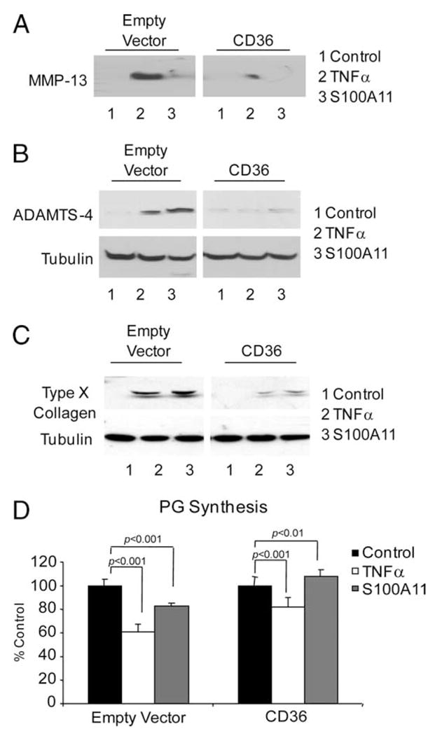
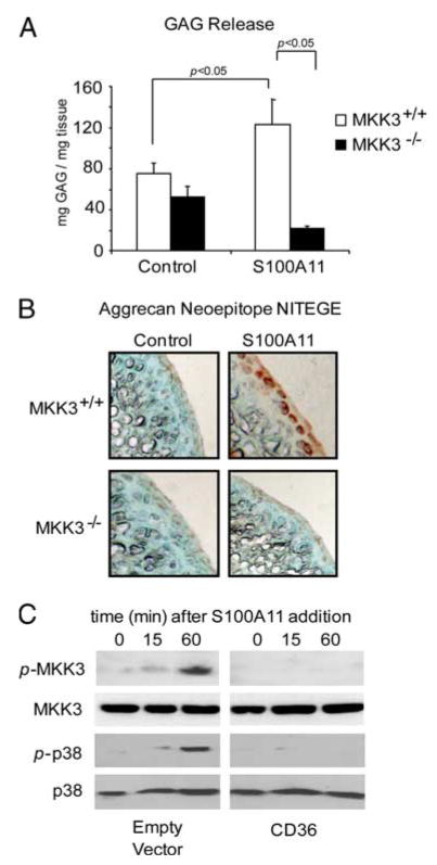
Multiple inflammatory mediators in osteoarthritis (OA) cartilage, including S100/calgranulin ligands of receptor for advanced glycation end products (RAGE), promote chondrocyte hypertrophy, a differentiation state associated with matrix catabolism. In this study, we observed that RAGE knockout was not chondroprotective in instability-induced knee OA in 8-wk-old mice. Hence, we tested the hypothesis that expression of the alternative S100/calgranulin and patterning receptor CD36, identified here as a marker of growth plate chondrocyte hypertrophy, mediates chondrocyte inflammatory and differentiation responses that promote OA. In rat knee joint destabilization-induced OA, RAGE expression was initially sparse throughout cartilage but increased diffusely by 4 wk after surgery. In contrast, CD36 expression focally increased at sites of cartilage injury and colocalized with developing chondrocyte hypertrophy and aggrecan cleavage NITEGE neoepitope formation. However, CD36 transfection in normal human knee-immortalized chondrocytes (CH-8 cells) was associated with decreased capacity of S100A11 and TNF-alpha to induce chondrocyte hypertrophy and ADAMTS-4 and matrix metalloproteinase 13 expression. S100A11 lost the capacity to inhibit proteoglycans synthesis and gained the capacity to induce proteoglycan synthesis in CD36-transfected CH-8 cells. Moreover, S100A11 required the p38 MAPK pathway kinase MKK3 to induce NITEGE development in mouse articular cartilage explants. However, CH-8 cells transfected with CD36 demonstrated decreased S100A11-induced MKK3 and p38 phosphorylation. Therefore, RAGE and CD36 patterning receptor expression were linked with opposing effects on inflammatory, procatabolic responses to S100A11 and TNF-alpha in chondrocytes.


Conflict of interest statement

Disclosures

The authors have no financial conflict of interest.

Figures

FIGURE 1
FIGURE 1
RAGE knockout is not chondroprotective for knee OA in mice. A, Two months after a “stab” procedure for minimally invasive disruption of the ACL in both knees done at 2 mo of age, congenic RAGE+/+ and RAGE−/− mouse knee joints were stained with toluidine blue and analyzed by immunohistochemistry for type X collagen. The extent of knee instability and OA were graded histologically. Knee joints with a grade of 0 for instability (due to insufficient ACL trauma by the needle penetration) served as controls and grade 1 is designated as mild OA, with grades 2 and 3 representing severe OA in A and B, respectively. Loss of blue staining (arrows) indicates loss of PG. These data are representative of at least five different mouse donors (original magnification, ×100). B, The total joint score was measured in euthanized RAGE+/+ and RAGE−/− mouse knee joints 2 mo after the stab procedure, as described in Materials and Methods. Scores for cartilage degeneration in the whole joint are represented as box and whisker plots showing the 25th through 75th percentiles boxed and the median joint score as the central horizontal line. For RAGE+/+, control n = 50, mild n = 7, and severe n = 10, and for RAGE−/−, control n = 49, mild n = 11, and severe n = 9.
FIGURE 2
FIGURE 2
RAGE and CD36 expression increase during the evolution of surgical instability-induced knee OA in adult rats. A, OA was induced by ACLT and partial anteromedial meniscectomy in adult rats. Sham (control) and OA knee joints isolated at 2, 4, and 8 wk after surgery were fixed, paraffin embedded, and sectioned through the medial joint compartment. Histological knee joint sections were analyzed by immunohistochemistry for RAGE and CD36. Two panels of CD36 expression are shown to indicate the variability of CD36 expression (arrows). B, Histological sections of the growth plate at 4 wk after surgery were analyzed by immunohistochemistry for type X collagen (arrows), RAGE, and CD36 (arrows).
FIGURE 3
FIGURE 3
CD36 colocalizes with both type X collagen (Col X) and aggrecan cleavage as rat knee OA progresses. Sham (control) and OA knee joints isolated at 4 and 8 wk after surgery were fixed, paraffin embedded, and sectioned through the medial joint compartment. A, Histological sections were analyzed by immunofluorescence for CD36 (red) and type X collagen (green). Nuclei were counterstained with TOTO-3 iodide (blue). B, Histological sections analyzed by immunofluorescence for CD36 (green) and the aggrecan neoepitope of aggrecanase cleavage NITEGE (red), with nuclei counterstained with TOTO-3 iodide (blue signal). Scale, 50 μm.
FIGURE 4
FIGURE 4
Both IGF-I and the PPARγ agonist GW1929 markedly increase CD36 expression. A, Human immortalized normal knee chondrocytes (CH-8 cells) were stimulated with 100 ng/ml S100A11, 10 ng/ml IL-1β, 10 ng/ml IGF-I, and 100 nM GW1929 for 8 h. Quantitative real-time PCR analysis of CD36 expression (as described in Materials and Methods) is shown pooled from three separate experiments. The p values indicated are compared with control, nonstimulated CH-8 cells. B, Mouse femoral head cartilage explants were stimulated for 48 h with the agonists described in A, and frozen sections were examined by immunohistochemistry for CD36 (arrows) as described in Materials and Methods. Data are representative of five different mouse donors.
FIGURE 5
FIGURE 5
TNF-α- and S100A11-induced hypertrophic differentiation and procatabolic responses of chondrocytic cells are inhibited by transfection of CD36. CH-8 cells were plated in 96-well plates containing poly-HEME and treated with 10 ng/ml TNF-α or 100 ng/ml S100A11. After 24 h, conditioned medium was examined for MMP-13 secretion (A) and SDS-PAGE/Western blotting of cell lysates was performed to assess induction of ADAMTS-4 (B) and, at 3 days, type X collagen was assessed in cell lysates by SDS-PAGE/Western blotting (C). Tubulin was visualized as a loading control. Data are representative of three or more separate experiments for each parameter. D, CH-8 cells were plated in 24-well plates and treated with 10 ng/ml TNF-α or 100 ng/ml S100A11 for 2 days in replicates of six. One microCurie of 35S and 0.5 μCi of [3H]proline were added, and sulfated PG synthesis was assayed as described in Materials and Methods. Data are representative of three separate experiments.
FIGURE 6
FIGURE 6
S100A11-stimulated MKK3 signaling essential to S100A11-induced cartilage matrix catabolism is inhibited in CH-8 cells transfected with CD36. A, Congenic MMK3−/− and MKK3+/+ adult mouse femoral head cartilage explants were treated with 100 ng/ml S100A11. A, At 48 h, GAG release was measured in conditioned medium. Data are pooled from five different mouse donors of each genotype in replicates of three. B, Frozen sections of mouse femoral head cartilage explants were analyzed by immunohistochemistry for the aggrecanase neoepitope NITEGE at 72 h in culture. Data are representative of five different mouse donors of each genotype. C, CH-8 cells were plated in 96-well plates containing polyHEME and serum starved overnight. The cells were then treated with 100 ng/ml S100A11 for the duration indicated and SDS-PAGE/Western blotting was performed for phosphorylated and total MMK3 and p38. These data are representative of results from six separate experiments.

References

    1. Pelletier JP, Martel-Pelletier J, Abramson SB. Osteoarthritis, an inflammatory disease: potential implication for the selection of new therapeutic targets. Arthritis Rheum. 2001;44:1237–1247. - PubMed
    1. Kobayashi M, Squires GR, Mousa A, Tanzer M, Zukor DJ, Antoniou J, Feige U, Poole AR. Role of interleukin-1 and tumor necrosis factor α in matrix degradation of human osteoarthritic cartilage. Arthritis Rheum. 2005;52:128–135. - PubMed
    1. Borzì RM, Mazzetti I, Marcu KB, Facchini A. Chemokines in cartilage degradation. Clin Orthop Relat Res. 2004;427:S53–S61. - PubMed
    1. Loeser RF. Molecular mechanisms of cartilage destruction: mechanics, inflammatory mediators, and aging collide. Arthritis Rheum. 2006;54:1357–1360. - PMC - PubMed
    1. Yammani RR, Carlson CS, Bresnick AR, Loeser RF. Increase in production of matrix metalloproteinase 13 by human articular chondrocytes due to stimulation with S100A4: role of the receptor for advanced glycation end products. Arthritis Rheum. 2006;54:2901–2911. - PubMed

Publication types

MeSH terms