Crystallization and preliminary X-ray diffraction studies of vitamin D3 hydroxylase, a novel cytochrome P450 isolated from Pseudonocardia autotrophica
- PMID: 19342783
- PMCID: PMC2664763
- DOI: 10.1107/S1744309109007829
Crystallization and preliminary X-ray diffraction studies of vitamin D3 hydroxylase, a novel cytochrome P450 isolated from Pseudonocardia autotrophica
Abstract
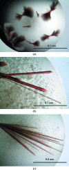
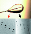
Vitamin D(3) hydroxylase (Vdh) is a novel cytochrome P450 monooxygenase isolated from the actinomycete Pseudonocardia autotrophica and consisting of 403 amino-acid residues. Vdh catalyzes the activation of vitamin D(3) via sequential hydroxylation reactions: these reactions involve the conversion of vitamin D(3) (cholecalciferol or VD3) to 25-hydroxyvitamin D(3) [25(OH)VD3] and the subsequent conversion of 25(OH)VD3 to 1alpha,25-dihydroxyvitamin D(3) [calciferol or 1alpha,25(OH)(2)VD3]. Overexpression of recombinant Vdh was carried out using a Rhodococcus erythropolis expression system and the protein was subsequently purified and crystallized. Two different crystal forms were obtained by the hanging-drop vapour-diffusion method at 293 K using polyethylene glycol as a precipitant. The form I crystal belonged to the trigonal space group P3(1), with unit-cell parameters a = b = 61.7, c = 98.8 A. There is one Vdh molecule in the asymmetric unit, with a solvent content of 47.6%. The form II crystal was grown in the presence of 25(OH)VD3 and belonged to the orthorhombic system P2(1)2(1)2(1), with unit-cell parameters a = 63.4, b = 65.6 c = 102.2 A. There is one Vdh molecule in the asymmetric unit, with a solvent content of 46.7%. Native data sets were collected to resolutions of 1.75 and 3.05 A for form I and form II crystals, respectively, using synchrotron radiation. The structure solution was obtained by the molecular-replacement method and model refinement is in progress for the form I crystal.
Figures


Similar articles
-
Structural evidence for enhancement of sequential vitamin D3 hydroxylation activities by directed evolution of cytochrome P450 vitamin D3 hydroxylase.J Biol Chem. 2010 Oct 8;285(41):31193-201. doi: 10.1074/jbc.M110.147009. Epub 2010 Jul 27. J Biol Chem. 2010. PMID: 20667833 Free PMC article.
-
Purification, characterization, and directed evolution study of a vitamin D3 hydroxylase from Pseudonocardia autotrophica.Biochem Biophys Res Commun. 2009 Jul 24;385(2):170-5. doi: 10.1016/j.bbrc.2009.05.033. Epub 2009 May 18. Biochem Biophys Res Commun. 2009. PMID: 19450562
-
A single mutation at the ferredoxin binding site of P450 Vdh enables efficient biocatalytic production of 25-hydroxyvitamin D(3).Chembiochem. 2013 Nov 25;14(17):2284-91. doi: 10.1002/cbic.201300386. Epub 2013 Sep 24. Chembiochem. 2013. PMID: 24115473
-
Metabolism of vitamin D3 by cytochromes P450.Front Biosci. 2005 Jan 1;10:119-34. doi: 10.2741/1514. Print 2005 Jan 1. Front Biosci. 2005. PMID: 15574355 Review.
-
Cytochrome P450 enzymes in the bioactivation of vitamin D to its hormonal form (review).Int J Mol Med. 2001 Feb;7(2):201-9. doi: 10.3892/ijmm.7.2.201. Int J Mol Med. 2001. PMID: 11172626 Review.
Cited by
-
The role of vitamin D in pulmonary disease: COPD, asthma, infection, and cancer.Respir Res. 2011 Mar 18;12(1):31. doi: 10.1186/1465-9921-12-31. Respir Res. 2011. PMID: 21418564 Free PMC article. Review.
-
Structural evidence for enhancement of sequential vitamin D3 hydroxylation activities by directed evolution of cytochrome P450 vitamin D3 hydroxylase.J Biol Chem. 2010 Oct 8;285(41):31193-201. doi: 10.1074/jbc.M110.147009. Epub 2010 Jul 27. J Biol Chem. 2010. PMID: 20667833 Free PMC article.
-
Structural insights into the mechanism of the drastic changes in enzymatic activity of the cytochrome P450 vitamin D3 hydroxylase (CYP107BR1) caused by a mutation distant from the active site.Acta Crystallogr F Struct Biol Commun. 2017 May 1;73(Pt 5):266-275. doi: 10.1107/S2053230X17004782. Epub 2017 Apr 26. Acta Crystallogr F Struct Biol Commun. 2017. PMID: 28471358 Free PMC article.
-
Cytochrome P450-mediated metabolism of vitamin D.J Lipid Res. 2014 Jan;55(1):13-31. doi: 10.1194/jlr.R031534. Epub 2013 Apr 6. J Lipid Res. 2014. PMID: 23564710 Free PMC article. Review.
-
Identification of a cyclosporine-specific P450 hydroxylase gene through targeted cytochrome P450 complement (CYPome) disruption in Sebekia benihana.Appl Environ Microbiol. 2013 Apr;79(7):2253-62. doi: 10.1128/AEM.03722-12. Epub 2013 Jan 25. Appl Environ Microbiol. 2013. PMID: 23354713 Free PMC article.
References
-
- Cupp-Vickery, J. R., Garcia, C., Hofacre, A. & MacGee-Estrada, K. (2001). J. Mol. Biol.311, 101–110. - PubMed
-
- Cupp-Vickery, J. R. & Podust, T. L. (1995). Nature Struct. Biol.2, 144–153. - PubMed
-
- Hayashi, K., Sugimoto, H., Shinkyo, R., Yamada, M., Ikeda, S., Ikushiro, S., Kamakura, M., Shiro, Y. & Sakaki, T. (2008). Biochemistry, 47, 11964–11972. - PubMed
-
- Jones, G., Strugnell, S. A. & DeLuka, H. F. (1998). Physiol. Rev.78, 1193–1231. - PubMed
Publication types
MeSH terms
Substances
LinkOut - more resources
Full Text Sources

