. 2009;4(4):e5049.
doi: 10.1371/journal.pone.0005049. Epub 2009 Apr 3.

Ste20-related proline/alanine-rich kinase (SPAK) regulated transcriptionally by hyperosmolarity is involved in intestinal barrier function

Affiliations

Ste20-related proline/alanine-rich kinase (SPAK) regulated transcriptionally by hyperosmolarity is involved in intestinal barrier function

Yutao Yan et al. PLoS One. 2009.

Abstract

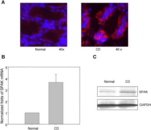
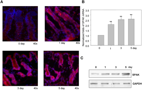
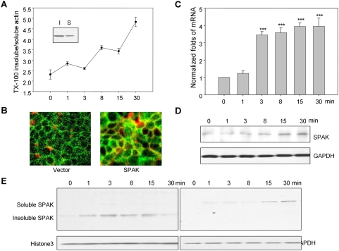
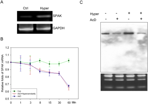
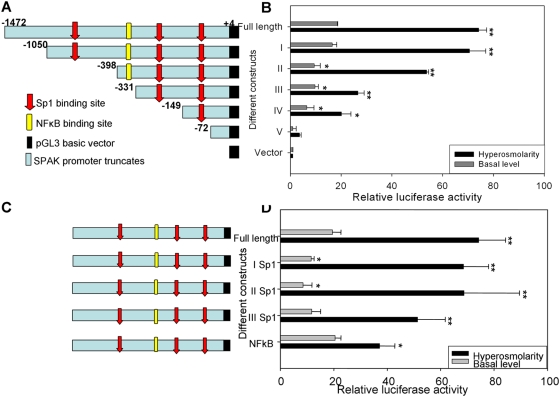
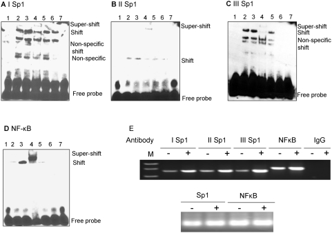
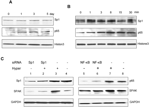
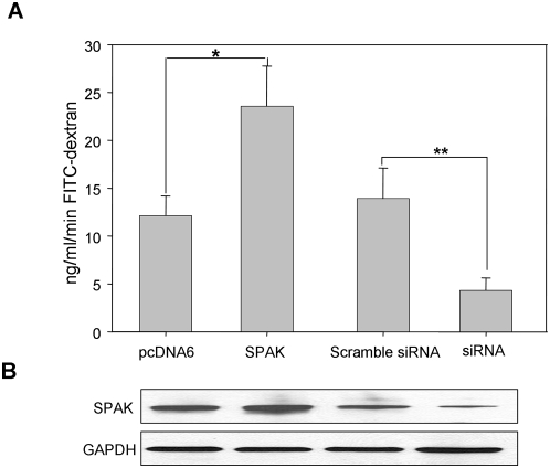
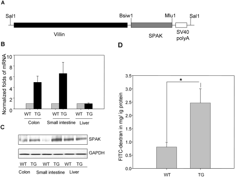
The Ste20-related protein proline/alanine-rich kinase (SPAK) plays important roles in cellular functions such as cell differentiation and regulation of chloride transport, but its roles in pathogenesis of intestinal inflammation remain largely unknown. Here we report significantly increased SPAK expression levels in hyperosmotic environments, such as mucosal biopsy samples from patients with Crohn's disease, as well as colon tissues of C57BL/6 mice and Caco2-BBE cells treated with hyperosmotic medium. NF-kappaB and Sp1-binding sites in the SPAK TATA-less promoter are essential for SPAK mRNA transcription. Hyperosmolarity increases the ability of NF-kappaB and Sp1 to bind to their binding sites. Knock-down of either NF-kappaB or Sp1 by siRNA reduces the hyperosmolarity-induced SPAK expression levels. Furthermore, expression of NF-kappaB, but not Sp1, was upregulated by hyperosmolarity in vivo and in vitro. Nuclear run-on assays showed that hyperosmolarity increases SPAK expression levels at the transcriptional level, without affecting SPAK mRNA stability. Knockdown of SPAK expression by siRNA or overexpression of SPAK in cells and transgenic mice shows that SPAK is involved in intestinal permeability in vitro and in vivo. Together, our data suggest that SPAK, the transcription of which is regulated by hyperosmolarity, plays an important role in epithelial barrier function.


Conflict of interest statement

Competing Interests: The authors have declared that no competing interests exist.

Figures

Figure 1
Figure 1. SPAK expression profile in colon tissue from patients with ulcerative colitis.
A. Immunostaining of SPAK in normal human colon tissue and Crohn's disease (CD) patient colon tissue from mucosal biopsies. SPAK expression (red); nuclear staining by DAPI (blue); SPAK is primarily expressed in epithelial cells. B. The expression of SPAK mRNA in normal human and Crohn's disease (CD) patient colon tissues from mucosal biopsies were quantified by real-time PCR, ** p<0.01. C. 30 μg of protein from normal human colon and Crohn's disease (CD) patient colon from mucosal biopsies were examined by western blot with SPAK antibody, colon tissue from CD patients demonstrated significantly higher levels of SPAK expression (upper part) vs. healthy colon, with GAPDH as the internal loading control.
Figure 2
Figure 2. SPAK expression profile in colon tissue from mice treated with hyperosmolarity.
A. Immunostaining of SPAK in colon sections of mice treated with hyperosmolarity at 0, 1, 3, and 5 days. SPAK (red); nuclear staining by DAPI (blue). B. Real time PCR analysis of SPAK mRNA expression in mucosa from colon tissue of hyperosmolarity treated mice. ** p<0.01. C. Western blot analysis of SPAK (upper part) expression in mucosa from colon tissue of hyperosmolarity treated mice, with GAPDH as the internal loading control.
Figure 3
Figure 3. SPAK expression in hyperosmolarity treated Caco2-BBE cells.
A. Graph of ratio of triton x-100 insoluble actin v.s. soluble actin and western blot. B. Immunostaining of SPAK in colonic Caco2-BBE cells treated with hyperosmolarity at 0 and 15 min. SPAK (green); actin by rhodamine (red). (C) Real time PCR and (D) Western blot demonstrated that treatment of hyperosmolarity increases SPAK expression with GAPDH as internal loading control, ** p<0.01, *** p<0.001. E. Western blot showed that SPAK expression is increased by hyperosmolarity and is recruited to the triton-100 insoluble pool at 0, 1, 3, 8, 15 and 30 min.
Figure 4
Figure 4. Hyperosmolarity regulates SPAK expression at the transcriptional level.
A. Nuclear run-on assay indicated the increase of SPAK mRNA transcription under the treatment of hyperosmolarity, with the mRNA transcription of GAPDH as internal control. B. Hyperosmolarity does not change SPAK mRNA stability; the percentage of remaining SPAK mRNA is shown at the different time point. Solid circle represents the value of real time PCR with the samples from Caco2-BBE cells without treatment, open circle represents the value of real time PCR with the samples from Caco2-BBE treated with actinomycin D, solid triangle represents real time PCR with the samples from Caco2-BBE treated with actinomycin D and hyperosmolarity. C. Northern blot analysis of total RNA from Caco2-BBE cells, Lane 1, no treatment, Lane 2, AcD, Lane 3, hyperosmolarity, and Lane 4, AcD and hyperosmolarity. The lower RNA electrophesis shows equal loading of each condition.
Figure 5
Figure 5. Characterization of SPAK promoter.
A. Schematic representation of human SPAK promoter constructs. the full-length SPAK promoter (nt-1472 to +4); construct I (nt −1050 to +4); construct II (nt −398 to +4); construct III (nt −331 to +4); construct IV (nt −149 to +4) and construct V (nt −72 to +4). Numbers are given in relation to the translational start codon (+1) and indicate 5′-ends of the deletion constructs. The location of the identified positive regulatory region is indicated by a light blue box. Positions of the putative Sp1 (Red) and NF-κB (Yellow) sites are indicated by arrows. B. Promoter activities of the 5′ deleted constructs in un-treated or hyperosmolarity-stimulated Caco2-BBE cells normalized to Renilla Luc activities driven by the phRL-CMV control vector. Activities are expressed as fold inductions over cells transfected with the empty pGL3-basic vector. Each value represents the mean±SD of at least 3 independent sets of transfection experiments performed in triplicate, *p<0.05; **p<0.01. C. Schematic representation of mutated SPAK promoter constructs: the full-length SPAK promoter; I Sp1 binding site (−496); II Sp1 binding site (−303); III Sp1 binding site (−114) and NF-κB binding site (−354). The digits are given in relation to the translational start codon (+1). The location of the identified positive regulatory region is indicated by a light blue box. Positions of the putative Sp1 sites are indicated by arrows and NF-κB is indicated by rectangle. The corresponding mutated transcription factor binding site is indicated by black arrow or black rectangle. D. Effects of mutations of Sp1 or NF-κB binding sites on SPAK promoter activity. The various mutated constructs were transiently transfected into Caco2-BBE cells under the basal (gray bar) or hyperosmolarity conditions. Promoter activity of the full-length wild-type construct was set to 100% (control). Values represent means±SD of at least 3 independent sets of transfection experiments performed in triplicate, *p<0.05, **p<0.01.
Figure 6
Figure 6. EMSA of (A) I Sp1 (−496), (B) II Sp1 (−303), (C) III Sp1 (−114), (D) NF-κB (−354).
Lane 1, biotin-labeled oligonucleotide alone; lane 2, biotin-labeled oligonucleotides incubated with 5 μg Caco2-BBE nuclear extracts; lane 3, biotin-labeled oligonucleotides incubated with 5 μg hyperosmolarity-treated Caco2-BBE nuclear extracts; lane 4, biotin-labeled oligonucleotides incubated with 5 μg Caco2-BBE nuclear extracts in the presence of anti-Sp1 (A–C) or NF-kB (p65) (D) antibodies; lane 5, biotin-labeled oligonucleotides incubated with 5 μg Caco2-BBE nuclear extracts in the presence of non-specific IgG; lane 6, biotin-labeled oligonucleotides incubated with 5 μg Caco2-BBE nuclear extracts in the presence of a 50-fold excess of cold competitor oligonucleotide; lane 7, biotin-labeled binding site-mutated oligonucleotides incubated with 5 μg Caco2-BBE nuclear extracts. E. Chromatin immunoprecipitation (ChIP) assay: the antibodies indicated were incubated with cross-linked DNA isolated from Caco2-BBE cells treated with (+) or without (−) hyperosmolarity, IgG antisera acts as control. Sp1 (I, II, and III) and NF-κB promoter elements in the immunoprecipitates were detected by PCR. The lower panel shows DNA input as template for internal control.
Figure 7
Figure 7. Western blots of transcription factors Sp1 and NF-κB (p65).
A. Western blots of Sp1 and NF-κB (p65) demonstrating hyperosmolarity effect on Sp1 and NF-κB protein levels in vivo. Histone3 acts as a control. B. Western blots of Sp1 and NF-κB (p65) demonstrating hyperosmolarity effect on Sp1 and NF-κB protein levels in vitro. Histone3 acts as a control. C. Reduction of NF-κB but not Sp1 expression reduced SPAK protein expression in unstimulated and in hyperosmolarity-stimulated Caco2-BBE cells. Cells were harvested and subjected to western blot analysis using Sp1, NF-κB (p65), and SPAK antibodies as described in materials and methods. GAPDH acts as a loading control.
Figure 8
Figure 8. SPAK is involved in epithelial barrier function in vitro.
A. In vitro permeability assay in Caco2-BBE cells transfected with pcDNA6, SPAK/pcDNA6, con siRNA or SPAK siRNA with 4 kDa FITC-Dextran. Fluorescence was quantified in lower chamber at 2 hours after the administration of FITC-dextran (λ ex = 492 nm, λ em = 510), *p<0.05, **p<0.01. B. Western blot of Caco2-BBE cells protein scraped from filter after the in vitro permeability assay.
Figure 9
Figure 9. SPAK is involved in epithelial barrier function in vivo.
A. Schematic diagram of villin/SPAK transgene construct, full length SPAK cDNA was cloned into villin vector by Bsiw1/Mlu1 sites; villin/SPAK was digested with Sal1 before microinjection. B. SPAK is tissue-specifically over-expressed in intestine by real time PCR with samples from small intestine, colon and liver. C. SPAK is specifically over-expressed in intestine by western blot with samples from small intestine, colon and liver. D. in vivo permeability assay in villin/SPAK transgenic mice, WT: wide type; TG: transgenic mice, ** p<0.01.

Similar articles

Cited by

References

    1. Schilli R, Breuer RI, Klein F, Dunn K, Gnaedinger A, et al. Comparison of the composition of faecal fluid in Crohn's disease and ulcerative colitis. Gut. 1982;23:326–332. - PMC - PubMed
    1. Katz KD, Hollander D, Vadheim CM, McElree C, Delahunty T, et al. Intestinal permeability in patients with Crohn's disease and their healthy relatives. Gastroenterology. 1989;97:927–931. - PubMed
    1. Sandle GI, Higgs N, Crowe P, Marsh MN, Venkatesan S, et al. Cellular basis for defective electrolyte transport in inflamed human colon. Gastroenterology. 1990;99:97–105. - PubMed
    1. Vernia P, Gnaedinger A, Hauck W, Breuer RI. Organic anions and the diarrhea of inflammatory bowel disease. Dig Dis Sci. 1988;33:1353–1358. - PubMed
    1. Cheromcha DP, Hyman PE. Neonatal necrotizing enterocolitis. Inflammatory bowel disease of the newborn. Dig Dis Sci. 1988;33:78S–84S. - PMC - PubMed

Publication types

MeSH terms