. 2009 Apr;5(4):e1000360.
doi: 10.1371/journal.ppat.1000360. Epub 2009 Apr 3.

Soluble CD4 and CD4-mimetic compounds inhibit HIV-1 infection by induction of a short-lived activated state

Affiliations

Soluble CD4 and CD4-mimetic compounds inhibit HIV-1 infection by induction of a short-lived activated state

Hillel Haim et al. PLoS Pathog. 2009 Apr.

Abstract

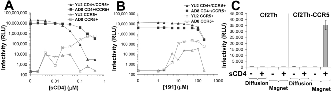
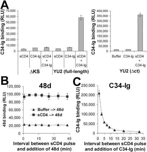
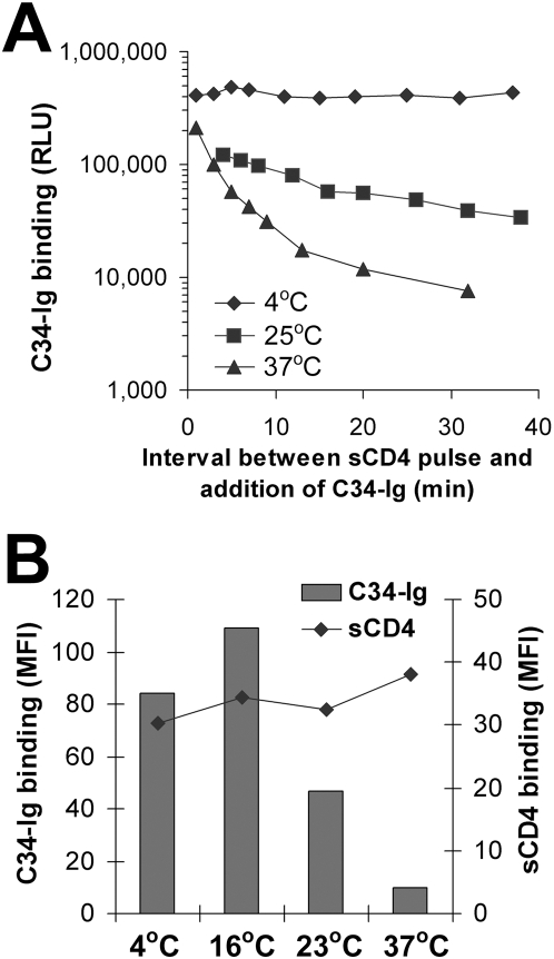
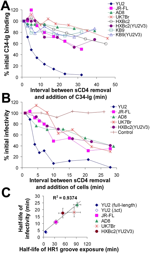
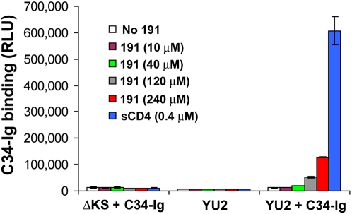
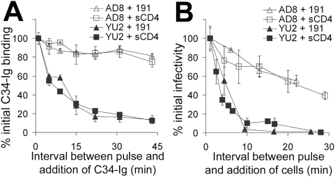
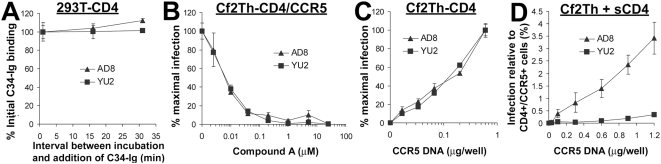
Binding to the CD4 receptor induces conformational changes in the human immunodeficiency virus (HIV-1) gp120 exterior envelope glycoprotein. These changes allow gp120 to bind the coreceptor, either CCR5 or CXCR4, and prime the gp41 transmembrane envelope glycoprotein to mediate virus-cell membrane fusion and virus entry. Soluble forms of CD4 (sCD4) and small-molecule CD4 mimics (here exemplified by JRC-II-191) also induce these conformational changes in the HIV-1 envelope glycoproteins, but typically inhibit HIV-1 entry into CD4-expressing cells. To investigate the mechanism of inhibition, we monitored at high temporal resolution inhibitor-induced changes in the conformation and functional competence of the HIV-1 envelope glycoproteins that immediately follow engagement of the soluble CD4 mimics. Both sCD4 and JRC-II-191 efficiently activated the envelope glycoproteins to mediate infection of cells lacking CD4, in a manner dependent on coreceptor affinity and density. This activated state, however, was transient and was followed by spontaneous and apparently irreversible changes of conformation and by loss of functional competence. The longevity of the activated intermediate depended on temperature and the particular HIV-1 strain, but was indistinguishable for sCD4 and JRC-II-191; by contrast, the activated intermediate induced by cell-surface CD4 was relatively long-lived. The inactivating effects of these activation-based inhibitors predominantly affected cell-free virus, whereas virus that was prebound to the target cell surface was mainly activated, infecting the cells even at high concentrations of the CD4 analogue. These results demonstrate the ability of soluble CD4 mimics to inactivate HIV-1 by prematurely triggering active but transient intermediate states of the envelope glycoproteins. This novel strategy for inhibition may be generally applicable to high-potential-energy viral entry machines that are normally activated by receptor binding.


Conflict of interest statement

The authors have declared that no competing interests exist.

Figures

Figure 1
Figure 1. Inhibition and activation of infection by sCD4 and the CD4-mimetic compound 191.
Recombinant HIV-1(YU2) or HIV-1(AD8) strains were incubated with CD4CCR5+ or CD4+CCR5+ Cf2Th cells for 48 hours in the presence of sCD4 (A) or 191 (B). Infectivity was determined by measurement of luciferase activity. (C) HIV-1(YU2) was incubated for three minutes in the presence or absence of sCD4 (40 µg/ml, 0.8 µM) and then adsorbed by diffusion or magnetically to cultures of the indicated cell type. Measured luciferase activity is presented as mean relative light units (RLU)±standard error of the mean (s.e.m.) of three replicate samples.
Figure 2
Figure 2. Soluble CD4-induced changes in the gp120 coreceptor–binding region and gp41 HR1 region.
(A) A cell-based ELISA was used to measure the binding of C34-Ig, which detects the HR1 gp41 region ,, to the trimeric HIV-1 envelope glycoproteins. COS-1 cells were transfected with the negative control ΔKS plasmid or plasmids expressing the full-length YU2 envelope glycoproteins (left) or the cytoplasmic tail-deleted (Δct) YU2 envelope glycoproteins (right). Two days later, cells were incubated with C34-Ig (20 µg/ml), in the presence or absence of sCD4 (20 µg/ml). C34-Ig binding was measured using a secondary horseradish peroxidase-conjugated antibody. (B,C) Change over time in the exposure of CD4-induced epitopes. Cells that express the YU2(Δct) envelope glycoproteins were pulsed for three minutes with sCD4 (40 µg/ml). Cells were then washed three times and incubated for different time periods at 37°C. The monoclonal antibody 48d (B) or C34-Ig (C) was then added. The bound 48d or C34-Ig molecules were detected using a horseradish peroxidase-conjugated anti-human IgG Fc antibody, as described in Materials and Methods. Values represent the mean RLU (±s.e.m.) of two replicate samples.
Figure 3
Figure 3. Temperature dependence of the decay of HR1 groove exposure.
(A) Decay of HR1 groove exposure at different temperatures after pulse activation with sCD4 (40 µg/ml; 0.8 µM) was measured by a cell-based ELISA, as described in Materials and Methods. The indicated values for C34-Ig binding were obtained by subtracting the values of C34-Ig binding in the absence of sCD4 from the binding measured at each time point after the sCD4 pulse. Mean background levels of C34-Ig binding (in the absence of sCD4) were 10316, 21413, and 19028 RLU for the experiments performed at 4°C, 25°C, and 37°C, respectively. (B) Binding of C34-Ig and sCD4 to the HIV-1 envelope glycoproteins was measured at different temperatures. Cells that express the YU2(Δct) envelope glycoproteins were incubated simultaneously with both C34-Ig (20 µg/ml) and sCD4 (20 µg/ml; 0.4 µM) at the indicated temperature. Binding of sCD4 and C34-Ig was detected by FACS analysis using a fluorescein-conjugated anti-CD4 antibody and a phycoerythrin-conjugated goat anti-human IgG antibody, respectively (see Materials and Methods). The mean fluorescence intensity (MFI) of the cells in both channels is shown.
Figure 4
Figure 4. Relationship between infectivity decay and loss of HR1 groove exposure.
(A) Decay of HR1 groove exposure of different HIV-1 envelope glycoproteins was studied at 25°C after pulse activation with sCD4. COS-1 cells expressing the indicated HIV-1 envelope glycoproteins were pulsed with sCD4 (40 µg/ml; 0.8 µM) for 3 minutes, followed by assessment of HR1 groove exposure using the cell-based ELISA method, as described in the Figure 2 legend. (B) Recombinant HIV-1 virions carrying the indicated envelope glycoproteins were pulsed with sCD4 (40 µg/ml; 0.8 µM) for 3 minutes and incubated at 25°C. After the indicated times, CD4CCR5+ Cf2Th cells were added to the viruses. For the sample marked as control, HIV-1(YU2) was pulsed with buffer and then CD4+CCR5+ cells were added. Two days later, virus infectivity was assessed by measuring luciferase activity in the target cells. (C) The relationship between the decay rate of HR1 groove exposure and the decay rate of infectivity, both measured at 25–27°C after pulse activation with sCD4, is shown for the panel of HIV-1 envelope glycoproteins. Half-lives were determined by fitting a model function to the data using nonlinear regression. Values represent half-lives (±s.e.m. for both variables) derived from two to four separate experiments for each envelope glycoprotein variant.
Figure 5
Figure 5. Effect of compound 191 on exposure of the gp41 HR1 groove.
COS-1 cells transfected with a plasmid expressing the YU2(Δct) envelope glycoproteins or with the control ΔKS plasmid, which does not express an envelope glycoprotein on the cell surface, were incubated with the indicated molecules in the presence or absence of C34-Ig (20 µg/ml). The data represent the means (±s.e.m.) of two replicate samples.
Figure 6
Figure 6. Longevity of the HIV-1 envelope glycoprotein intermediate at room temperature after activation by sCD4 or 191.
(A) The decay of HR1 groove exposure for cell-surface–expressed YU2 and AD8 envelope glycoproteins is compared after pulse activation with 191 (360 µM) or sCD4 (40 µg/ml, 0.8 µM). (B) The decay of the ability to infect CD4CCR5+ Cf2Th cells after pulse activation with 191 or sCD4 is compared.
Figure 7
Figure 7. HIV-1 activation by native CD4 and sCD4.
(A) The stability of HR1 groove exposure was measured after activation of the YU2 or AD8 envelope glycoproteins by cell-surface CD4. Background was defined as C34-Ig binding to cells transfected with the YU2-GS8 construct, which engages sCD4 with an affinity similar to that of the wild-type YU2 envelope glycoproteins but does not expose the HR1 groove (see Figure S6). The background was subtracted from all measurements, which are presented as percent (±s.e.m.) of C34-Ig binding measured at the initial time point. (B) The effect of the CCR5 antagonist “Compound A” , on infectivity of recombinant HIV-1 pseudotyped with the indicated envelope glycoproteins was investigated. Cf2Th-CD4/CCR5 cells were infected with HIV-1(AD8) or HIV-1(YU2) in the presence of increasing concentrations of Compound A. Data are presented as the percentage (±s.e.m.) of infection measured in the absence of the compound. (C) The effect of CCR5 expression on the infection of CD4-expressing cells by HIV-1(AD8) or HIV-1(YU2) was examined. Cf2Th-CD4 cells were transfected with different amounts of a plasmid that expresses human CCR5. Two days later, transfected cells were incubated with HIV-1(AD8) or HIV-1(YU2). Infectivity is expressed as the percentage (±s.e.m.) of infectivity measured for cells transfected with the highest amount of the CCR5-expressing plasmid. (D) The graph shows infection by cell-bound virus of CD4 Cf2Th cells transfected with different amounts of the CCR5-expressing plasmid in the presence of 20 µg/ml sCD4. Infectivity is expressed as the percentage (±s.e.m.) of infection measured in cells transfected with plasmids expressing CD4 and CCR5 (0.6 and 0.9 µg, respectively, of each plasmid per well).

Similar articles

Cited by

References

    1. Wyatt R, Sodroski J. The HIV-1 envelope glycoproteins: fusogens, antigens, and immunogens. Science. 1998;280:1884–1888. - PubMed
    1. Myszka DG, Sweet RW, Hensley P, Brigham-Burke M, Kwong PD, et al. Energetics of the HIV gp120-CD4 binding reaction. Proc Natl Acad Sci U S A. 2000;97:9026–9031. - PMC - PubMed
    1. Choe H, Farzan M, Sun Y, Sullivan N, Rollins B, et al. The beta-chemokine receptors CCR3 and CCR5 facilitate infection by primary HIV-1 isolates. Cell. 1996;85:1135–1148. - PubMed
    1. Deng H, Liu R, Ellmeier W, Choe S, Unutmaz D, et al. Identification of a major co-receptor for primary isolates of HIV-1. Nature. 1996;381:661–666. - PubMed
    1. Dragic T, Litwin V, Allaway GP, Martin SR, Huang Y, et al. HIV-1 entry into CD4+ cells is mediated by the chemokine receptor CC-CKR-5. Nature. 1996;381:667–673. - PubMed

Publication types

MeSH terms

Substances