. 2009 Apr;5(4):e1000361.
doi: 10.1371/journal.ppat.1000361. Epub 2009 Apr 3.

Caspase-7 activation by the Nlrc4/Ipaf inflammasome restricts Legionella pneumophila infection

Affiliations

Caspase-7 activation by the Nlrc4/Ipaf inflammasome restricts Legionella pneumophila infection

Anwari Akhter et al. PLoS Pathog. 2009 Apr.

Abstract

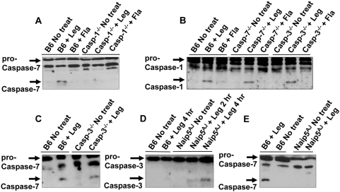
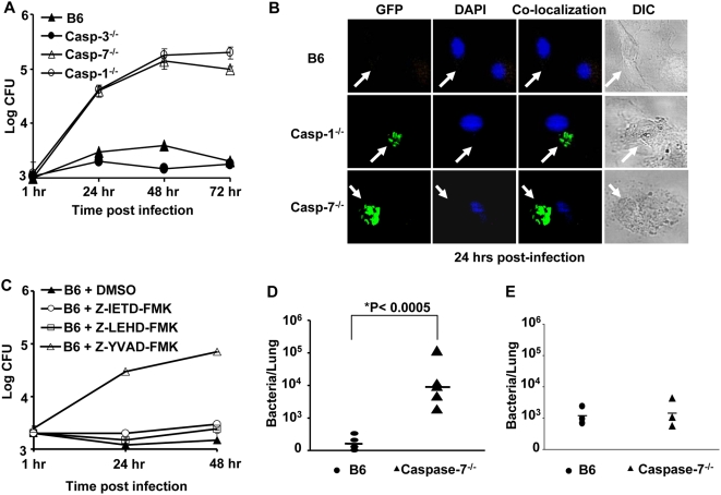
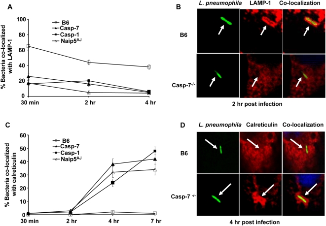
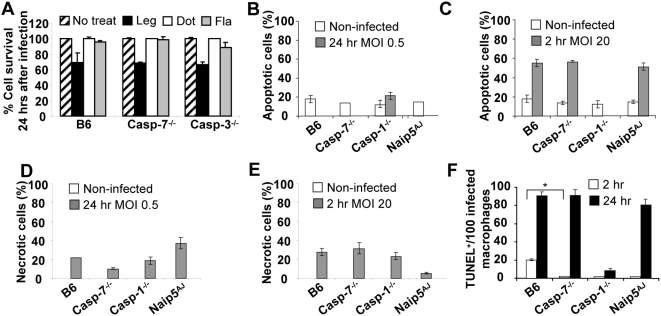
Legionella pneumophila (L. pneumophila), the causative agent of a severe form of pneumonia called Legionnaires' disease, replicates in human monocytes and macrophages. Most inbred mouse strains are restrictive to L. pneumophila infection except for the A/J, Nlrc4(-/-) (Ipaf(-/-)), and caspase-1(-/-) derived macrophages. Particularly, caspase-1 activation is detected during L. pneumophila infection of murine macrophages while absent in human cells. Recent in vitro experiments demonstrate that caspase-7 is cleaved by caspase-1. However, the biological role for caspase-7 activation downstream of caspase-1 is not known. Furthermore, whether this reaction is pertinent to the apoptosis or to the inflammation pathway or whether it mediates a yet unidentified effect is unclear. Using the intracellular pathogen L. pneumophila, we show that, upon infection of murine macrophages, caspase-7 was activated downstream of the Nlrc4 inflammasome and required caspase-1 activation. Such activation of caspase-7 was mediated by flagellin and required a functional Naip5. Remarkably, mice lacking caspase-7 and its macrophages allowed substantial L. pneumophila replication. Permissiveness of caspase-7(-/-) macrophages to the intracellular pathogen was due to defective delivery of the organism to the lysosome and to delayed cell death during early stages of infection. These results reveal a new mechanism for caspase-7 activation downstream of the Nlrc4 inflammasome and present a novel biological role for caspase-7 in host defense against an intracellular bacterium.


Conflict of interest statement

The authors have declared that no competing interests exist.

Figures

Figure 1
Figure 1. The host protein Nlrc4 and bacterial flagellin are required for the induction of caspase-7 activation by L. pneumophila.
(A) Wild-type C57BL/6 (B6) derived macrophages were not treated (No treat) or infected with wild-type L. pneumophila (Leg) at different multiplicities of infection (MOI) (3-0.5) or with L. pneumophila type IV secretion mutant (Dot) at an MOI of 1 for 2 hrs. (B) B6 derived macrophages were infected with Leg at an MOI of 0.5 for different durations. (C,D) B6 and Nlrc4−/− macrophages were infected with Leg, the Dot mutant, the mutant lacking flagellin (Fla) (MOI of 0.5) or treated with streptolysin O (SLO) alone, flagelin alone (flagel), or SLO and purified flagel (C), or treated with SLO and bacterial lipoprotein (LP), bacterial RNA, bacterial DNA, or bacterial lipopolysaccharide (LPS) (D). (A–D) Immunoblots were developed with anti-caspase-7 antibody and are representative of more than three independent experiments.
Figure 2
Figure 2. Caspase-1 and Naip5 are required for caspase-7 activation by L. pneumophila.
(A) Wild-type C57BL/6 (B6) and caspase-1−/− (casp-1−/−) derived macrophages were not treated (No treat) or infected with L. pneumophila (Leg), or the mutant lacking flagellin (Fla) for 2 hr. Cell lysates were analyzed by western blot with anti–caspase-7 antibodies. (B) B6, caspase-7−/− (casp-7−/−), and caspase-3−/− (casp-3−/−) macrophages were treated with wild-type Leg or with Fla mutant, then cell lysates were analyzed by western blot with anti–caspase-1 antibodies. (C) B6 and casp-3−/− were infected with Leg, then cell lysates were examined by western blots with anti–caspase-7 antibodies. (D,E) B6– and A/J–derived macrophages (Naip5AJ) were infected or not with Leg for times indicated, then cell lysates were examined by western blot with anti–caspase-3 (D) or –caspase-7 (E) antibodies.
Figure 3
Figure 3. Caspase-7 restricts L. pneumophila replication in macrophages and in mice.
(A) Wild-type C57BL/6 (B6), caspase-3−/− (casp-3−/−), caspase-7−/− (casp-7−/−), or caspase-1−/− (casp-1−/−) macrophages were infected with wild-type L. pneumophila, then colony forming units (CFU) were scored at indicated time points. The results represent the mean of four independent experiments ±SD. (B) B6, casp-1−/−, or casp-7−/− macrophages were infected with GFP–expressing L. pneumophila for 24 hrs and examined by fluorescence microscopy. (C) B6 macrophages were treated or not with 50 µM caspase-1 inhibitor (YVAD), caspase-8 inhibitor (IETD), caspase-9 inhibitor (LEHD), or caspase-3 inhibitor or dimethylsulfoxide alone (DMSO) 45 min before infection with L. pneumophila. Cells were lysed and the number of colony forming units (CFU) was quantified at 1, 24, and 48 hrs post infection. The results represent the mean of three independent experiments ±SD. (D) B6 and caspase-7−/− mice received 106 wild-type L. pneumophila intra-tracheally. Lungs were homogenized and plated for CFUs at 96 hrs post-infection. Data were analyzed by Student's t-test. *, P value≤0.05. (E) B6 and caspase-7−/− mice were infected with L. pneumophila for 6 hrs and lungs were examined for bacterial load as described in D.
Figure 4
Figure 4. L. pneumophila–containing phagosomes avoid the fusion with the lysosome in caspase-7−/−, caspase-1−/−, and A/J–derived macrophages but not in wild-type macrophages.
Macrophages from wild-type C57BL/6 (B6), caspase-7−/− (casp-7−/−), caspase-1−/− (casp-1−/−), or A/J mice were seeded on cover slips and infected with L. pneumophilla. The internalized bacteria were quantified for co-localization with the late endosomal marker LAMP-1 (A and B), or for the localization with the endoplasmic reticulum marker calreticulin at times indicated (C and D). Data represent the mean of three independent experiments ±SD.
Figure 5
Figure 5. The role of caspase-1 and -7 in L. pneumophila–induced apoptosis at different multiplicities of infection (MOI).
(A) Wild-type C57BL/6 (B6), casp-7−/−, and casp-3−/− macrophages were not treated (striped bars), or treated with wild-type L. pneumophila (black bars), type IV secretion mutant (white bars), or flagellin mutant (grey bars) at MOI of 0.5 for 24 hrs, then percent cell survival was measured by LDH release from overall population of macrophages. Analysis of apoptosis (B and C) or necrosis (D and E) of macrophages not infected (white bars) or infected (grey bars) with wild-type L. pneumophila at an MOI of 0.5 for 24 hrs (B and D) or MOI of 20 for 2 hrs (C and E) by ELISA photometric enzyme immunoassay analysis of the cytoplasmic (apoptosis) and extracellular (necrosis) histone-associated-DNA-fragments. (F) Microscopic single cell analysis of apoptosis by TUNEL staining after 2 hrs (white bars) and 24 hrs (black bars) of infection with wild-type L. pneumophila at MOI of 0.5. The y axis represents the number of TUNEL positive (TUNEL+) macrophages among 100 infected macrophages. *, P value≤0.05. (A–F) The results represent the mean of three independent experiments ±SD.

Similar articles

Cited by

References

    1. Li J, Yuan J. Caspases in apoptosis and beyond. Oncogene. 2008;27:6194–6206. - PubMed
    1. Martinon F, Tschopp J. Inflammatory caspases: linking an intracellular innate immune system to autoinflammatory diseases. Cell. 2004;117:561–574. - PubMed
    1. Kuribayashi K, Mayes PA, El-Deiry WS. What are caspases 3 and 7 doing upstream of the mitochondria? Cancer Biology & Therapy. 2006;5:763–765. - PubMed
    1. Stennicke HR, Salvesen GS. Biochemical characteristics of caspases-3, -6, -7, and -8. Journal of Biological Chemistry. 1997;272:25719–25723. - PubMed
    1. Thornberry NA, Molineaux SM. Interleukin-1 beta converting enzyme: a novel cysteine protease required for IL-1 beta production and implicated in programmed cell death. Protein Science. 1995;4:3–12. - PMC - PubMed

Publication types

MeSH terms