. 2009;4(4):e5033.
doi: 10.1371/journal.pone.0005033. Epub 2009 Apr 3.

The promoter of the pri-miR-375 gene directs expression selectively to the endocrine pancreas

Affiliations

The promoter of the pri-miR-375 gene directs expression selectively to the endocrine pancreas

Tali Avnit-Sagi et al. PLoS One. 2009.

Abstract

microRNAs (miRNAs) are known to play an essential role in controlling a broad range of biological processes including animal development. Accordingly, many miRNAs are expressed preferentially in one or a small number of cell types. Yet the mechanisms responsible for this selectivity are not well understood. The aim of this study was to elucidate the molecular basis of cell-specific expression of the pri-miR-375 gene, which is selectively expressed in pancreatic islets, and has been implicated both in the development of islets, and the function of mature pancreatic beta cells. An evolutionarily conserved 768 bp region of DNA upstream of the pri-miR-375 gene was linked to GFP and luciferase reporter genes, and expression monitored in transgenic mice and transfected cultured cells. Deletion and targeted mutagenesis analysis was used to evaluate the functional significance of sequence blocks within the upstream fragment. 5'-RACE analysis was used for mapping the pri-miR-375 gene transcription start site. The conserved 768 bp region was able to direct preferential expression of a GFP reporter gene to pancreatic islets in transgenic mice. Deletion analysis using a luciferase reporter gene in transfected cultured cell lines confirmed the cell specificity of the putative promoter region, and identified several key cis-elements essential for optimal activity, including E-boxes and a TATA sequence. Consistent with this, 5'-RACE analysis identified a transcription start site within this DNA region, 24 bp downstream of the TATA sequence. These studies define the promoter of the pri-miR-375 gene, and show that islet-specific expression of the pri-miR-375 gene is controlled at the transcriptional level. Detailed analysis of the transcriptional mechanisms controlling expression of miRNA genes will be essential to permit a comprehensive understanding of the complex role of miRNAs such as miR-375 in developmental processes.


Conflict of interest statement

Competing Interests: The authors have declared that no competing interests exist.

Figures

Figure 1
Figure 1. miR-375 gene regulatory region.
Genomic locus of the mouse miR-375 gene and upstream conserved regions from UCSC browser (upper figure) with a graphic representation of the conserved regions upstream of the pre-miR-375 sequence (lower figure). The location of conserved blocks 1–4, and pre-miR-375 is indicated.
Figure 2
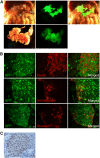
Figure 2. Expression of GFP in pancreas and islets of miR-375-EGFP transgenic mice.
A. Abdominal organs of a 6 week old transgenic mouse (bright field - upper left, GFP - upper middle, merged -upper right. I, intestine. C, colon. P, pancreas. Sp, spleen. St, stomach). The pancreas was dissected and photographed (bright field - lower left, GFP – lower right). B. Immunofluorescence analysis of pancreas sections showing GFP (green), and insulin, somatostatin or glucagon (red). C. Immunohistochemical analysis using anti-GFP antibodies, counter-stained with hematoxylin. Scale bar indicated in B and C represents 10 µm.
Figure 3
Figure 3. Mapping of transcription start site of miR-375 gene by 5′-RACE.
A. DNase-treated total RNA from HIT cells transfected with the pGL3-375a construct was reverse transcribed, using a primer complementary to the luciferase sequence. The cDNA was poly-(dG) tailed and amplified by PCR using a poly-C primer and a nested primer complementary to block 4 (lane 1). Lane 2, no RT control; lane 3, no template control. Marker sizes are indicated. The location of the band in lane 1 is indicated by an arrow. B. Alignment of miR-375 upstream sequences (from −96 to +244, relative to transcription start site) of mouse, rat and human. The primer used for PCR amplification is located at the middle of block 4. The TATA sequence is marked by a box. The large arrow-head indicates the major transcription start site. Small arrow-heads indicate the start site of shorter species detected by 5′-RACE. C. Representation of the pGL3-375a construct. Conserved regions 1–4 are indicated. The luciferase gene is indicated as an unmarked open box. The arrow indicates the transcription start site revealed by 5′-RACE analysis. Numbers above the bar indicate location relative to the transcription start site.
Figure 4
Figure 4. Activity of the miR-375 promoter in cultured cells.
The putative promoter region of the miR-375 gene (construct 375b) was ligated upstream to the firefly luciferase reporter gene in the promoter-less pGL3-basic vector. As a positive control, the insulin promoter was fused upstream to the firefly luciferase reporter gene. The herpes simplex virus thymidine kinase (TK) promoter fused upstream to the firefly luciferase reporter gene served as non cell-specific control promoter. Promoter activity of each construct was determined following transfection to the beta cell line HIT or the non-beta cell line CHO. Values were normalized for transfection efficiency according to the activity of a co-transfected renilla luciferase plasmid and expressed relative to the activity of pGL3-basic vector. Each data point represents the mean±SEM of at least three independent transfection experiments.
Figure 5
Figure 5. Mapping of transcriptional regulation regions by deletion analysis.
A. Portions of the miR-375 upstream region were deleted according to the location of the conserved blocks (Fig. 1) and ability to drive expression of firefly luciferase reporter gene was determined in the context of the promoter-less pGL3-basic vector. Values shown are mean±SEM of at least 3 independent transfection experiments. B. Construct 375a or 375g were tested for enhancer activity. The different regions were ligated (in both orientations) upstream to the TK promoter driving expression of the firefly luciferase reporter gene. Normalised luciferase activity was expressed relative to the activity of pGL3-basic vector (A) or TK-LUC (B). Values shown are mean±SEM of at least 3 independent transfection experiments.
Figure 6
Figure 6. Mutagenesis of putative transcription factor-binding sites.
A and B. Nucleotide sequence of conserved regions of mouse miR-375 promoter. Stars indicate nucleotides conserved among mouse, rat, and human miR-375 genes. Location relative to the transcription start site is marked by numbers at the beginning and end of the sequence. A. Alignment of Block 1. E boxes are indicated. B. Alignment of block 2. Sites of binding sites for putative transcription factors, E boxes, and mutations are marked on sequence. C. Specific sites were mutated and reporter plasmids containing the mutations were tested for luciferase activity as before. Values shown are mean±SEM of at least 4 independent transfection experiments. *, p<0.05, as compared with wild-type. D. The indicated constructs were transfected to HIT cells in the presence or absence of Id2 or Id3 expression vectors. Normalised luciferase activity was expressed relative to the activity of pGL3-basic vector. Values shown are mean±SEM of at least 4 independent transfection experiments. *, p<0.05, as compared with transfections performed in the absence of Id expression vector.

References

    1. Bartel DP. MicroRNAs: genomics, biogenesis, mechanism, and function. Cell. 2004;116:281–297. - PubMed
    1. Stefani G, Slack FJ. Small non-coding RNAs in animal development. Nat Rev Mol Cell Biol. 2008;9:219–230. - PubMed
    1. Griffiths-Jones S, Grocock RJ, van Dongen S, Bateman A, Enright AJ. miRBase: microRNA sequences, targets and gene nomenclature. Nucleic Acids Res. 2006;34:D140–144. - PMC - PubMed
    1. Saini HK, Griffiths-Jones S, Enright AJ. Genomic analysis of human microRNA transcripts. Proc Natl Acad Sci U S A. 2007;104:17719–17724. - PMC - PubMed
    1. Lee Y, Kim M, Han J, Yeom KH, Lee S, et al. MicroRNA genes are transcribed by RNA polymerase II. Embo J. 2004;23:4051–4060. - PMC - PubMed

Publication types