Review
. 2009:48:225-42.
doi: 10.1007/400_2009_1.

Axonal protein synthesis and the regulation of local mitochondrial function

Affiliations
Review

Axonal protein synthesis and the regulation of local mitochondrial function

Barry B Kaplan et al. Results Probl Cell Differ. 2009.

Abstract

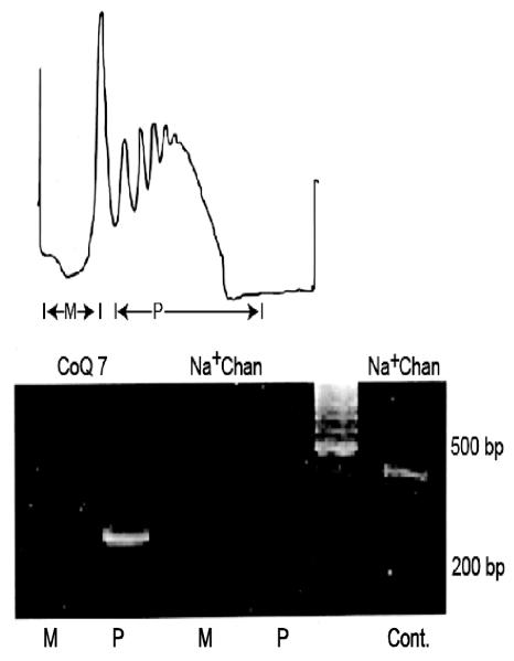
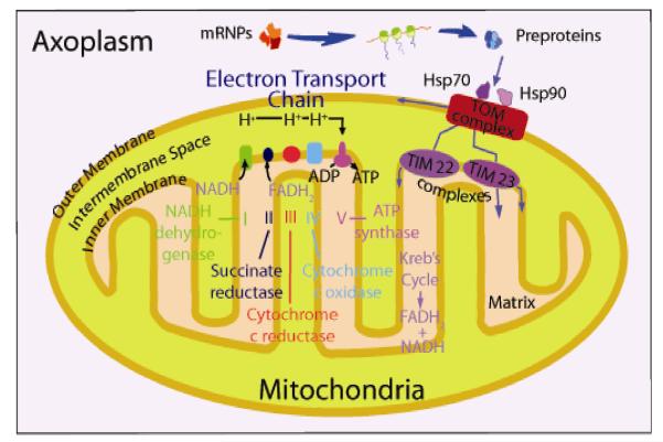
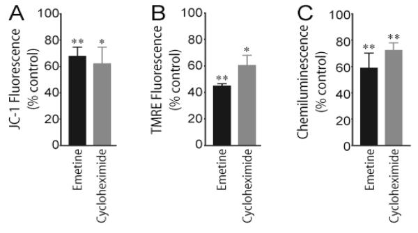
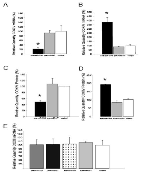
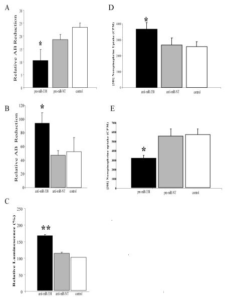
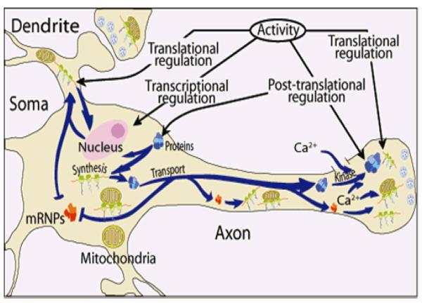
Axons and presynaptic nerve terminals of both invertebrate and mammalian SCG neurons contain a heterogeneous population of nuclear-encoded mitochondrial mRNAs and a local cytosolic protein synthetic system. Nearly one quarter of the total protein synthesized in these structural/functional domains of the neuron is destined for mitochondria. Acute inhibition of axonal protein synthesis markedly reduces the functional activity of mitochondria. The blockade of axonal protein into mitochondria had similar effects on the organelle's functional activity. In addition to mitochondrial mRNAs, SCG axons contain approximately 200 different microRNAs (miRs), short, noncoding RNA molecules involved in the posttranscriptional regulation of gene expression. One of these miRs (miR-338) targets cytochrome c oxidase IV (COXIV) mRNA. This nuclear-encoded mRNA codes for a protein that plays a key role in the assembly of the mitochondrial enzyme complex IV and oxidative phosphorylation. Over-expression of miR-338 in the axon markedly decreases COXIV expression, mitochondrial functional activity, and the uptake of neurotransmitter into the axon. Conversely, the inhibition of endogeneous miR-338 levels in the axon significantly increased mitochondrial activity and norepinephrine uptake into the axon. The silencing of COXIV expression in the axon using short, inhibitory RNAs (siRNAs) yielded similar results, a finding that indicated that the effects of miR-338 on mitochondrial activity and axon function were mediated, at least in part, through local COXIV mRNA translation. Taken together, recent findings establish that proteins requisite for mitochondrial activity are synthesized locally in the axon and nerve terminal, and call attention to the intimacy of the relationship that has evolved between the distant cellular domains of the neuron and its energy generating systems.


Figures

Fig. 1
Fig. 1
Nuclear-encoded m RNAs are actively translated in squid brain synaptosomes. (A) Polysomes were prepared from squid optic lobe synaptosomes and were displayed on linear sucrose density gradients. Gradients were divided into monosome (M) and polysome (P) fractions. UV absorbance of RNA was monitored continuously at 254 nm (B) RT-PCR analysis of monosome and polysome fractions using gene-specific primer sets for the nuclear-encoded mitochondrial protein CoQ7 and Na+ channel used as an internal control. PCR products were fractionated on agarose gels and visualized by ethidium bromide staining. The absence of amplicons generated from the Na+ channel primers indicates that the polysome fraction is devoid of RNA contamination from the neuronal cell soma. Cont, Na+ channel amplicons obtained from total RNA prepared from the optic lobe; bp, base-pairs; MW, molecular weight. Reproduced from Gioio et al. (2001).
Fig. 2
Fig. 2
SCG axons contain a heterogeneous population of mRNA. (A) RT-PCR analyses performed on total RNA isolated from SCG axons and somas. Note the shift in abundance of β-tubulin mRNA relative to H+-ATP synthase and DNA Polymerase γ mRNAs between axon and soma. (B) PCR products for β- tubulin and COXIV, a nuclear-encoded mitochondrial protein, were size-fractionated by agarose gel electrophoresis and visualized by ethidium bromide staining. MW, molecular weight. (C) COX IV subunit mRNA is present in SCG axons, as demonstrated with in situ hybridization histochemistry. Representative phase contrast photomicrographs of SCG axons after fixation and hybridization with antisense (COX IV antisense) and sense (Control; COX IV sense) riboprobes. Bar = 10 μm. From Hillefors et al. (2007).
Fig. 3
Fig. 3
Model of a mitochondrion showing the outer and inner membrane, intramembrane space, and matrix. The Kreb’s cycle takes place in the matrix and the electron transport chain, generating ATP, is located in the inner membrane. Molecular chaperones such as Hsp70 and Hsp90 help facilitate the import of pre-proteins synthesized in the axoplasm into the organelle via the translocase of outermembrane (TOM) complex, which is located on the mitochondrion outer membrane.
Fig. 4
Fig. 4
Inhibition of local protein synthesis decreases axonal mitochondrial membrane potential and generation of ATP. Axons in one side compartment of the Campenot cell culture chamber were exposed to the protein synthesis inhibitors emetine and cycloheximide for 3 hours and mitochondrial membrane potential assessed by subsequent treatment with the mitochondrial-specific fluorescent dyes JC-1 (A) or TMRE (B). Distal axons present in the contralateral side compartment served as vehicle-treated controls. Values for mitochondrial membrane potentials, as determined with each dye, are given as Mean ± S.E.M. (*P < 0.05, **P < 0.01) (C) Protein synthesis inhibitors impede the recovery of depolarization-induced decreases in axonal ATP levels. Depolarization was induced by exposing untreated SCG axons and axons exposed to emetine or cycloheximide for 3 hours followed by a 5 min exposure to 50 mM KCl. Axonal ATP levels were measured using ATPlite 1step kit and a microplate reader. Data shown are provided as the Mean ± S.E.M. (**P < 0.01). Reproduced with modification from Hillefors et al. (2007).
Fig. 5
Fig. 5
Introduction of miRNA-338 into distal SCG axons reduces COXIV expression . Quantification of COXIV mRNA levels in the distal axons of SCG neurons transfected with pre-miR-338 (A), or anti-miR-338 (B), as determined by quantitative RT-PCR 24 hours after oligonucleotide transfection. COXIV mRNA levels are expressed relative to β-actin mRNA. Values given are the Mean ± S.E.M. (*P<0.05). The introduction into the distal axons of pre-miR-338 (C) or anti-miR-338 (D) also altered axon COXIV protein levels 24 hours after transfection as determined by immunoblot analyses. Values shown are Mean ±S.E.M. (*P<0.05). Transfection of SCG axons with either precursor or anti-miR-338 oligonucleotides did not affect the relative abundance of COXII mRNA in the axon as shown by quantitative RT-PCR (E). Reproduced from Aschrafi et al. (2008) with modification.
Fig. 6
Fig. 6
MiR-338 mediated alteration in COXIV levels modulates metabolic activity of mitochondria and neurotransmitter uptake into the distal axons of sympathetic neurons. SCG axons were transfected with either precursor miR-338 (A), anti-miR-338 (B), or non-targeting short oligonucleotides and metabolic activity of the axons assessed using the redox dye Alomar Blue (AB). Data represent Mean ±S.E.M. (* P < 0.05). ATP levels were measured in anti-miR-338 transfected axons using the luciferase cell viability assay (C). Values are given in arbitrary luminescence units and represent the Mean ±S.E.M. (** P <0.0001). Distal axons were also transfected with either anti-miR-338 (D), pre-miR-338 (E), or non-targeting short oligonucleotides (NT) and were subsequently exposed to radiolabeled norepinephrine (NE). NE uptake into distal treated axons was measured by liquid scintillation spectrometry. Data represent Mean ±S.E.M. (* P < 0.002). Reproduced with modification from Aschrafi et al. (2008).
Fig. 7
Fig. 7. Knock-down of local COXIV levels decreases axonal respiration and ATP levels, and diminishes NE uptake into distal axons
Two independent siRNA oligonucleotides targeted against COXIV mRNA were introduced into distal axons of SCG neurons by lipofection. COXIV mRNA (A) and protein levels (B) were quantitated 24 hours later by quantitative RT-PCR and immunoblot analyses, respectively. Knock-down of axonal COXIV expression reduced basal oxygen consumption (C), ATP levels (D), and [3H]NE uptake (E). Values are expressed as Mean ±S.E.M and statistical significance evaluated by One-Way ANOVA (* P<0.03). From Aschrafi et al. (2008).
Fig. 8
Fig. 8
Model of neuronal protein synthesis. Protein synthesis occurs in multiple compartments within neurons to include the dendrite, axon, and nerve terminal. Key features of the model include the rapid and selective transport of stable messenger ribonucleoprotein complexes (mRNPs) to the neuronal periphery and the local regulation of mRNA translation in response to neuronal activity. The model also shows that synthesis of proteins occurs in the vicinity or on the surface of mitochondria in the distal parts of the neuron. The activity in the neuron can be modulated by transcriptional regulation in the soma, as well as translational and post-translational regulation in dendrites, axons, and nerve terminals. The down regulation of gene expression in the distal structural/functional domains of the neuron can also be effected by microRNAs. From Hillefors et al. (2007).

Similar articles

Cited by

References

    1. Amiri M, Hollenbeck PJ. Mitochondrial biogenesis in the axons of vertebrate peripheral neurons. Dev Neurobiol. 2008;68:1348–1361. - PMC - PubMed
    1. Aschrafi A, Schwechter AD, Mameza MG, Natera-Naranjo O, Gioio AE, Kaplan BB. MicroRNA-338 regulates local cytochrome c oxidase IV mRNA levels and oxidative phosphorylation in the axons of sympathetic neurons. J Neurosci. 2008;28:12581–12590. - PMC - PubMed
    1. Bauer MF, Hofman S. Import of mitochondrial proteins. In: Shapira AHV, editor. Mitochondrial function and dysfunction. Academic Press; San Diego, CA: 2006. pp. 57–90.
    1. Beaumont V, Zhong N, Fletcher R, Froemke RC, Zucker RS. Phosphorylation and local presynaptic protein synthesis in calcium- and calcineurin-dependent induction of crayfish long-term facilitation. Neuron. 2001;32:489–501. - PubMed
    1. Bleher R, Martin R. Ribosomes in the squid giant axon. Neuroscience. 2001;107:527–534. - PubMed

Publication types

LinkOut - more resources