Review
. 2009:78:673-99.
doi: 10.1146/annurev.biochem.78.080207.092132.

Proton-coupled electron transfer in biology: results from synergistic studies in natural and model systems

Affiliations
Review

Proton-coupled electron transfer in biology: results from synergistic studies in natural and model systems

Steven Y Reece et al. Annu Rev Biochem. 2009.

Abstract

Proton-coupled electron transfer (PCET) underpins energy conversion in biology. PCET may occur with the unidirectional or bidirectional transfer of a proton and electron and may proceed synchronously or asynchronously. To illustrate the role of PCET in biology, this review presents complementary biological and model systems that explore PCET in electron transfer (ET) through hydrogen bonds [azurin as compared to donor-acceptor (D-A) hydrogen-bonded networks], the activation of C-H bonds [alcohol dehydrogenase and soybean lipoxygenase (SLO) as compared to Fe(III) metal complexes], and the generation and transport of amino acid radicals [photosystem II (PSII) and ribonucleotide reductase (RNR) as compared to tyrosine-modified photoactive Re(I) and Ru(II) complexes]. In providing these comparisons, the fundamental principles of PCET in biology are illustrated in a tangible way.


Figures

Figure 1
Figure 1
Scheme for proton-coupled electron transfer (PCET). X is the electron transfer (ET) and proton transfer (PT) donor, and Y is the acceptor.
Figure 2
Figure 2
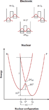
Potential energy well for an electron transfer reaction treated quantum mechanically. R and P are the reactant and product vibrational energy well, respectively. ΔG° and ΔG* are the free energies of reaction and activation, λ represents the total reorganization energy, and HAD is the electronic coupling between acceptor and donor. Q* is the coordinate of interest to the system at the transition state.
Figure 3
Figure 3
Three dimensional vibronic free-energy surfaces for reactants, I, μ (blue), and products, II, v(red), of a proton-coupled electron transfer reaction. From Reference .
Figure 4
Figure 4
Two-dimensional potential energy profiles for a concerted proton-coupled electron transfer reaction. The circles (pink) represent the vibrational energy surface for the transferring hydrogen atom at the lowest energy of the reactant and product well and at the transition state. Note that optimal overlap occurs at the reaction transition state where the lowest energy of the H-vibrational wavefunction is the same in the reactant and product well. From Reference .
Figure 5
Figure 5
Biological and model systems for examination of the role of the proton in electron transfer (ET) in proteins. (a) X-ray crystal structure of Pseudomonas aeruginosa oxidized azurin(Cu2+) with Ru(tpy)(phen)(His83)2+ label. Data from Reference , Protein Data Bank (PDB) code 1JZE. (b) Model systems developed to examine the role of the proton in mediating ET through hydrogen-bonded networks (54, 56). Numbers identify the compounds in the text.
Figure 6
Figure 6
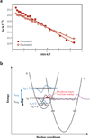
(a) T dependency of the rate of proton-coupled electron transfer in assembly 3 with a protonated (solid circles) and deuterated (open circles) in the solvent 2-methyltetrahydrofuran. (b) Model for interpretation of the inverted kinetic isotope effect. Proton and deuteron ground (v = 0) and excited-state (v = 1) reactant vibrational wavefunctions are illustrated in blue and black, respectively. Proton vibrational energies (ℏω) are greater than that for the deuteron. A vibrational wavefunction for the product well is illustrated in magenta. Vibrational wavefunction overlap (red) facilitates electron tunneling from the reactant (R) to the product (P) well.
Figure 7
Figure 7
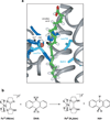
Biological and model systems for examination of the role of PCET in C–H activation of substrates: (a) Model of linoleic acid (LA) in the crystal structure of soybean lipoxygenase-1 (SLO). Fe3+ and its protein-derived ligands (dark blue); the hydroxo-ligand (red/white); LA (green/red/white), with the pro-S hydrogen (black) of C-11; and Leu546, Leu754, and Ile553 (light blue) are illustrated. From Reference . (b) The concerted proton-coupled electron transfer reaction of FeIII(Hbim) with dihydroanthracene (DHA), which models the C–H activation reaction of SLO.
Figure 8
Figure 8
Biological and model systems for examination of the proton-coupled electron transfer (PCET) mechanism of tyrosine radical generation and transport: (a) (top) Crystal structure of photosystem II–oxidizing cofactors (93). Purple and green spheres represent a model for electron density corresponding to Mn and Ca, respectively, in the oxygen-evolving complex (OEC) and red sphere represents O. PDB code: 2AXT. (bottom) Conserved residues of class I ribonucleotide reductase that compose the putative PCET pathway for radical transport from Y122 in β2 to C439 in the α2 active site (100, 102). Residues where the radical has been directly observed or trapped via site-specific replacement with nonnatural amino acid analogs are green. Y356 is not located in either the β2 or α2 crystal structures. (b) Structures of model complexes RuY, RuesterY, and Re(P–Y) discussed herein for the study of PCET mechanisms of Y oxidation.
Scheme 1
Scheme 1
The language of proton-coupled electron transfer (PCET). Abbreviations: ET, electron transfer; PT, proton transfer. The numbers in parentheses correspond to references listed in the Literature Cited section.
Scheme 2
Scheme 2
The guanidinium of an Arg-Asp salt bridge can assume multiple two-point binding conformations; only one two-point conformation may be assumed upon replacing guanidinium with amidinium.
Scheme 3
Scheme 3
Mechanisms of Y generation employed in model complexes discussed herein.

Similar articles

Cited by

References

    1. Reece SY, Hodgkiss JM, Stubbe J, Nocera DG. Proton-coupled electron transfer: The mechanistic underpinning for radical transport and catalysis in biology. Philos. Trans. R. Soc. Lond. Ser. B. 2006;361:1351–1364. - PMC - PubMed
    1. Cukier RI, Nocera DG. Proton-coupled electron transfer. Annu. Rev. Phys. Chem. 1998;49:337–369. - PubMed
    1. Mayer JM. Proton-coupled electron transfer: a reaction chemist’s view. Annu. Rev. Phys. Chem. 2004;55:363–390. - PubMed
    1. Huynh MHV, Meyer TJ. Proton-coupled electron transfer. Chem. Rev. 2007;107:5004–5064. - PMC - PubMed
    1. Pourbaix M. Atlas of Electrochemical Equilibria in Aqueous Solutions. Oxford: Pergamon; 1966.

Publication types

LinkOut - more resources