Review
. 2009:78:929-58.
doi: 10.1146/annurev.biochem.77.032207.120833.

Collagen structure and stability

Affiliations
Review

Collagen structure and stability

Matthew D Shoulders et al. Annu Rev Biochem. 2009.

Abstract

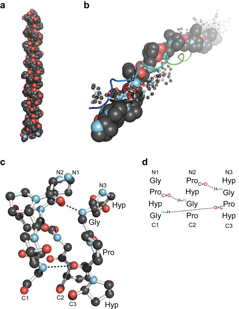
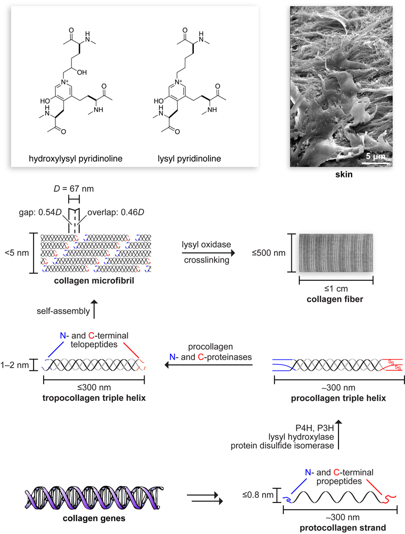
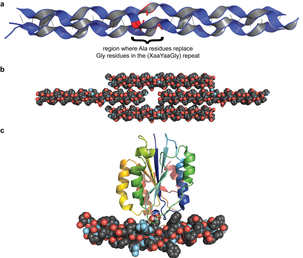
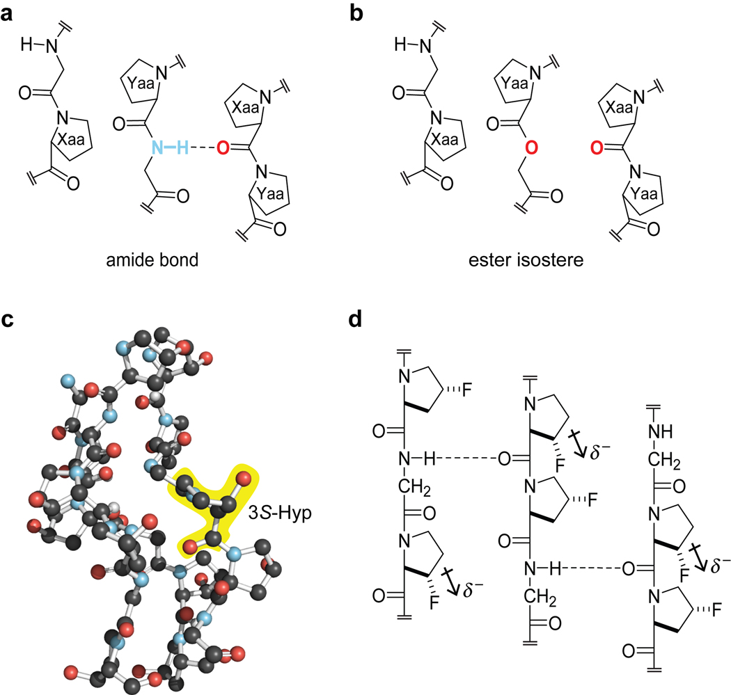
Collagen is the most abundant protein in animals. This fibrous, structural protein comprises a right-handed bundle of three parallel, left-handed polyproline II-type helices. Much progress has been made in elucidating the structure of collagen triple helices and the physicochemical basis for their stability. New evidence demonstrates that stereoelectronic effects and preorganization play a key role in that stability. The fibrillar structure of type I collagen-the prototypical collagen fibril-has been revealed in detail. Artificial collagen fibrils that display some properties of natural collagen fibrils are now accessible using chemical synthesis and self-assembly. A rapidly emerging understanding of the mechanical and structural properties of native collagen fibrils will guide further development of artificial collagenous materials for biomedicine and nanotechnology.


Figures

Figure 1
Figure 1
Overview of the collagen triple helix. (a) First high-resolution crystal structure of a collagen triple helix, formed from (ProHypGly)4–(ProHypAla)–(ProHypGly)5 [Protein Data Bank (PDB) entry 1cag] (19). (b) View down the axis of a (ProProGly)10 triple helix [PDB entry 1k6f (7)] with the three strands depicted in space-filling, ball-and-stick, and ribbon representation. (c) Ball-and-stick image of a segment of collagen triple helix [PDB entry 1cag (19)], highlighting the ladder of interstrand hydrogen bonds. (d) Stagger of the three strands in the segment in panel c.
Figure 2
Figure 2
Biosynthetic route to collagen fibers (110), which are the major component of skin. Size and complexity is increased by posttranslational modifications and self-assembly. Oxidation of lysine side chains leads to the spontaneous formation of hydroxylysyl pyridinoline and lysyl pyridinoline cross-links.
Figure 3
Figure 3
Snapshots of interesting crystal structures of collagen triple helices. (a) Impact of a Gly→Ala substitution on the structure of a collagen triple helix formed from the collagen-related peptide (CRP) (ProHypGly)4–(ProHypAla)–(ProHypGly)5 [Protein Data Bank (PDB) entry 1cag (19)]. The Ala residues (red) disturb the structure. Mutations leading to such structural irregularities are common in osteogenesis imperfecta and can be lethal. (b) Depiction of the effect of a single GluLysGly triplet on the packing of neighboring triple-helical CRPs in crystalline (ProHypGly)4–(GluLysGly)–(ProHypGly)5 [PDB entry 1qsu (21)]. The axial stagger of the individual triple helices, which is presumably compelled by deleterious Coulombic interactions between charged residues, is reminiscent of the D-periodic structure in collagen fibrils. Similar interactions could contribute to the morphology of collagen fibrils. (c) Triple-helical CRP containing the integrin-binding domain GFOGER in complex with the I domain of integrin α2β1 [PDB entry 1dzi (22)]. The bend in the triple helix is thought to arise from the protein-protein interaction. A Glu residue in the middle strand of the triple helix coordinates to Co(II) (blue) bound in the I domain of integrin α2β1.
Figure 4
Figure 4
Importance of interstrand hydrogen bonds for collagen triple-helix stability. (a) A segment of a (ProProGly)10 triple helix. (b) Comparison of the stability of a triple helix formed from (ProProGly)4–ProProOGly–(ProProGly)5, wherein one Pro–Gly amide bond is replaced with an ester, with that in panel a revealed that each interstrand hydrogen bond contributes ΔG = −2.0 kcal/mol to triple-helix stability (30). (c) Crystal structure of a triple helix formed from a collagen-related peptide that mimics a common sequence in type IV collagen, (GlyProHyp)3–(3S-HypHypGly)2–(GlyProHyp)4, showing that 3S-Hyp in the Xaa position yields a prototypical collagen triple helix [PDB entry 2g66 (78)]. (d) (2S,3S)-3-Fluoroproline in the Xaa position destabilizes a collagen triple helix, perhaps by withdrawing electron density from the proximal Xaa carbonyl and thereby reducing the strength of the interstrand hydrogen bond (79).
Figure 5
Figure 5
Pro cistrans isomerization. Unlike other proteinogenic amino acids, Pro forms tertiary amide bonds, resulting in a significant population of the cis conformation.
Figure 6
Figure 6
Reaction catalyzed by prolyl 4-hydroxylase (P4H). Pro residues in the Yaa position of collagen strands are converted into Hyp prior to triple-helix formation.
Figure 7
Figure 7
Ring conformations of Pro and Pro derivatives. The Cγ-endo conformation is favored strongly by stereoelectronic effects when R1 = H, R2 = F (flp) or Cl (clp), and by steric effects when R1 = Me (mep) or SH (mcp), R2 = H. The Cγ-exo conformation is favored strongly by stereoelectronic effects when R1 = OH (Hyp), F (Flp), OMe (Mop), or Cl (Clp), R2 = H, and by steric effects when R1 = H, R2 = Me (Mep) or SH (Mcp). The Cγ-endo:Cγ-exo ratio is ~2 when R1 = R2 = H (56).
Figure 8
Figure 8
Stereoelectronic effects that stabilize the collagen triple helix. (a) A gauche effect and an n→π* interaction preorganize main chain torsion angles and enhance triple-helix stability. (b) A gauche effect, elicited by an electron-withdrawing group (EWG) in the 4R position, stabilizes the Cγ-exo ring pucker. (c) An n→π* interaction stabilizes the trans isomer of the peptide bond but is substantial only when Pro derivatives are in the Cγ-exo ring pucker (e.g., R1 = OH or F, R2 = H). (d) Depiction of overlap between n and π* natural bond orbitals (NBOView©) in a Pro residue with Cγ-exo pucker.
Figure 9
Figure 9
Heterotrimeric synthetic collagen triple helices. (ac) Steric approach. Space-filling models of triple-helix segments constructed from the structure of a (ProHypGly)n triple helix [PDB entry 1cag (19)] with the program SYBYL (Tripos, St. Louis, MO). In panel a, rF⋯F = 2.4 Å in a (flpFlpGly)n triple helix (79). In panel b, rCl⋯Cl = 1.9 Å (61) in a (clpClpGly)n triple helix. In panel c, the methyl groups in a (mepMepGly)n triple helix are radial and distal. (d) Coulombic approach. Favorable Coulombic interactions drive the preferential assembly of triple helices having a 1:1:1 ratio of (ProLysGly)10:(AspHypGly)10:(ProHypGly)10 (96).
Figure 10
Figure 10
Strategies for the self-assembly of long, synthetic collagen triple helices and fibrils. (a) Disulfide bonds enforce a strand register with sticky ends that self-assemble (131). (b) Stacking interactions between electron-poor pentafluorophenyl rings and electron-rich phenyl rings lead to self-assembly (133, 134). (c) Coulombic forces between cationic and anionic blocks encourage self-assembly. TEM image of a resulting fiber shows D-periodicity with D = 17.9 nm (137). Natural type I collagen has D = 67 nm.

References

LITERATURE CITED

    1. Brinckmann J. Collagens at a glance. Top. Curr. Chem. 2005;247:1–-6.
    1. Veit G, Kobbe B, Keene DR, Paulsson M, Koch M, Wagener R. Collagen XXVIII, a novel von Willebrand factor A domain-containing protein with many imperfections in the collagenous domain. J. Biol. Chem. 2006;281:3494–3504. - PubMed
    1. Schweitzer MH, Suo Z, Avci R, Asara JM, Allen MA, et al. Analyses of soft tissue from Tyrannosaurus rex suggest the presence of protein. Science. 2007;316:277–280. - PubMed
    1. Asara JM, Schweitzer MH, Freimark LM, Phillips M, Cantley LC. Protein sequences from mastodon and Tyrannosaurus rex revealed by mass spectrometry. Science. 2007;316:280–285. - PubMed
    1. Buckley M, Walker A, Ho SYW, Yang Y, Smith C, et al. Comment on “Protein sequences from mastodon and Tyrannosaurus rex revealed by mass spectrometry.”. Science. 2008;319:333. - PMC - PubMed

RELATED RESOURCES

    1. Dalgleish R. A database of osteogenesis imperfecta and type III collagen mutations. 2009. http://www.le.ac.uk/genetics/collagen/
    1. Khoshnoodi J, Cartailler J-P, Alvares K, Veis A, Hudson BG. Computer-generated animation of assembly of type I and type IV collagen for Reference 38. 2006. http://www.mc.vanderbilt.edu/cmb/collagen/
    1. Ricard-Blum S, Ruggiero F, van der Rest M. The collagen superfamily. Top. Curr. Chem. 2005;247:35–84.
    1. Koide T, Nagata K. Collagen biosynthesis. Top. Curr. Chem. 2005;247:85–114.
    1. Greenspan DS. Biosynthetic processing of collagen molecules. Top. Curr. Chem. 2005;247:149–183.

Publication types

LinkOut - more resources