. 2009 Apr;9(4):709-18.
doi: 10.1111/j.1600-6143.2009.02578.x.

Preferential priming of alloreactive T cells with indirect reactivity

Affiliations

Preferential priming of alloreactive T cells with indirect reactivity

T V Brennan et al. Am J Transplant. 2009 Apr.

Abstract

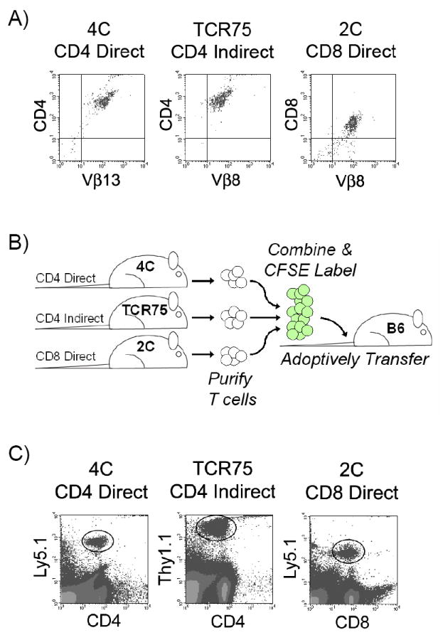
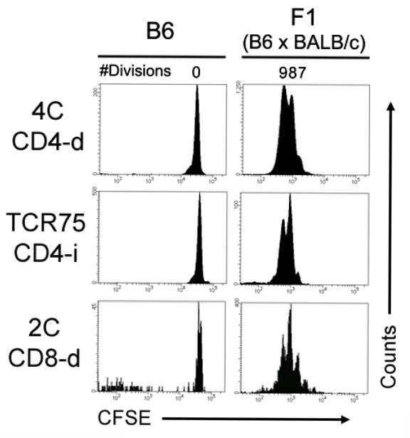
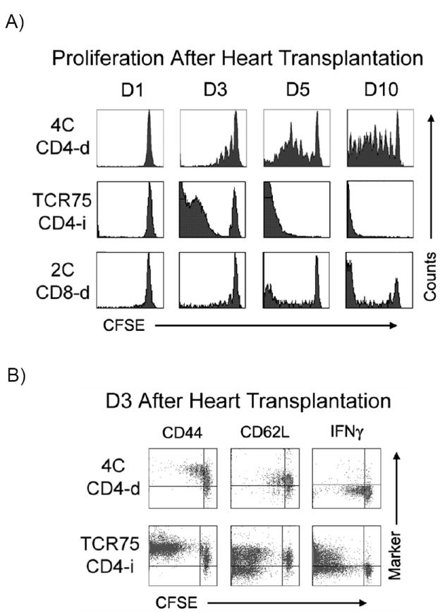
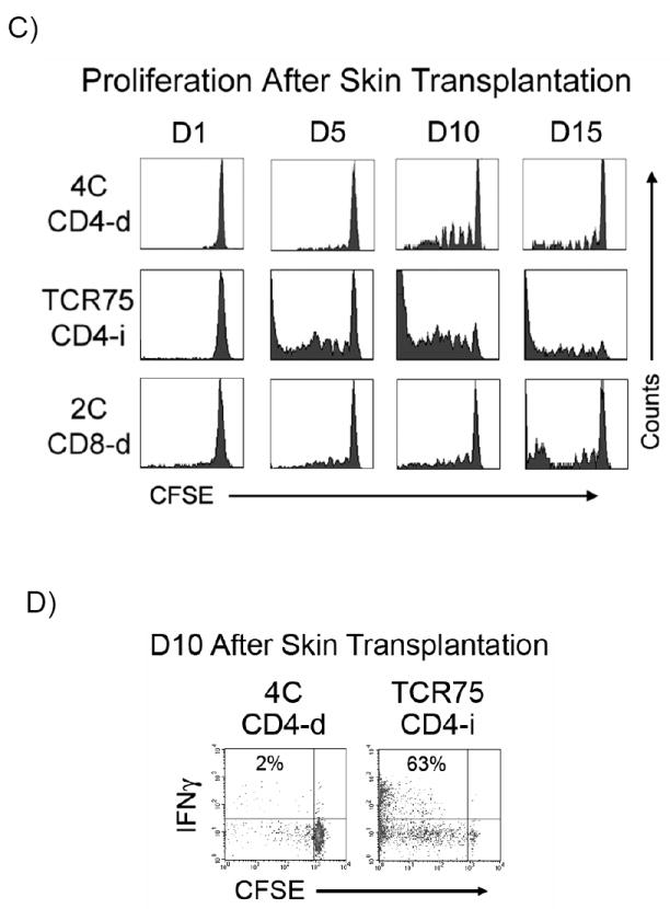
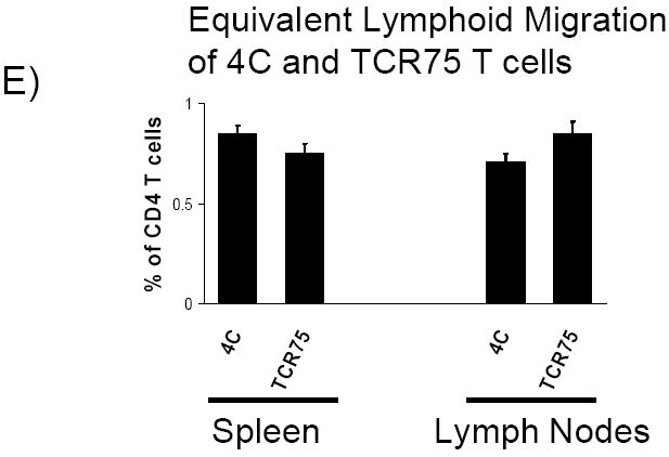
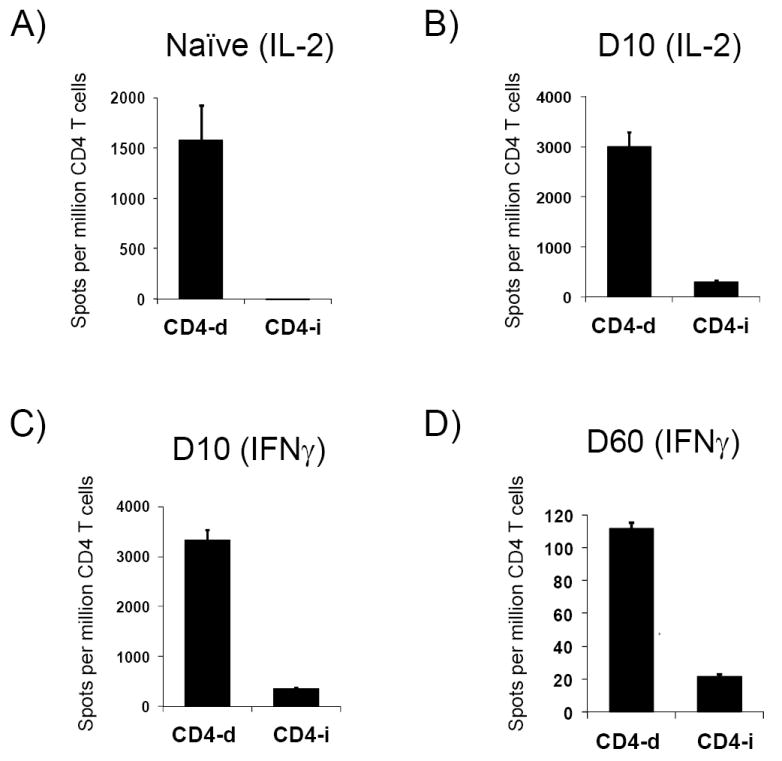
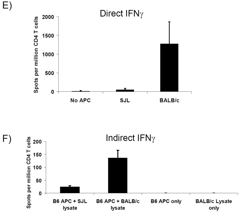
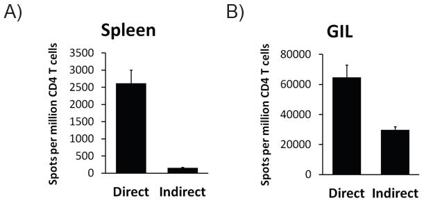
The relative contributions of the direct and indirect pathways in alloimmune responses have not been fully elucidated. We report a novel murine TCR transgenic system that can simultaneously track the CD4-direct (CD4-d), CD4-indirect (CD4-i) and CD8-direct (CD8-d) pathways after transplantation. Using this system, we have observed a profoundly greater proliferation of CD4-i T cells relative to CD4-d and CD8-d T cells after transplantation. Furthermore, a much larger proportion of CD4-i T cells attain an effector phenotype. We also analyzed endogenous, wild-type T cells using enzyme-linked immunospot analysis. In naïve mice, T cells with indirect reactivity were undetectable, but T cells with direct reactivity were abundant. However, 10 days after skin or heterotopic heart transplantation, CD4-i T cells comprised approximately 10% of the CD4+ response. Consistent with increased priming of the CD4-i pathway, we observed that the CD4-i T cells were further enriched in the effector cells migrating to the allograft and in memory-like T cells persisting after rejection. Thus, priming of the CD4-i pathway is favored after transplantation, allowing a rare population to rapidly become a major component of the CD4+ T-cell response in acute allograft rejection. The generalizability of this observation to other models remains to be determined.


Figures

Figure 1
Figure 1. Adoptive transfer of TCR-tg T cells allows for simultaneous tracking of three subtypes of alloreactive T cells
A) TCR-tg T cells representing the CD4-d (4C), CD4-i (TCR75) and CD8-d (2C) pathways of alloantigen recognition were purified by magnetic bead isolation and tested for purity by TCR-Vβ transgene expression and CD4 or CD8 expression using FACS analysis. B) Schematic of T cell purification, labeling with intravital dye (CFSE), and intravenous adoptive transfer into a syngeneic host. C) After transplantation, TCR-tg T cells could be identified amongst host T cells by the congenic cell surface markers, Ly5.1 and Thy1.1.
Figure 2
Figure 2. TCR-tg T cells have equivalent proliferative capabilities in vivo when Ag is abundant
CFSE-labeled TCR-tg T cells representing the CD4-d, CD4-i and CD8-d pathways of alloantigen recognition were co-adoptively transferred into syngeneic (B6) or semi-allogeneic F1 (BALB/c × B6) recipients. After 3 days, recipient splenocytes were harvested and their proliferation was assessed by CFSE dilution detected by FACS. Results are representative of two independent experiments with 2-3 mice per group.
Figure 3
Figure 3. CD4-i TCR-tg T cells are preferentially primed after transplantation
A) At various time-points after BALB/c heart transplantation, recipient splenocytes were isolated and the proliferation of adoptively transferred TCR-tg T cells was determined by CFSE dilution. Data is representative of at least 3 independent experiments, with at least 2-3 mice per experiment. B) CD44, CD62L and IFNγ expression by adoptively transferred CD4-d and CD4-i TCR-tg T cells were assessed by FACS three days (D3) following cardiac allograft transplantation. C) Proliferation of TCR-tg T cells following allogeneic skin transplantation at the draining axillary lymph nodes demonstrated by CFSE dilution. Data are representative of 3 independent experiments, 3 mice per experiment. D) Comparison of intracellular IFNγ production by CD4-d and CD4-i TCR-tg T cells 10 days after BALB/c skin transplantation. E) Equivalent migration of 4C and TCR75 TCR-tg T cells to spleen and lymph node. 2.5 × 10⁶ T cells from each TCR-tg were co-injected intravenously into B6 mice. 7 days later, spleen and lymph node were isolated and analyzed by FACS.
Figure 4
Figure 4. ELISPOT analysis of non-transgenic CD4-d and CD4-i T cells
After BALB/c → B6 skin transplantation, CD4+ T cells were purified from recipient axillary LN. CD4-d and CD4-i T cells were then identified by IL-2 and IFNγ ELISPOT assays by their response to direct or indirect alloantigen presentation. No TCR-tg T cells were utilized in these experiments. A) Naive B6 mouse (no transplant), IL-2; B) day (D) 10 after skin transplant, IL-2; C) D10 after skin transplant, IFNγ; D) D60 after skin transplant, IFNγ. Data are representative of two independent experiments with 2-3 mice per data point. E, F) IFNγ ELISPOT of draining lymph node CD4+ T cells 10 days after skin transplant with 3rd party controls.
Figure 5
Figure 5. CD4-i T cells are enriched in graft infiltrating lymphocytes
On postoperative day 7 after BALB/c → B6 heart transplantation, cardiac allografts were excised and CD4+ T cells were isolated from the graft infiltrating lymphocytes (GIL) using magnetic beads. Purified CD4+ T cells from GIL and spleen were tested for direct and indirect alloreactivity by IFNγ ELISPOT assay. Data represent 3 heart transplant recipients.
Figure 6
Figure 6. Differential priming of CD4-d and CD4-i pathways in fresh vs. healed grafts
Kaplan-Meier survival plot of BALB/c skin transplants on B6 Rag1-/- hosts. A) Two million CD4-i TCR-tg T cells were adoptively transferred at the time of skin transplantation (fresh skin) or 100 days after skin transplantation (healed skin) (p = NS). B) Similarly, two million CD4-d TCR-tg T cells were adoptively transferred into recipients of fresh skin or healed skin transplants. 107 BALB/c DCs were injected at the time of CD4-d TCR-tg T cell transfer in recipients with healed BALB/c allografts.

Comment in

Similar articles

Cited by

References

    1. Rogers NJ, Lechler RI. Allorecognition. Am J Transplant. 2001 Jul;1(2):97–102. - PubMed
    1. Auchincloss H, Jr, Lee R, Shea S, Markowitz JS, Grusby MJ, Glimcher LH. The role of “indirect” recognition in initiating rejection of skin grafts from major histocompatibility complex class II-deficient mice. Proc Natl Acad Sci U S A. 1993 Apr 15;90(8):3373–7. - PMC - PubMed
    1. Suchin EJ, Langmuir PB, Palmer E, Sayegh MH, Wells AD, Turka LA. Quantifying the frequency of alloreactive T cells in vivo: new answers to an old question. J Immunol. 2001 Jan 15;166(2):973–81. - PubMed
    1. Baker RJ, Hernandez-Fuentes MP, Brookes PA, Chaudhry AN, Cook HT, Lechler RI. Loss of direct and maintenance of indirect alloresponses in renal allograft recipients: implications for the pathogenesis of chronic allograft nephropathy. J Immunol. 2001 Dec 15;167(12):7199–206. - PubMed
    1. Vella JP, Vos L, Carpenter CB, Sayegh MH. Role of indirect allorecognition in experimental late acute rejection. Transplantation. 1997 Dec 27;64(12):1823–8. - PubMed

Publication types

MeSH terms