. 2009 Apr 3:9:32.
doi: 10.1186/1472-6750-9-32.

Noncytotoxic orange and red/green derivatives of DsRed-Express2 for whole-cell labeling

Affiliations

Noncytotoxic orange and red/green derivatives of DsRed-Express2 for whole-cell labeling

Rita L Strack et al. BMC Biotechnol. .

Abstract

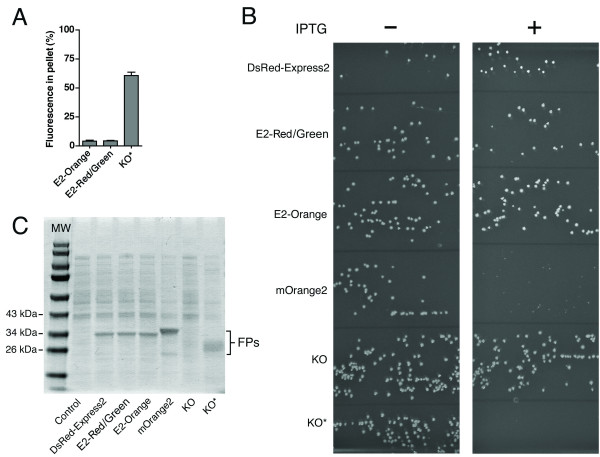
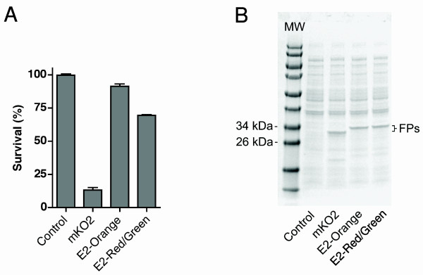
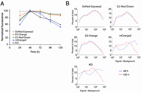
Background: Whole-cell labeling is a common application of fluorescent proteins (FPs), but many red and orange FPs exhibit cytotoxicity that limits their use as whole-cell labels. Recently, a tetrameric red FP called DsRed-Express2 was engineered for enhanced solubility and was shown to be noncytotoxic in bacterial and mammalian cells. Our goal was to create derivatives of this protein with different spectral properties.

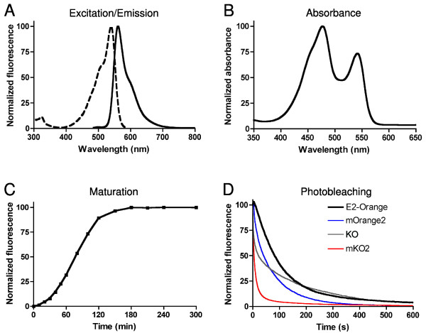
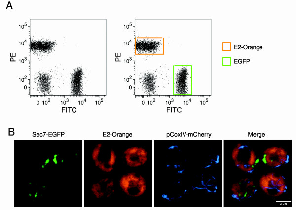
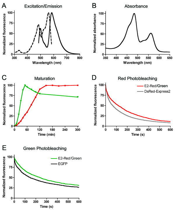
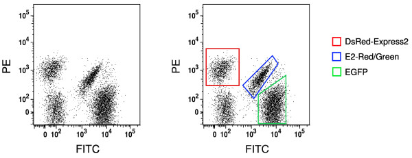
Results: Building on previous studies of DsRed mutants, we created two DsRed-Express2 derivatives: E2-Orange, an orange FP, and E2-Red/Green, a dual-color FP with both red and green emission. We show that these new FPs retain the low cytotoxicity of DsRed-Express2. In addition, we show that these new FPs are useful as second or third colors for flow cytometry and fluorescence microscopy.

Conclusion: E2-Orange and E2-Red/Green will facilitate the production of healthy, stably fluorescent cell lines and transgenic organisms for multi-color labeling studies.


Figures

Figure 1
Figure 1
Fluorescence properties of E2-Orange. Shown are (A) excitation and emission and (B) absorbance spectra of E2-Orange. (C) Maturation kinetics of E2-Orange fluorescence. For these measurements the FPs were excited at 520 ± 10 nm excitation and emission was recorded at 560 ± 10 nm. (D) Photobleaching kinetics of E2-Orange (black line), mOrange2 (blue line), KO (gray line), and mKO2 (red line) during widefield fluorescence microscopy. Data were collected using a Texas Red filter set. For (C) and (D), data represent the mean of three independent measurements.
Figure 2
Figure 2
E2-Orange is useful as a second or third color. (A) Shown is a dot plot generated by flow cytometry of S. cerevisiae cells expressing E2-Orange, EGFP, or no FP. The three strains were grown separately, then pooled at equivalent cell concentrations and analyzed. Cells were excited using a 488-nm laser, and orange and green fluorescence signals were detected using PE and FITC filter sets, respectively. On the right are the same data with E2-Orange-expressing cells boxed in orange and EGFP-expressing cells boxed in green. (B) A three-color yeast strain was generated with GFP-labeled Golgi, E2-Orange-labeled cytosol, and mCherry-labeled mitochondria. Cells were imaged using confocal microscopy. E2-Orange and GFP were immediately resolvable, and E2-Orange and mCherry were resolvable after linear unmixing, yielding clear three color images as shown in the overlay.
Figure 3
Figure 3
Fluorescence properties of E2-Red/Green. Shown are (A) excitation and emission and (B) absorbance spectra of E2-Red/Green. (C) Maturation kinetics of green (green line) and red (red line) fluorescence of E2-Red/Green. For these measurements the green species was excited with 480 ± 10 nm light and emission was recorded at 515 ± 10 nm, and the red species was excited with 540 ± 10 nm light and emission was recorded at 590 ± 10 nm. Also shown are photobleaching kinetics for the red fluorescence of E2-Red/Green and DsRed-Express2 (D) and for the green fluorescence of E2-Red/Green and EGFP (E). The red and green photobleaching measurements were recorded using Texas Red and Endow GFP filter sets, respectively.
Figure 4
Figure 4
E2-Red/Green is a useful third color for flow cytometry. Shown is a dot plot generated by flow cytometry of S. cerevisiae cells expressing DsRed-Express2, E2-Red/Green, EGFP, or no FP. Cells were grown individually, pooled at equivalent cell concentrations, and analyzed by flow cytometry. Fluorescence was excited using a 488-nm laser, and red and green signals were detected using PE and FITC filter sets, respectively. On the right are the same data with DsRed-Express2-expressing cells boxed in red, E2-Red/Green-expressing cells boxed in blue, and EGFP-expressing cells boxed in green.
Figure 5
Figure 5
E2-Orange and E2-Red/Green do not form higher-order aggregates and are noncytotoxic to bacteria. (A) To assay higher-order aggregation of oligomeric FPs, the percent fluorescence in the pellet fraction of a lysate from E. coli cells expressing E2-Orange, E2-Red/Green, or KO* was measured for eight independent replicates. Error bars represent s.e.m. (B) To measure bacterial cytotoxicity of FPs, E. coli DH10B cells harboring the pREP4 repressor plasmid were transformed with pQE-60NA encoding DsRed-Express2, E2-Red/Green, E2-Orange, mOrange2, KO, or KO*. Equal volumes of transformation mixtures were plated onto adjacent sectors of plates under either repressing (no IPTG) or derepressing (1 mM IPTG) conditions. Large colonies under derepressing conditions (right panel) indicate low cytotoxicity. (C) Quantitation of FP expression under derepressing conditions. Cells were grown to an OD600 of ~ 0.6 and then treated with 1 mM IPTG for 4 h. Whole-cell lysates were separated using SDS-PAGE followed by staining with Coomassie Blue. Control cells were transformed with empty pQE-60NA.
Figure 6
Figure 6
E2-Orange and E2-Red/Green show relatively mild phototoxicity. (A) E. coli cells were treated for 4 h with 1 mM IPTG to express either no FP (Control) or the indicated FP. Cells were then illuminated through a Texas Red (535–585 nm) filter for 15 min. In parallel, identical samples were not illuminated. Cells were then plated and grown overnight, and the percent survival was calculated based on colony number for the illuminated versus non-illuminated samples. Error bars represent s.e.m. (B) Quantitation of FP expression under the conditions of the phototoxicity experiment. Immediately before light treatment, aliquots of cells were taken for expression analysis. Whole-cell lysates were separated using SDS-PAGE followed by staining with Coomassie Blue.
Figure 7
Figure 7
E2-Orange and E2-Red/Green are noncytotoxic to HeLa cells under conditions of standard high-level expression. (A) HeLa cells were transiently transfected in 24-well plates for constitutive high-level expression of the indicated FP. Three wells per FP per day were analyzed by flow cytometry, in parallel with untransfected cells, and the average brightness of the viable fluorescent cells was measured. The highest signal for a given FP was normalized to 100 units. Error bars represent s.e.m. (B) Fluorescence intensity distributions for the same data were analyzed for 48 h (blue) and 120 h (red) post-transfection. Each data point is a binned value that represents the percentage of cells with fluorescence in a range centered about the data point.

References

    1. Shaner NC, Patterson GH, Davidson MW. Advances in fluorescent protein technology. J Cell Sci. 2007;120:4247–4260. doi: 10.1242/jcs.005801. - DOI - PubMed
    1. Tao W, Evans BG, Yao J, Cooper S, Cornetta K, Ballas CB, Hangoc G, Broxmeyer HE. Enhanced green fluorescent protein is a nearly ideal long-term expression tracer for hematopoietic stem cells, whereas DsRed-Express fluorescent protein is not. Stem Cells. 2007;25:670–678. doi: 10.1634/stemcells.2006-0553. - DOI - PubMed
    1. Hadjantonakis AK, Macmaster S, Nagy A. Embryonic stem cells and mice expressing different GFP variants for multiple non-invasive reporter usage within a single animal. BMC Biotechnol. 2002;2:11. doi: 10.1186/1472-6750-2-11. - DOI - PMC - PubMed
    1. Hirrlinger PG, Scheller A, Braun C, Quintela-Schneider M, Fuss B, Hirrlinger J, Kirchhoff F. Expression of reef coral fluorescent proteins in the central nervous system of transgenic mice. Mol Cell Neurosci. 2005;30:291–303. doi: 10.1016/j.mcn.2005.08.011. - DOI - PubMed
    1. Strack RL, Strongin DE, Bhattacharyya D, Tao W, Berman A, Broxmeyer HE, Keenan RJ, Glick BS. A noncytotoxic DsRed variant for whole-cell labeling. Nat Methods. 2008;5:955–957. doi: 10.1038/nmeth.1264. - DOI - PMC - PubMed

Publication types

Substances