

Expression of PDGF receptor-alpha in corneal myofibroblasts in situ
- PMID: 19344713
- PMCID: PMC2720438
- DOI: 10.1016/j.exer.2009.03.017
Expression of PDGF receptor-alpha in corneal myofibroblasts in situ
Abstract
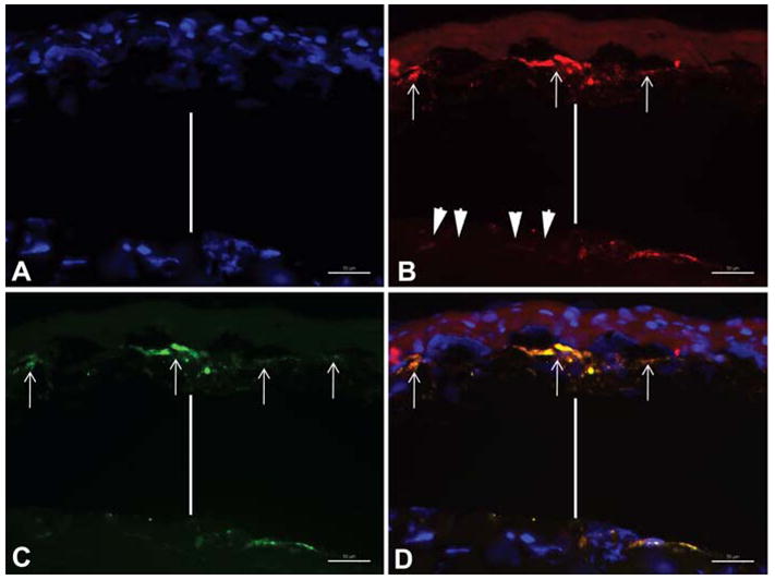
The purpose of this study was to investigate the expression of platelet-derived growth factor receptor-alpha (PDGFR-alpha) in the myofibroblasts of corneas with stromal haze. Central corneal sections from rabbit eyes that had -9 diopter PRK were analyzed by immunocytochemistry (IHC) for the expression of PDGFR-alpha at 4 week after surgery. PDGFR-alpha was expressed immediately beneath the epithelial basement membrane in the anterior stroma. Double IHC studies revealed the expression of PDGFR-alpha in the anterior stroma co-localized with alpha-smooth muscle actin (SMA) marker for myofibroblasts. In vitro studies have suggested that PDGF is important in the development and viability of myofibroblasts after corneal injury. Expression of PDGFR-alpha in myofibroblasts supports these findings.
Figures
Similar articles
-
Corneal myofibroblast biology and pathobiology: generation, persistence, and transparency.Exp Eye Res. 2012 Jun;99(1):78-88. doi: 10.1016/j.exer.2012.03.018. Epub 2012 Apr 20. Exp Eye Res. 2012. PMID: 22542905 Free PMC article. Review.
-
Dynamics of the expression of intermediate filaments vimentin and desmin during myofibroblast differentiation after corneal injury.Exp Eye Res. 2009 Aug;89(2):133-9. doi: 10.1016/j.exer.2009.02.022. Epub 2009 Mar 11. Exp Eye Res. 2009. PMID: 19285070 Free PMC article.
-
Transforming growth factor beta-3 localization in the corneal response to epithelial-stromal injury and effects on corneal fibroblast transition to myofibroblasts.Exp Eye Res. 2023 Oct;235:109631. doi: 10.1016/j.exer.2023.109631. Epub 2023 Aug 25. Exp Eye Res. 2023. PMID: 37633325
-
Corneal epithelial basement membrane assembly is mediated by epithelial cells in coordination with corneal fibroblasts during wound healing.Mol Vis. 2023 May 20;29:68-86. eCollection 2023. Mol Vis. 2023. PMID: 37287640 Free PMC article.
-
The corneal fibrosis response to epithelial-stromal injury.Exp Eye Res. 2016 Jan;142:110-8. doi: 10.1016/j.exer.2014.09.012. Exp Eye Res. 2016. PMID: 26675407 Free PMC article. Review.
Cited by
-
Induction of corneal myofibroblasts by lens-derived transforming growth factor beta1 (TGFbeta1): a transgenic mouse model.Brain Res Bull. 2010 Feb 15;81(2-3):287-96. doi: 10.1016/j.brainresbull.2009.10.019. Epub 2009 Nov 6. Brain Res Bull. 2010. PMID: 19897021 Free PMC article.
-
Corneal Regeneration After Photorefractive Keratectomy: A Review.J Optom. 2015 Jul-Sep;8(3):149-69. doi: 10.1016/j.optom.2014.09.001. Epub 2014 Oct 23. J Optom. 2015. PMID: 25444646 Free PMC article. Review.
-
Corneal myofibroblast biology and pathobiology: generation, persistence, and transparency.Exp Eye Res. 2012 Jun;99(1):78-88. doi: 10.1016/j.exer.2012.03.018. Epub 2012 Apr 20. Exp Eye Res. 2012. PMID: 22542905 Free PMC article. Review.
-
Genome-wide meta-analysis of five Asian cohorts identifies PDGFRA as a susceptibility locus for corneal astigmatism.PLoS Genet. 2011 Dec;7(12):e1002402. doi: 10.1371/journal.pgen.1002402. Epub 2011 Dec 1. PLoS Genet. 2011. PMID: 22144915 Free PMC article.
-
A method to generate enhanced GFP+ chimeric mice to study the role of bone marrow-derived cells in the eye.Exp Eye Res. 2013 Nov;116:366-70. doi: 10.1016/j.exer.2013.10.007. Epub 2013 Oct 17. Exp Eye Res. 2013. PMID: 24140502 Free PMC article.
References
-
- Hart KC, Galvin BD, Donoghue DJ. Structure and function of the platelet derived growth factor family and their receptors. Genet Eng. 1995;17:181–208. - PubMed
-
- Jester JV, Huang J, Barry-Lane PA, Kao WW, Petroll WM, Cavanagh HD. Transforming growth factor (beta)- mediated corneal myofibroblast differentiation requires actin and fibronectin assembly. Invest Ophthalmol Vis Sci. 1999;40:1959–1967. - PubMed
-
- Jester JV, Huang J, Petroll WM, Cavanagh HD. TGF beta induced myofibroblast differentiation of rabbit keratocytes requires synergistic TGF beta, PDGF and integrin signaling. Exp Eye Res. 2002;75:645–657. - PubMed
Publication types
MeSH terms
Substances
Grants and funding
LinkOut - more resources
Full Text Sources
Miscellaneous
