. 2009 Jun;34(3):535-44.
doi: 10.1016/j.nbd.2009.03.012. Epub 2009 Apr 1.

Preconditioning-induced protection from oxidative injury is mediated by leukemia inhibitory factor receptor (LIFR) and its ligands in the retina

Affiliations

Preconditioning-induced protection from oxidative injury is mediated by leukemia inhibitory factor receptor (LIFR) and its ligands in the retina

Srinivas Chollangi et al. Neurobiol Dis. 2009 Jun.

Abstract

Preconditioning with moderate oxidative stress (e.g., moderate bright light or mild hypoxia) can induce changes in retinal tissue that protect photoreceptors from a subsequent dose of lethal oxidative stress. The mechanism underlying this induced protection is likely a general mechanism of endogenous protection which has been demonstrated in heart and brain using ischemia and reperfusion. While multiple factors like bFGF, CNTF, LIF and BDNF have been hypothesized to play a role in preconditioning-induced endogenous neuroprotection, it has not yet been demonstrated which factors or receptors are playing an essential role. Using quantitative PCR techniques we provide evidence that in the retina, LIFR activating cytokines leukemia inhibitory factor (LIF), cardiotrophin-1 (CT-1) and cardiotrophin like cytokine (CLC) are strongly upregulated in response to preconditioning with bright cyclic light leading to robust activation of signal transducer and activator of transcription-3 (STAT3) in a time-dependent manner. Further, we found that blocking LIFR activation during preconditioning using a LIFR antagonist (LIF05) attenuated the induced STAT3 activation and also resulted in reduced preconditioning-induced protection of the retinal photoreceptors. These data demonstrate that LIFR and its ligands play an essential role in endogenous neuroprotective mechanisms triggered by preconditioning-induced stress.


Figures

Figure 1
Figure 1
Schematic diagram of bright cyclic light preconditioning
Figure 2
Figure 2
Functional and morphological evaluation of preconditioning-induced protection of retinal photoreceptors. A) Representative ERG traces and B) a-wave amplitudes of BALB/cJ mice that have been light damaged (LD) (4000 lux for 4 h) with (○) or without (□) prior bright cyclic light preconditioning (600 lux; 6 AM to 6 PM) for 6 days. Control eyes are represented in solid squares (■). n=6; value = mean ± SD. (*p<0.001, vs. LD eyes, paired t-test). C) Representative sections and D) quantification of number of rows of photoreceptor nuclei in the outer nuclear layer (ONL) along the vertical meridian of the retina. n=6; value = mean ± SD (*p<0.001, vs. LD eyes, paired t-test). ONL – Outer nuclear layer; INL – Inner nuclear layer; GCL – Ganglion cell layer; ON – Optic nerve.
Figure 3
Figure 3
Preconditioning with bright cyclic light leads to strong upregulation of neuroprotective factors. A) Time-dependent quantitative evaluation of cytokine mRNA upregulation using real time RT-PCR in response to bright cyclic light preconditioning (600lux; 6AM to 6PM). Members of IL-6 family cytokines (LIF, OSM, CT-1 and CLC) are upregulated 166, 74, 21 and 47 fold in response to bright cyclic light preconditioning. While CNTF did not exhibit a similar upregulation, BDNF exhibited an 11-fold increase in its mRNA levels by the end of the 6th day of preconditioning. Ribosomal protein RPL19 served as a control. n=4; value = mean ± SD. (*p<0.01, paired t-test). B) Representative Western blots and C) quantification of STAT3 activation. n=3; value = mean ± SD. We observed a robust preconditioning-dependent STAT3 activation which reached peak activation by the end of 4 days of preconditioning and remained elevated throughout the preconditioning exposure.
Figure 4
Figure 4
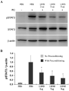
LIF05 inhibited preconditioning-induced STAT3 activation. A) After 2 days of bright cyclic light preconditioning (600lux; 6Am to 6PM), mice were injected with PBS in the left eye (control) and LIF05 (2μg, 5μg or 8μg) in the right eye and returned to their cages for additional bright cyclic light preconditioning. After the 6th day of preconditioning, retinas were harvested and STAT3 activation was analyzed using Western blotting. PBS injected eyes without preconditioning served as controls for basal levels of STAT3 activation. PC – Preconditioning. B) Bands were quantified by conventional digital image analysis using a KODAK Image Station 4000R. n=3; value = mean ± SD. (*p<0.05, vs. PBS injected eyes with preconditioning, one way ANOVA and post hoc Holm-Sidak test for multiple comparisons).
Figure 5
Figure 5
LIF05 inhibited preconditioning-induced protection of retinal photoreceptors. A) Representative ERG traces and B) quantification of a-wave amplitudes of BALB/cJ mice that are light damaged (LD) (4000lux for 4h) with (○,▲,◆) or without (△, □) prior bright cyclic light preconditioning (600lux; 6AM to 6PM for 6 days). Compared to uninjected eyes with preconditioning (○), LIF05 injected eyes lost 44% at the brightest flash intensity. n=6; value = mean ± SD (*p<0.05, vs. PC+LD eyes, paired t-test). C) Representative sections from superior retina and D) quantification of the number of rows of photoreceptor nuclei in the outer nuclear layer (ONL) along the vertical meridian of the retina. n=6; value = mean ± SD (*p<0.05, vs. PC+LD eyes, paired t-test). ONL – Outer nuclear layer; INL – Inner nuclear layer; GCL – Ganglion cell layer; ON – Optic nerve.
Figure 6
Figure 6
Evaluation of LIF05 toxicity towards retinal function and morphology. A) Representative ERG traces and B) quantification of a-wave amplitudes of BALB/cJ mice 8 days after injection with PBS (■) or LIF05 (◇). C) Representative sections from superior retina and D) quantification of the number of rows of photoreceptor nuclei in the outer nuclear layer (ONL). n=3, value = mean ± SD. ONL – Outer nuclear layer; INL – Inner nuclear layer; GCL – Ganglion cell layer; ON – Optic nerve.
Figure 7
Figure 7
LIF05 inhibits LIF stimulated STAT3 activation in vivo. A) 0.1μg of LIF was injected intravitreally with 0, 2, 5, or 8 μg of the antagonist LIF05. After 2 days of injection, retinas were harvested and STAT3 activation levels were analyzed using Western blot analysis. Staining for total STAT3 and β-actin served as loading controls. A) Bands were quantified by conventional digital image analysis using a KODAK Image Station 4000R. n=3; value = mean ± SD (*p<0.05, vs. LIF only injected eyes (LIF 0.1 μg), one way ANOVA and post hoc Holm-Sidak test for multiple comparisons).
Figure 8
Figure 8
Kinetic analysis of LIFR and gp130 interaction towards LIF and LIF05. Soluble LIFR at concentrations of 1.5 nM, 3 nM, 6.25 nM, 12.5 nM, 25 nM or 50 nM were injected over an SPR sensor chip with anti-GST immobilized A) GST-LIF or B) GST-LIF05 at flow rates of 25 μl/min. Responses obtained were corrected for background signal using a control flow cell. Association and dissociation rates are derived by global analysis of the response curves fit to a 1:1 kinetic model using QDat software (BioLogic Software, Ltd. Knoxville TN and Nomadics, Inc. Stillwater, OK) using 1:1 stoichiometry. Models are indicated in smooth gray line overlaid over response curve traces. Soluble gp130 (100 nM) was injected over C) GST-LIF or D) GST-LIF05 either as a mixture with soluble LIFR (10nM) or separately at flow rates of 25 μl/min.

References

    1. Amaravadi R, Thompson CB. The survival kinases Akt and Pim as potential pharmacological targets. J. Clin. Invest. 2005;115:2618–2624. - PMC - PubMed
    1. Bird AC. Retinal photoreceptor dystrophies LI. Edward Jackson Memorial Lecture. Am. J. Ophthalmol. 1995;119:543–562. - PubMed
    1. Cao W, Li F, Steinberg RH, Lavail MM. Development of normal and injury-induced gene expression of aFGF, bFGF, CNTF, BDNF, GFAP and IGF-I in the rat retina. Exp. Eye Res. 2001;72:591–604. - PubMed
    1. Cao W, Wen R, Li F, Lavail MM, Steinberg RH. Mechanical injury increases bFGF and CNTF mRNA expression in the mouse retina. Exp. Eye Res. 1997;65:241–248. - PubMed
    1. Chaum E. Retinal neuroprotection by growth factors: a mechanistic perspective. J. Cell Biochem. 2003;88:57–75. - PubMed

Publication types

MeSH terms