. 2009 Jun;41(2):258-73.
doi: 10.1016/j.mcn.2009.03.005. Epub 2009 Apr 1.

Characterization of trans-neuronal trafficking of Cbln1

Affiliations

Characterization of trans-neuronal trafficking of Cbln1

Peng Wei et al. Mol Cell Neurosci. 2009 Jun.

Abstract

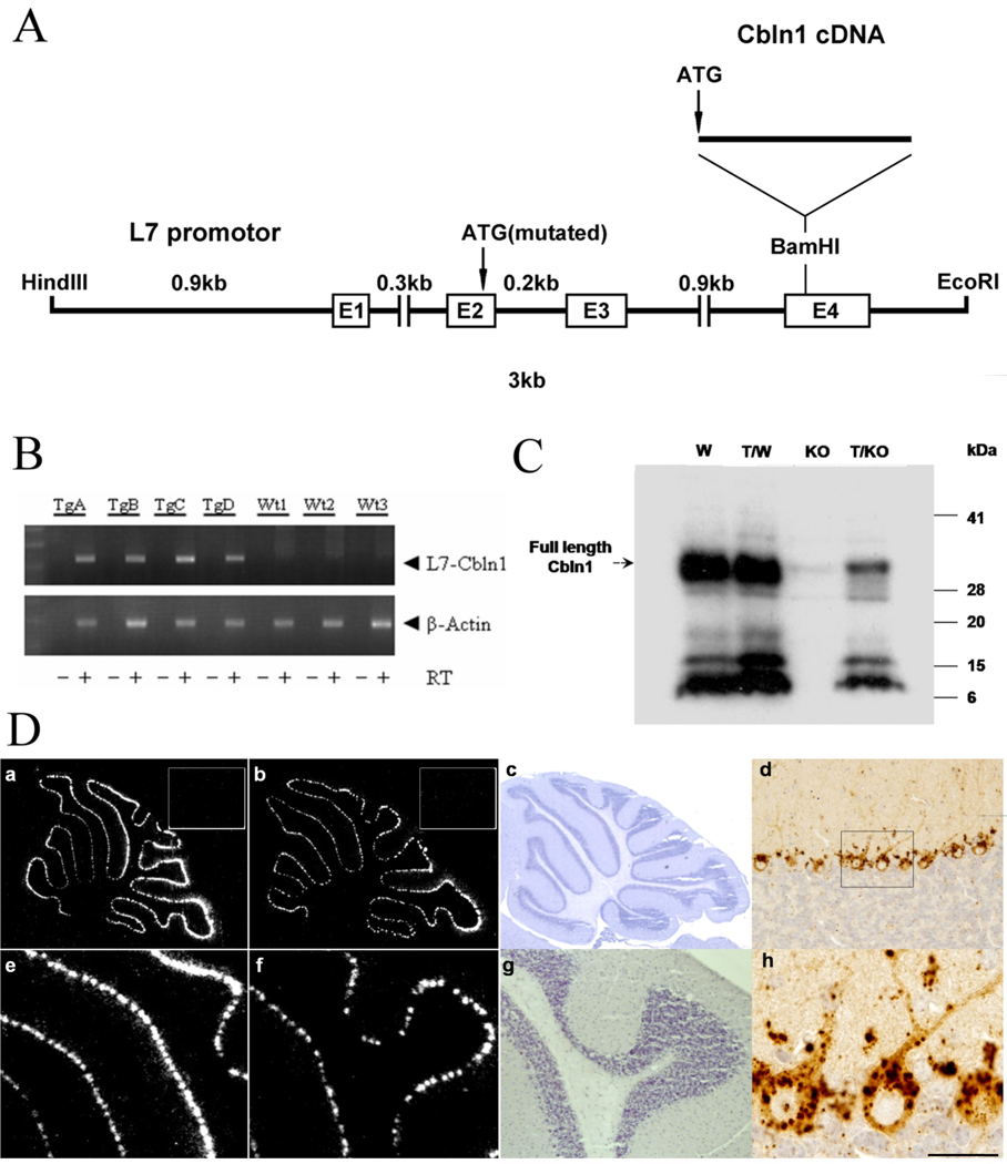
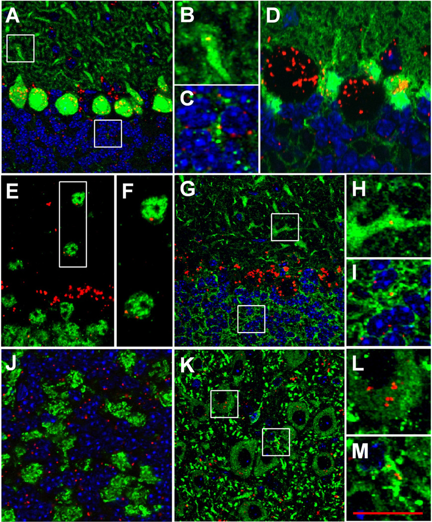
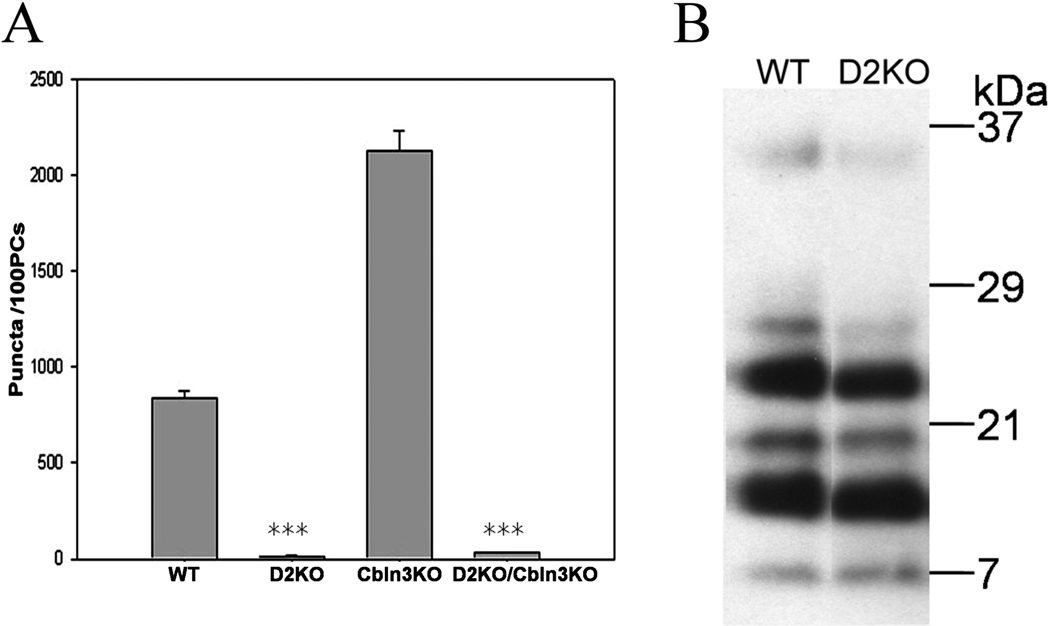
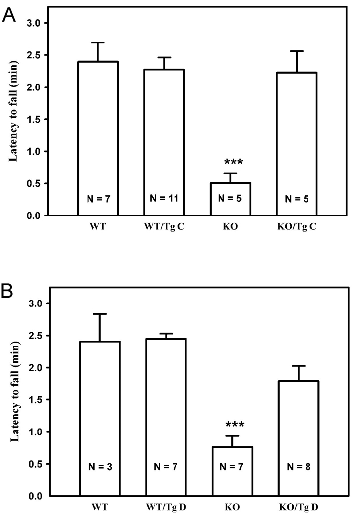
Cbln1, a glycoprotein secreted from granule cells and GluRdelta2 in the postsynaptic densities of Purkinje cells are components of an incompletely understood pathway essential for integrity and plasticity of parallel fiber-Purkinje cell synapses. We show that Cbln1 undergoes anterograde transport from granule cells to Purkinje cells and Bergmann glia, and enters the endolysosomal trafficking system, raising the possibility that Cbln1 exerts its activity on or within Purkinje cells and Bergmann glia. Cbln1 is absent in Purkinje cells and Bergmann glia of GluRdelta2-null mice, suggesting a mechanistic convergence on Cbln1 trafficking. Ectopic expression of Cbln1 in Purkinje cells of L7-cbln1 transgenic mice reveals Cbln1 undergoes anterograde and retrograde trans-neuronal trafficking even across synapses that lack GluRDelta2, indicating that it is not universally essential for Cbln1 transport. The L7-cbln1 transgene also ameliorates the locomotor deficits of cbln1-null mice, indicating that the presence and/or release of Cbln1 from the postsynaptic neuron has functional consequences.


Figures

Fig. 1
Fig. 1
Cbln1-like immunoreactivity (CLI) in adult mouse cerebellum. Immunohistochemistry with the E3 epitope antiserum revealed punctate CLI in wild type cerebellum (A) whereas there was no staining in cerebellum from cbln1-null mice (B). Panels (C) and (D) show enlarged views of the boxed regions in (A) and (B), respectively. Panel (E) reveals enhanced expression of CLI in an adult cbln3-null mouse cerebellum whereas there is no CLI in a cbln1-, cbln3-double null mouse (F). Scale bars: A and B, 50 µm; C–F, 15 µm.
Fig. 2
Fig. 2
Localization of Cbln1-like immunoreactivity (CLI) to Purkinje cells and Bergmann glia. Double immunofluorescence labeling was performed on cbln3-null (A, B, E, F) or cbln1-null (C, D, G, H) mouse cerebellum using the E3 Cbln1 antiserum (red) and either the Purkinje cell marker, calbindin-D28K (green, A–D) or the glia marker, S100 (green, E–H). Merged confocal images revealed that punctate CLI is co-localized with both calbindin- (A, B) and S100-positive (E, F) cells, indicative of its presence in Purkinje cells and Bergmann glia, respectively. There is no detectable CLI in cbln1-null cerebellum (C,D,G,H). DAPI is used to visualize nuclei. Scale bar: 20 µm.
Fig. 3
Fig. 3
Localization of Cbln1-like immunoreactivity (CLI) to lysosomes and endosomes in Purkinje cells. Triple immunofluorescence labeling was performed on cbln3-null mouse cerebellum using the anti-E3 Cbln1 antiserum (red, A, E, I and M), anti-calbindin-D28K (blue, D, H, L and P), and markers (green) of endoplasmic reticulum (KDEL, B), Golgi apparatus (GM130, F), late endosomes (mannose-6-phosphate receptor, J), and lysosomes (cathepsin-D, N). Merged confocal images reveal that CLI is almost always co-localized with the lysosome marker (O). The majority of CLI is co-localized with this endosome marker (K) suggesting that CLI is present in endosomes and persists through their maturation into lysosomes. CLI is not present in ER (C) or Golgi (G). Merged confocal images (D, H, L, P) with calbindin-D28K show the localization of CLI in Purkinje cells. Scale bar, 10 µm.
Fig. 4
Fig. 4
Generation and characterization of L7-cbln1 transgenic mice. (A) Structure of the L7-cbln1 transgene. A cbln1 cDNA including its native initiator ATG and signal sequence was inserted into a unique BamHI site in the fourth exon of the L7 gene (see Materials and Methods for details). (B) RT-PCR of mRNA from cerebellum of wild type (Wt) and four transgenic (TgA-D) mouse lines showing reverse transcriptase-dependent (+/−RT) expression of the L7-cbln1 recombinant mRNA. Lower panel shows actin loading control. (C) Immunoblotting of protein extracts from cerebella of wild type (W), transgenic line C on a wild type background (T/W), cbln1-null (KO), and transgenic line C on a cbln1-null background (T/KO) mice using an anti-Cbln1 E3 antiserum. The arrow shows the location of full length Cbln1. (D) In situ hybridization (a, b, e, f) and immunohistochemistry (d, h) showing localization of L7-cbln1 mRNA and protein, respectively, to cerebellar Purkinje cells of transgenic line C on a cbln1-null background. An L7-specific probe (a, e) and a cbln1-specific probe (b,f) were used for in situ hybridization. The inserted panels in (a) and (b) show the negative control using the respective sense probes and have no specific hybridization signal. (e) and (f) show enlarged views of (a) and (b), respectively. Cresyl violet stained sagittal sections (c, g) show the location of the positive signals in Purkinje cells in a cbln1−/−L7-cbln1 mouse. The L7 probe detects both endogenous L7 mRNA as well as recombinant RNA, and is used to detect ectopic expression of the transgene. As a cbln1-null mouse was used the cbln1 probe can only detect recombinant mRNA generated from the transgene. (d) Using immunohistochemistry, intense, punctate CLI is detected in Purkinje cells. (h) An enlarged view of the boxed region in (d) confirms punctate cytoplasmic CLI in Purkinje cells and also reveals additional punctate CLI in the granule cell layer. Scale bar: d, 150 µm; h, 50µm.
Fig. 5
Fig. 5
Localization of Cbln1-like immunoreactivity (CLI) in the cbln1−/−/L7-cbln1 mouse cerebellum. To identify the cell types containing CLI in cbln1−/−/L7-cbln1 mice double immunofluorescence labeling was performed. The anti-E3 Cbln1 antiserum (red) was used in combination with the markers (green) of Purkinje cells (calbindin-D28K, A–C), glia (S100, D), neurons (NeuN, E,F), dendrites (MAP2, G–I and K–M), and presynaptic terminals (synaptophysin, J). (A) shows co-localization of CLI with the soma of Purkinje cells. Enlarged views (B and C) of the boxed regions in (A) showing CLI associated with Purkinje cell dendrite (B) and axon (C). (D) co-localization of CLI with S100 in Bergmann glia. (E) shows co-localization of CLI with NeuN-positive nuclei. (F) is enlarged view of the boxed region in (E) showing CLI in neurons in the molecular layer. (G) shows co-localization of CLI with MAP2 in molecular and internal granule cell layers. Enlarged view (H) of boxed region in G shows CLI in a Purkinje cell dendrite and enlarged view (I) of boxed region in (G) shows CLI co-localized with presumptive dendrites of granule neurons. (J) shows abundant punctate CLI in the granule cell layer that is not co-localized with the presynaptic marker, synaptophysin, which predominantly labels mossy fiber terminals. (K) shows co-localization of CLI with MAP2 in a deep cerebellar nucleus. (L) is an enlarged view of box in (M) and shows association of CLI with the cell body of a large nuclear neuron and panel (M) is an enlarged view of box in (K) showing association of CLI with a dendrite of a small nuclear neuron. DAPI staining is used throughout to provide additional orientation. Scale bar: A,G,K, 50 µm; B,C,F,H,I,L,M, 7µm; D,E,J, 35 µm.
Fig. 6
Fig. 6
Presence of Cbln1-like immunoreactivity (CLI) in the inferior olive of cbln1−/−/L7-cbln1 mice. (A) Immunohistochemistry reveals CLI in inferior olive neurons. (B) Enlarged view of boxed region in A showing punctate cytoplasmic CLI within inferior olive neurons. CLI staining is not detected in inferior olive neurons of wild type (C) or cbln1-null (D) mice. (E) Cresyl violet stained sagittal section shows the location of the inferior olive (circled region) in a cbln1−/−/L7-cbln1 mouse. (F) Serial sagittal section processed for in situ hybridization with a cbln1-specificic probe does not detect cbln1 mRNA in the inferior olive (circled region). Scale bars: A, 70 µm; B–D, 30 µm.
Fig. 7
Fig. 7
Subcellular localization of Cbln1-like immunoreactivity (CLI) in Purkinje cells and inferior olive neurons of cbln1−/−/L7-cbln1 mice. (A–F) Purkinje cells, (G–L) inferior olive neurons. Double immunofluorescence labeling was performed using the E3 Cbln1 antiserum (red, A, D, G and J) and the markers, cathepsin D (B and H) for lysosomes or mannose-6-phosphate receptor (E and K) for late endosomes. Merged confocal images show that the majority of the CLI is co-localized with both cathepsin D (C and I), and mannose-6-phosphate receptor (F and L) in both classes of neurons. DAPI is used to visualize nuclei in the merged images. Scale bar: A–F, 10 µm; G–L, 5 µm.
Fig. 8
Fig. 8
Cbln1-like immunoreactivity (CLI) is dramatically decreased in the Purkinje cell layer of adult GluRδ2-deficient mice. Prominent CLI is detected in the Purkinje cell layer of wild type (A) but not GluRδ2-null cerebellum (B) using the E3 antiserum. (C) and (D) show enlarged views of the boxed regions in (A) and (B), respectively. Punctate CLI is present in the granule cell layers of wild type (C) and GluRδ2-null cerebellum (D), but little or no staining is evident in presumptive Purkinje cells and Bergmann glia of GluRδ2-deficient mice (D). To obtain a stronger signal for CLI, immunohistochemistry was performed on GluRδ2-null mice bred onto a cbln3-null background. Prominent CLI was present in the granule cell layers of cbln3-null (E) and cbln3/GluRδ2-double null (F) mice. However, no CLI was evident in presumptive Bergman glia or Purkinje cells of cbln3/GluRδ2-double null (F) mice. Scale bar: A and B, 50 µm; C–F, 20 µm.
Fig. 9
Fig. 9
Cbln1-like immunoreactivity (CLI) is absent in Purkinje cells and Bergmann glia of GluRδ2-null mice. Double immunofluorescence labeling was performed on sections from cbln3-null (A–C and G–I) and cbln3/GluRδ2-double null (D–F and J–L) mice using the E3 Cbln1 antiserum (red, A, D, G and J) and antisera to either the Purkinje cell marker calbindin D-28 (green, B and E) or the glia marker S100 (green, H and K). Merged confocal images show that the CLI associated with Purkinje cells (C) and Bergmann glia (I) in cbln3-null mice is absent in the equivalent cell populations (F and L, respectively) in cbln3/GluRδ2-double null mice. DAPI is used to visualize nuclei. Scale bar: 10 µm.
Fig. 10
Fig. 10
GluRδ2 influences Cbln1 trafficking but not Cbln1 protein levels. (A) To quantify the influence of GluRδ2 on CLI, the number of CLI-positive puncta were counted in the cell bodies of identified Purkinje neurons from 3 wild type (WT), GluRδ2-null (D2KO), cbln3-null (Cbln3KO), and cbln3/GluRδ2-double null mice. Data are presented as mean and SEM of the number of CLI-positive puncta per 100 Purkinje cells (PCs). Immunoreactive puncta decreased significantly (***p<0.0001, n=3) in the GluRδ2-null strains compared to their respective control littermates. (B) Immunoblotting of cerebellar extracts from wild type (WT) and GluRδ2-null (D2KO) mice with the E3 antiserum revealed no obvious difference in the levels or pattern of Cbln1 and its degradation products.
Fig. 11
Fig. 11
Ectopic expression of Cbln1 in Purkinje cells of cbln1-null mice improves rota-rod performance. Gender balanced littermates of each genotype aged between 35–45 days old were tested on a standardized accelerating rota-rod. To reduce variability due to genetic background and integration site effects, two independent lines of L7-cbln1 transgenic mice (TgC, and TgD, see Figure 4B for details) were crossed onto a cbln1-null background) and offspring analyzed separately. Panel (A) shows results for trangenic line C (TgC) and panel (B) the data for line D (TgD). Wild type (WT) mice, wild type mice harboring the respective transgene (WT/TgC/D), cbln1-null (KO) mice and cbln1-null animals carrying the transgene (KO/TgC/D) were tested on 5 consecutive days. The latency to fall in minutes for all animals of a given genotype was summed over the 5 trials and data presented as mean and SEM (error bars). The number of animals in each group (N) is shown. Multiple comparisons between groups using the Bonferroni post-hoc test revealed that the presence of TgC (panel A, *** p=0.002) and TgD (panel B, ***p=0.004) significantly improved the performance of cbln1-null animals. One-way ANOVA showed that only the cbln1-null group differed significantly from all other groups.

Similar articles

Cited by

References

    1. Altar CA, DiStefano PS. Neurotrophin trafficking by anterograde transport. Trends Neurosci. 1998;21:433–437. - PubMed
    1. Bao D, Pang Z, Morgan JI. The structure and proteolytic processing of Cbln1 complexes. J. Neurochem. 2005;95:618–629. - PubMed
    1. Bao D, Pang Z, Morgan MA, Parris J, Rong Y, Li L, Morgan JI. Cbln1 is essential for interaction-dependent secretion of Cbln3. Mol. Cell. Biol. 2006;26:9327–9337. - PMC - PubMed
    1. Bartz JC, Kincaid AE, Bessen RA. Rapid prion neuroinvasion following tongue infection. J. Virol. 2003;77:583–591. - PMC - PubMed
    1. Bellamy TC. Interactions between Purkinje neurones and Bergmann glia. Cerebellum. 2006;5:116–126. - PubMed

Publication types