. 2009 Apr 3;137(1):123-32.
doi: 10.1016/j.cell.2009.01.040.

Separase is recruited to mitotic chromosomes to dissolve sister chromatid cohesion in a DNA-dependent manner

Affiliations

Separase is recruited to mitotic chromosomes to dissolve sister chromatid cohesion in a DNA-dependent manner

Yuxiao Sun et al. Cell. .

Abstract

Sister chromatid separation is triggered by the separase-catalyzed cleavage of cohesin. This process is temporally controlled by cell-cycle-dependent factors, but its biochemical mechanism and spatial regulation remain poorly understood. We report that cohesin cleavage by human separase requires DNA in a sequence-nonspecific manner. Separase binds to DNA in vitro, but its proteolytic activity, measured by its autocleavage, is not stimulated by DNA. Instead, biochemical characterizations suggest that DNA mediates cohesin cleavage by bridging the interaction between separase and cohesin. In human cells, a fraction of separase localizes to the mitotic chromosome. The importance of the chromosomal DNA in cohesin cleavage is further demonstrated by the observation that the cleavage of the chromosome-associated cohesins is sensitive to nuclease treatment. Our observations explain why chromosome-associated cohesins are specifically cleaved by separase and the soluble cohesins are left intact in anaphase.


Figures

Figure 1
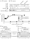
Figure 1. The in vitro cohesin cleavage assay is inhibited by ribonuclease activity
A. The in vitro cohesin cleavage by separase-PM2/4 is sensitive to S100 prepared from securin−/− HCT116 cells. The cleavage of SCC1 was determined by the disappearance of the full-length SCC1 (fl-SCC1) and the appearance of a cleaved SCC1 fragment (clvd-SCC1). The clvd-SCC1 is a product of the cleavage of the N-terminal site on SCC1 (Hauf et al., 2005). In lanes 3 and 4, the bands marked by an asterisk represent a protein from the cell lysate that cross-reacted with anti-SCC1. B. Schematic diagram of the purification scheme. The S100 was prepared from securin−/− HCT116 cells. The activity was eluted at 100 mM NaCl from the mono Q column. C. The inhibitory activity in the Superose 6 column fractions. The fraction numbers are indicated above the blot. The active fractions are underlined. The controls include the cohesin cleavage reactions in the column buffer (a), the cleavage buffer (b), and in the absence of separase (c). The protein standards are indicated below the blot. D. The inhibitory activity is resistant to heat. The peak fraction (#13) described in C was analyzed in a standard cohesin cleavage assay. Before adding into the cleavage reaction, 2 μl of fraction 13 was incubated at 65°C for 20 minutes. E. The inhibitory activity is sensitive to RNasin. Before adding into the cleavage reaction, 2 μl of fraction 13 was mixed with 10 units of RNasin.
Figure 2
Figure 2. Polynucleotide is required for the cleavage of cohesin by separase
A. Cohesin cleavage occurs in the presence of RNA or DNA. RNase-treated separase and cohesin were assembled in a standard cohesin cleavage assay. RNasin (20 units) was next added to inhibit the ribonuclease, followed by 0.5 μg of RNA (poly A, Roche) or a generic plasmid DNA. B. Comparison of cohesin cleavage in the presence and absence of RNA. Separase with concentrations from 2 nM to 200 nM was added in the cohesin cleavage assay in the absence (lanes 1–4) and presence (lanes 5–7) of RNase A. The cleavage of SCC1 was analyzed by immunoblot. C. Comparison of cohesin cleavage in the presence and absence of DNA. The experiments described in B were repeated, except that RNase was included in all reactions and 0.5 μg DNA (4000 bps) was added in lanes 2–7.
Figure 3
Figure 3. Separase and cohesin bind to DNA in a sequence-nonspecific manner
A. Separase binds to DNA-cellulose beads. DNA-cellulose beads and control beads were mixed with the purified securin-free separase-C2029S (catalytically inactive separase). After the pull-down assay, the amount of separase associated with beads and the amount left in the supernatant were analyzed by immunoblot. B. Measurement of the Kd of the separase-DNA interaction. The concentration of separase was determined at 15 ng/μl (Figure S5). Aliquots of 150 ng purified securin-bound separase were diluted in 10–500 μl of cohesin cleavage buffer. After the pull-down assay, the amount of separase associated with the beads was analyzed. To estimate the physiological concentration of separase in 293T and HeLa cells, 2.5 μl of cell pellets were lysed in SDS sample loading buffer and analyzed together. Indicated amounts of the same separase preparation were also analyzed as the standards on the same SDS-PAGE gel. The relationship between the signal strengths and the amount of separase was calculated by a simple linear regression (R2=0.998). We estimated that the amount of separase in the 293T and HeLa cell pellets is 144 ng and 109 ng, respectively. Assuming that the volume of the cell pellets represented the total volume of the cells, the physiological concentrations were estimated at 259 nM and 181 nM for 293T and HeLa cells, respectively. Because the cell pellets also included some residual buffer, the actual physiological concentrations should be higher than our estimates. C. The dissociation constant of the separase-DNA interaction. The amount of separase on the beads was determined using the standards described in B. The percentage of separase associated with the beads was calculated using the total amount of separase in the binding reaction as 100%. Two independent experiments were plotted. The concentrations of separase that exhibited a 50% binding were 21 nM and 24 nM. BSA was used as the negative control. The amounts of BSA were determined by immunoblot with anti-BSA. D. Binding of separase to the DNA beads was competed by free DNA as short as 25 bps. The DNA-beads pull-down assay contained 8 μg of various-length DNA. The 4000 bp fragment was generated by a single restriction digest of a generic plasmid. The 200 and 300 bp fragments were produced by PCR amplification of an arbitrarily chosen sequence. The 25 and 65 bp fragments were generated by annealing two pairs of synthesized oligonucleotides. The amounts of separase that associated with the beads and that remained in the supernatants were analyzed by immunoblot. E. Binding of cohesin to the DNA beads was competed by free DNA as short as 25 bps. The competition assay was performed as described in D, except that cohesin (200 nM) was analyzed instead of separase. The 100 bp fragment was generated by PCR.
Figure 4
Figure 4. The proteolytic activity of separase is independent of polynucleotide
Myc6-tagged wild-type separase and the S1126A mutant were transiently expressed in 293T cells and purified on myc-beads. The beads (2 μl) were next incubated with 10 μl various Xenopus egg extracts to degrade the associated securin. The preparation of the extracts containing low (Low Δ90) and high (High Δ90) levels of cyclin B1 was described previously (Stemmann et al., 2001). The RNA-free extract was prepared by pre-incubating with of RNase A (1 μg/μl final concentration) for 15 minutes at room temperature. DNA (0.1 μg/μl final concentration) was supplemented in some extracts as indicated above the panels. After incubation with the extract, the beads in lanes 5, 9 and 13 were also incubated with recombinant securin (5 ng/μl final concentration). After three washes, 1 μl of the beads was analyzed by immunoblot to determine the status of separase auto-cleavage, which converted the slow-migrating full-length separase (fl-separase) into a faster migrating N-terminal fragment (clvd-separase). The amount of securin on the beads was also examined. To test the cleavage activity towards cohesin, 1 μl of the remaining separase beads was incubated with cohesin in a standard cohesin cleavage assay.
Figure 5
Figure 5. In vitro cohesin cleavage requires DNA of a minimum length and at an optimal concentration
A. The response of cohesin cleavage reaction to dsDNA of various lengths. Double-stranded DNA fragments (30 ng/μl) with various lengths (4000-25 bps) were analyzed for their effectiveness in supporting cohesin cleavage (lanes 4–9). All dsDNA fragments were precipitated in ethanol and re-dissolved in H2O. Their concentration was normalized to about 300 ng/μl as determined by OD260. Negative controls (lanes 1 and 2) and a cleavage reaction (lane 3) mediated by heparin (30 ng/μl) were included. B. Measurement of the optimal concentration of dsDNA in the cohesin cleavage assay. Standard cohesin cleavage assays were performed in the presence of various concentrations of 600 bp dsDNA. Both the full-length and cleaved SCC1 were detected by immunoblot. C. The response curve of cohesin cleavage versus DNA concentration. We quantified the signals for both the full-length and the cleaved SCC1 shown in C. The percentages of cleaved SCC1 were calculated by dividing the signal for the cleaved SCC1 to the combined signal of both the full-length and cleaved SCC1. D. Comparison of the stimulation of the cohesin cleavage by dsDNA of various lengths at various concentrations. Using the same batch of separase, we compared the cohesin cleavage reaction to the increases in the concentration of various-length dsDNA. Double stranded DNA of 25 (diamond), 600 (square), and 4000 (circle) bps were analyzed. The experiments were performed as in B and the signals were analyzed as in C.
Figure 6
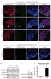
Figure 6. Separase localizes to chromosomes in mitosis
A. Immunofluorescent staining of mitotic chromosome spreads. Chromosome spreads were prepared from 293T cells stably expressing separase-V5. Separase-V5 was detected with anti-V5 (red). The kinetochores were illuminated with CREST serum (green). Chromosomal DNA was stained with DAPI. Interphase spreads are indicated by white arrows. A negative control (in the absence of ponasterone A) is shown at the bottom. B. Analysis of separase localization by cellular fractionation. HeLa cells arrested in interphase (G1+S) and mitosis (M) were fractionated into the cytoplasm (S2), nucleoplasm (S3) and chromatin (P3) fractions. The amount of separase in each fraction was analyzed. Topo IIα and GAPDH plus Erk2 were used as the controls for chromosome and cytoplasm fractions, respectively. C. Confirmation of the cell cycle stages of the samples used in B. Phospho-H3 Ser10-specific antibody was used to detect the mitotic-specific histone modification. GAPDH served as the loading control. D. Quantification of chromatid-bound separase, normalized to Topo IIα. The amount of separase relative to Topo IIα in interphase cells was defined as 1 unit. The error bar was calculated based on three independent experiments (p=0.001).
Figure 7
Figure 7. Chromosomal DNA is required for the cleavage of chromosome-associated cohesins
A. Separase cleaves chromosome-associated, but not soluble, mitotic cohesins. The chromosome-associated mitotic cohesin (C) was purified from mitotic-arrested HeLa cells, exactly as described (Waizenegger et al., 2000). This culture contained less than 4% of interphase cells (Figure S8). Nocodazole-arrest HeLa cells, stably expressing a myc6-SCC1 at a level about 20% of endogenous SCC1 (Figure S9), was used as the source of myc6-tagged soluble cohesin. The soluble myc6-SCC1 (S) was affinity purified on anti-myc beads, together with the rest of the cohesin subunits (Figure S9), and eluted with myc peptides. The cleaved products were detected by immunoblot using anti-SCC1 and anti-myc. B. Cleavage of chromosome-associated cohesins depends on the presence of chromosomal DNA. Aliquots of the aforementioned chromosome-associated cohesin were pretreated with micrococcal nuclease (MN) at 37°C for 15 minutes in the presence of 1 mM CaCl2 to remove chromosomal DNA. The rest of the pellet was mock treated. The cohesin cleavage buffer contained 3 mM EGTA to inactivate the nuclease. When indicated, DNA was added back to the reaction. C. A model to explain the selective cleavage of chromosome-associated cohesin in anaphase. Separase, inhibited by securin and S1126 phosphorylation, is preloaded onto the entire length of mitotic chromosomes as early as in prophase. Chromosomal DNA enables the interaction between separase and chromosome-associated cohesin. Thus, only chromosome-associated cohesins are cleaved at the onset of anaphase. The soluble cohesins, phosphorylated at the SA2 subunit, are not cleaved because of the lack the DNA cofactor.

Similar articles

Cited by

References

    1. Akhmedov AT, Gross B, Jessberger R. Mammalian SMC3 C-terminal and coiled-coil protein domains specifically bind palindromic DNA, do not block DNA ends, and prevent DNA bending. J Biol Chem. 1999;274:38216–38224. - PubMed
    1. Alexandru G, Uhlmann F, Mechtler K, Poupart MA, Nasmyth K. Phosphorylation of the cohesin subunit Scc1 by Polo/Cdc5 kinase regulates sister chromatid separation in yeast. Cell. 2001;105:459–472. - PubMed
    1. Bembenek JN, Richie CT, Squirrell JM, Campbell JM, Eliceiri KW, Poteryaev D, Spang A, Golden A, White JG. Cortical granule exocytosis in C. elegans is regulated by cell cycle components including separase. Development (Cambridge, England) 2007;134:3837–3848. - PMC - PubMed
    1. Chestukhin A, Pfeffer C, Milligan S, DeCaprio JA, Pellman D. Processing, localization, and requirement of human separase for normal anaphase progression. Proc Natl Acad Sci U S A. 2003;100:4574–4579. - PMC - PubMed
    1. Fan HY, Sun QY, Zou H. Regulation of Separase in Meiosis: Separase is Activated at the Metaphase I-II Transition in Xenopus Oocytes during Meiosis. Cell Cycle. 2006;5:198–204. - PubMed

Publication types

LinkOut - more resources