. 2009 Apr 3;137(1):146-58.
doi: 10.1016/j.cell.2009.02.044.

A systematic survey identifies prions and illuminates sequence features of prionogenic proteins

Affiliations

A systematic survey identifies prions and illuminates sequence features of prionogenic proteins

Simon Alberti et al. Cell. .

Abstract

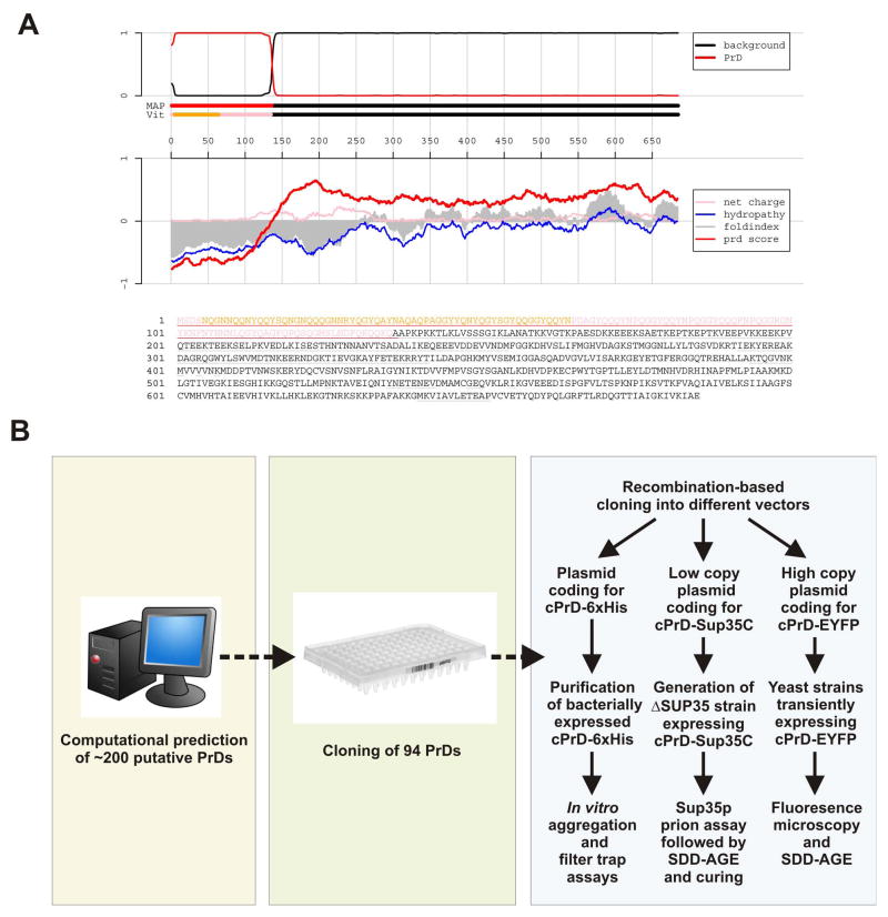
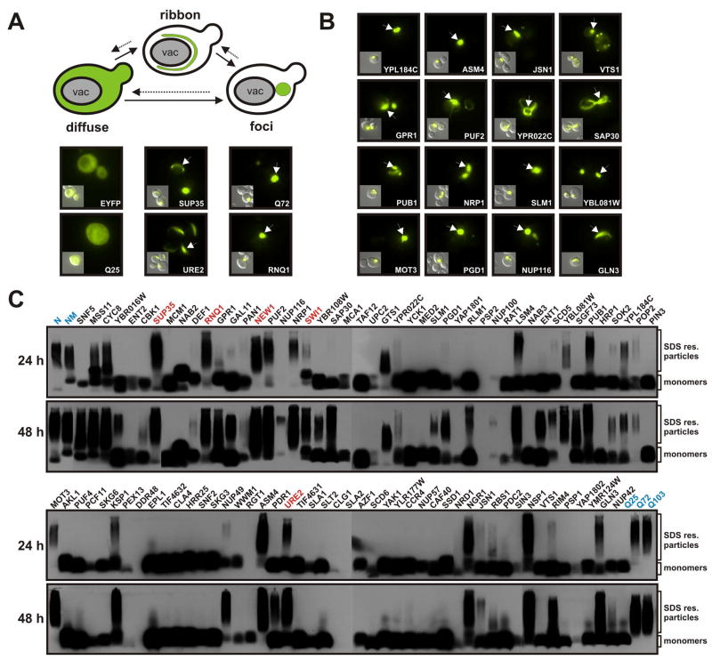
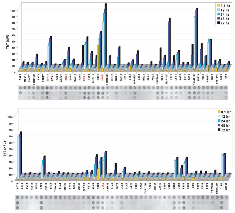
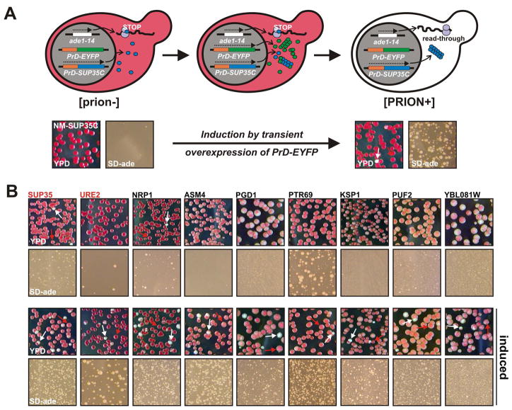
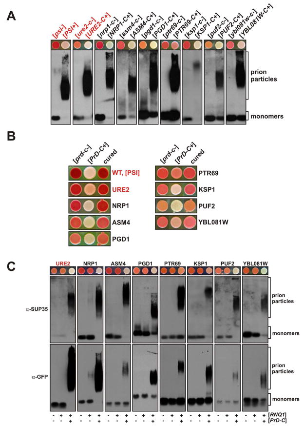
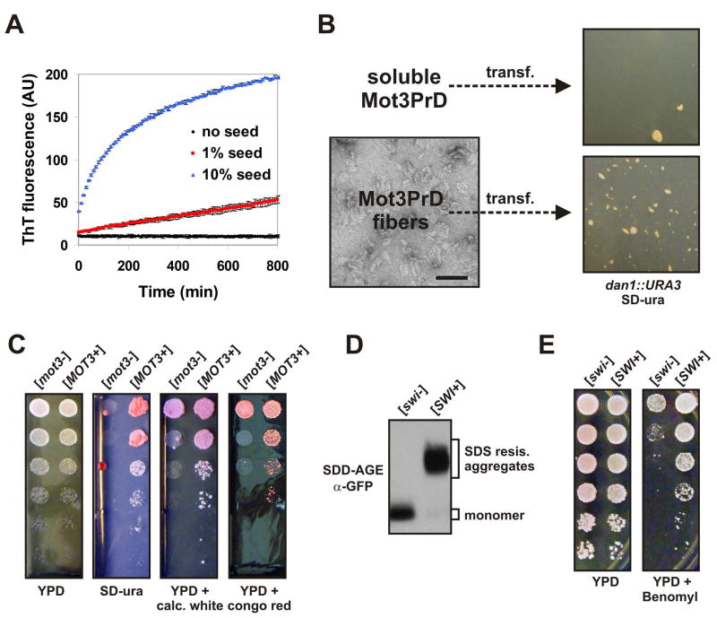
Prions are proteins that convert between structurally and functionally distinct states, one or more of which is transmissible. In yeast, this ability allows them to act as non-Mendelian elements of phenotypic inheritance. To further our understanding of prion biology, we conducted a bioinformatic proteome-wide survey for prionogenic proteins in S. cerevisiae, followed by experimental investigations of 100 prion candidates. We found an unexpected amino acid bias in aggregation-prone candidates and discovered that 19 of these could also form prions. At least one of these prion proteins, Mot3, produces a bona fide prion in its natural context that increases population-level phenotypic heterogeneity. The self-perpetuating states of these proteins present a vast source of heritable phenotypic variation that increases the adaptability of yeast populations to diverse environments.


Figures

Figure 1
Figure 1. Computational prediction and outline of the prion screen
(A) Output format of the cPrD prediction algorithm for Sup35p. The core region of the cPrD is highlighted in orange and additional predicted regions in pink. The top panel shows the probability of each residue belonging to the HMM state “cPrD” (red) and “background” (black); the tracks “MAP” and “Vit” illustrate the Maximum a Posteriori and the Viterbi parses of the protein into these two states. The lower panel shows sliding averages over a window of width 60 of net charge (pink), hydropathy (blue), and predicted disorder (gray) as in FoldIndex (Prilusky et al., 2005), along with a sliding average based on cPrD amino acid propensities (red). (B) Overview of the experimental procedures employed to screen for new Q/N-rich prions in yeast. Based on our computational prediction, we generated a cPrD library that was shuttled into a panel of expression vectors for analysis. Experiments were performed with cPrDs expressed in bacteria and yeast and included biochemical assays, cell biological assays and different aggregate visualization techniques.
Figure 2
Figure 2. Prion domains form intracellular aggregates detectable by microscopy and SDD-AGE
(A) Expression of amyloidogenic proteins in the yeast cytosol leads to the formation of ribbon and dot-like structures. cPrD-EYFP fusion proteins were expressed from a galactose-regulatable plasmid in yeast cells containing the [RNQ+] prion. The yeast cells were subjected to fluorescence microscopy after 24 h of expression. Representative fluorescence microscopy images are shown together with DIC images (insets). Arrows point to aggregates in the yeast cytosol. (B) A selected set of cPrD candidates forming fluorescent foci after 24 h of expression. Conditions were as described in (A). (C) Detection of SDS-resistant aggregates by SDD-AGE in cell lysates of yeast strains expressing cPrD-EYFP fusions. Expression of the proteins was induced for 24 h (top gels) or 48 h (bottom gels). Control proteins (highlighted in blue) were the N or NM domains of Sup35p (top left) and the huntingtin protein length variants Q25, Q72 and Q103 (bottom right). Proteins were detected with a GFP-specific antibody. Previously identified prions and the prion candidate New1p are highlighted in red.
Figure 3
Figure 3. Prion domains have diverse amyloid propensities
Amyloid formation of cPrD-M-His proteins was followed by ThT fluorescence (arbitrary units) measured at the indicated time points. Shown are means of three replicate assemblies (coefficients of variation were generally < 30; exceptions were Pan1, Nup116, and Yap1802, which each had highly variable lag phases). After the final measurement, reactions were analyzed for detergent-resistant aggregation. Aliquots from one experiment were spotted directly onto nitrocellulose (“total”), or treated with either 0.1% Tween 20 or 2% SDS and filtered through a non-binding membrane. Retained protein was visualized with Ponceau S. *These cPrDs were purified with a polyHis-tag only (no M). **These cPrDs were purified with the M-His tag at their N-terminus. Red labels indicate known prion proteins.
Figure 4
Figure 4. A SUP35-based prion assay is used to detect switching behavior
(A) A schematic overview of the genetic manipulations deployed to identify cPrDs with prion properties (top) and an example of the used selection procedure (bottom). In the bottom half, the prion state was induced by expressing NM-EYFP for 24 h and the cells were subsequently plated on complete (YPD) and adenine-deficient medium (SD-ade). The same strain grown under non-inducing conditions served as a control. Note that the number of cells plated on SD-ade plates was 200 times the number on YPD plates. Arrows point to colonies that switched to a white colony color. (B) A selected set of positive candidates identified with the SUP35C-based prion assay. Conditions were as described in (A).
Figure 5
Figure 5. The phenotypic switches of cPrD-Sup35C chimeras involve amyloid and are curable
(A) Cells lysates were prepared from [prd-c−] and [PrD-C+] strains and analyzed by SDD-AGE. The cPrD-Sup35C fusion proteins were detected by using a C domain-specific anti-Sup35p antibody. Corresponding [prd-c−] and [PrD-C+] strains growing on YPD are displayed above the SDD-AGE Western blots. (B) [PrD-C+] strains were passaged three times on YPD plates containing 5 mM GdnHCl and then spotted onto YPD plates (“cured”). The [prd-c−] and [PrD-C+] strains are shown for comparison. (C) cPrD-SUP35C strains with a coding sequence for Cerulean integrated at the 3′ end of the corresponding chromosomal gene were subjected to an SDD-AGE analysis in the respective [prd-c−] and [PrD-C+] states. cPrD-Sup35C particles were detected with a C domain-specific anti-Sup35p antibody and the particles containing Cerulean-tagged endogenous protein were detected with an anti-GFP antibody.
Figure 6
Figure 6. The transcription factor Mot3p is a prion
(A) A Mot3p-reporter strain is Ura+ when Mot3p is inactive. A dan1::URA3/DAN1 mot3::KanMX4/MOT3 diploid was sporulated and tetrads dissected. Shown are five-fold serial dilutions of four spores from a tetratype tetrad, plated onto SD-CSM and SD-ura. Spore genotypes are as indicated. (B) 5-fold serial dilutions of [mot3−] and [MOT3+] dan1::URA3 cells were spotted onto YPD and SD-ura. (C) Lysates of diploid [mot3−] and [MOT3+] cells were investigated by SDD-AGE and Western blotting. Mot3p was detected via its naturally occurring 6xHis motif using an anti-His antibody. (D) Wildtype HSP104 yeast cells and HSP104-deleted yeast cells, each carrying plasmids for galactose-inducible expression of either Mot3PrD-EYFP or control protein EYFP, were compared for [MOT3+] induction. Two transformants each were grown over night in galactose media, washed once in water, then plated at five fold serial dilutions to SD-CSM or SD-ura plates. (E) A [MOT3+] isolate was passaged three times on YPD plates or YPD plates containing GdnHCl, and then grown over night in liquid YPD prior to spotting onto SD-ura plates. (F) A [RNQ+] dan1::URA3 strain was converted to [rnq−] by four passages on GdnHCl-containing plates. The [RNQ+] and [rnq−] strains were transformed with a Mot3PrD-EYFP plasmid and assessed for [MOT3+] induction as in (D).
Figure 7
Figure 7. Mot3p and Swi1p form amyloid-based prions that can be beneficial
(A) Mot3PrD amyloid fibers used for fiber transformation experiments were added at 1% or 10% (w/w) to fresh 20 μM Mot3PrD amyloid assembly experiments and monitored for acquisition of ThT fluorescence. Shown are means +/− STDV. (B) Mot3PrD-M-His protein was polymerized and examined for a fibrillar amyloid morphology by transmission electron microscopy (bar = 100 nm). [mot3−] spheroplasts were transformed with either soluble (freshly diluted) or amyloid Mot3PrD-M-His protein and plated directly onto SD-ura plates containing 1 M sorbitol. (C) [MOT3+] and [mot3−] isolates were grown over night in YPD, then spotted (serial 5 fold dilutions) to SD-ura, YPD, or YPD containing calcofluor white (50 μg/ml) or congo red (500 μg/ml). (D) Lysates of diploid [swi−] and [SWI+] cells carrying an integration of the coding sequence for EGFP at one of the two chromosomal loci of the SWI1 gene were investigated by SDD-AGE and Western blotting. Swi1p-EGFP fusion proteins were detected using an anti-GFP antibody. (E) 5-fold serial dilutions of [swi−] and [SWI+] cells were spotted on YPD and YPD containing benomyl (5 mg/l).

Comment in

References

    1. Abramova N, Sertil O, Mehta S, Lowry CV. Reciprocal regulation of anaerobic and aerobic cell wall mannoprotein gene expression in Saccharomyces cerevisiae. Journal of bacteriology. 2001;183:2881–2887. - PMC - PubMed
    1. Aigle M, Lacroute F. Genetical aspects of [URE3], a non-mitochondrial, cytoplasmically inherited mutation in yeast. Mol Gen Genet. 1975;136:327–335. - PubMed
    1. Alberti S, Gitler AD, Lindquist S. A suite of Gateway cloning vectors for high-throughput genetic analysis in Saccharomyces cerevisiae. Yeast. 2007;24:913–919. - PMC - PubMed
    1. Alexandrov IM, Vishnevskaya AB, Ter-Avanesyan MD, Kushnirov VV. Appearance and propagation of polyglutamine-based amyloids in yeast: tyrosine residues enable polymer fragmentation. J Biol Chem. 2008;283:15185–15192. - PMC - PubMed
    1. Bagriantsev SN, Kushnirov VV, Liebman SW. Analysis of amyloid aggregates using agarose gel electrophoresis. Methods in enzymology. 2006;412:33–48. - PubMed

Publication types

MeSH terms