. 2009 Sep;89(3):333-43.
doi: 10.1016/j.exer.2009.03.019. Epub 2009 Apr 2.

Microtubule disassembly breaks down the barrier integrity of corneal endothelium

Affiliations

Microtubule disassembly breaks down the barrier integrity of corneal endothelium

Supriya S Jalimarada et al. Exp Eye Res. 2009 Sep.

Abstract

Increased contractility of the peri-junctional actomyosin ring (PAMR) breaks down the barrier integrity of corneal endothelium. This study has examined the effects of microtubule disassembly on Myosin Light Chain (MLC) phosphorylation, a biochemical marker of actomyosin contraction, and barrier integrity in monolayers of cultured bovine corneal endothelial cells (BCEC). Exposure to nocodazole, which readily induced microtubule disassembly, led to disruption of the characteristically dense assembly of cortical actin cytoskeleton at the apical junctional complex (i.e., PAMR) and dispersion of ZO-1 from its normal locus. Nocodazole also led to an increase in phosphorylation of MLC. Concomitant with these changes, nocodazole caused an increase in permeability to HRP and FITC dextran (10 kDa) and a decrease in trans-endothelial electrical resistance (TER). Y-27632 (a Rho kinase inhibitor) and forskolin (known to inhibit activation of RhoA through direct elevation of cAMP) opposed the nocodazole-induced MLC phosphorylation, decrease in TER, and dispersion of ZO-1. Thrombin, which breaks down the barrier integrity of BCEC monolayers, also induced microtubule disassembly and MLC phosphorylation. Pre-treatment with paclitaxel to stabilize microtubules opposed the thrombin effects. These results suggest that microtubule disassembly breaks down the barrier integrity of BCEC through activation of RhoA and subsequent disruption of the PAMR. The thrombin effect also highlights that signaling downstream of GPCRs can also influence the organization of microtubules.


Figures
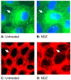
Fig. 1
Fig. 1
Effect of nocodazole on the organization of the cytoskeleton. Immunostaining of microtubule using antibody against α-tubulin in untreated cells (A) or cells treated with 10 μM nocodazole (NDZ) for 30 min (B). Note the disassembly of microtubules, as evident by the loss of the fibrillary extensions to the periphery and the apparent condensation of microtubules around the nucleus (indicated by arrows). Staining for F-ctin in untreated cells (C). The characteristic PAMR is indicated by the arrow. Exposure to nocodazole (D) induces disruption of the PAMR as shown by the arrow. The responses shown are typical of more than ten independent trials.
Fig. 2
Fig. 2
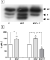
Effect of nocodazole on MLC phosphorylation. MLC phosphorylation was assayed using urea glycerol gel electrophoresis, followed by immunoblotting. Typical blot depicting the phophorylation status of MLC (A) (NP: Non-phosphorylated MLC, P: Mono-phosphorylated MLC; PP: Di-phosphorylated MLC). Treatment with 2 μM Nocodazole (NDZ) for 30 min enhances MLC phosphorylation, as indicated by the intense diphosphorylated (PP) MLC bands, when compared to control (n = 10). Co-treatment with 10 μM Y-27632 (Y) opposes nocodazole-induced response. Summary of densitometric analysis of the representative experiments (n = 5) expressed as %pMLC (B) shown in panel (A). * indicates statistical significance at P < 0.05.
Fig. 3
Fig. 3
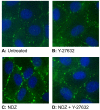
Localization of phosphorylated MLC (ppMLC). In untreated cells (A), ppMLC staining is diffuse and moderate in the cortical region, consistent with a low basal level of MLC phosphorylation. Treatment with 10 μM Y-27632 for 30 min reduces the ppMLC when compared to untreated cells (B). Treatment with 2 μM nocodazole (NDZ) for 30 min (C) increases the of ppMLC consistent with its ability to increase MLC phosphorylation. Co-treatment with nocodazole and Y-27632 opposes nocodazole response (D).
Fig. 4
Fig. 4
Effect of cytochalasin D on TER. The influence of cytochalasin D on TER was monitored by ECIS. Exposure to cytochalasin D induces an immediate, dose-dependent decline in TER with a maximum decline observed at 2 μg/mL. The responses shown are representative of three independent trials.
Fig. 5
Fig. 5
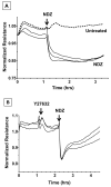
Effect of nocodazole on TER. The changes in TER in response to nocodazole (NDZ) were measured by ECIS (A). Treatment with 2 μM nocodazole induces a persistent reduction in TER with a peak decline occurring within 10 min (n = 8). Pretreatment with 2 μM Y-27632 for 1 hr attenuates the decline in TER in response to nocodazole (B). The decline is not persistent, unlike with nocodazole (Fig. 5A), and is characterized by recovery to baseline by around 3 hr. Histogram analysis of representative experiments (n = 8) showing the recovery of TER in response to Y-27632 pretreatment (C). The % reduction in TER over 2 hr period with nocodazole alone was greater compared to when cells were pretreated with Y-27632. * indicates statistical significance at P < 0.05.
Fig. 5
Fig. 5
Effect of nocodazole on TER. The changes in TER in response to nocodazole (NDZ) were measured by ECIS (A). Treatment with 2 μM nocodazole induces a persistent reduction in TER with a peak decline occurring within 10 min (n = 8). Pretreatment with 2 μM Y-27632 for 1 hr attenuates the decline in TER in response to nocodazole (B). The decline is not persistent, unlike with nocodazole (Fig. 5A), and is characterized by recovery to baseline by around 3 hr. Histogram analysis of representative experiments (n = 8) showing the recovery of TER in response to Y-27632 pretreatment (C). The % reduction in TER over 2 hr period with nocodazole alone was greater compared to when cells were pretreated with Y-27632. * indicates statistical significance at P < 0.05.
Fig. 6
Fig. 6
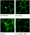
Effect of nocodazole on localization of ZO-1. Immunostaining of ZO-1 in untreated cells (A). Note the continuous ZO-1 localization throughout the cell periphery (shown by arrow). Exposure to 10 μM Y-27632 for 30 min (B) does not alter the localization of ZO-1 and was similar to control. Exposure to 2 μM nocodazole (NDZ) for 30 min (C) induces discontinuities in ZO-1 localization (shown by arrow), indicating barrier instability. Pretreatment with 2 μM Y-27632 for 1 hr (D) opposes the nocodazole-induced response. The responses shown are typical of three independent trials.
Fig. 7
Fig. 7
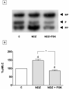
Effects of cAMP on nocodazole-induced MLC phosphorylation. Co-treatment with 10 μM forskolin (FSK) significantly inhibits the nocodazole-induced MLC phosphorylation (A), as evident by the loss of intense di-phosphorylated (PP) MLC bands compared to nocodazole (NDZ). Summary of densitometric analysis (B) expressed as %pMLC of the representative experiments (n = 4) shown in panel (A). * indicates statistical significance at P < 0.05.
Fig. 8
Fig. 8
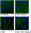
Influence of cAMP on phosphorylated MLC (ppMLC). In untreated cells (A), the intensity of ppMLC along cell periphery is moderate, indicating basal level of MLC phosphorylation. Treatment with 10 μM forskolin (FSK) and 50 μM rolipram (B) reduced the intensity of ppMLC when compared to untreated cells indicating MLC dephosphorylation. Exposure to 2 μM nocodazole for 30 min (C) increases ppMLC intensity in the cortical region, consistent with the observed increase in MLC phosphorylation. Co-treatment with 10 μM forskolin and 50 μM rolipram (D) opposes nocodazole response. The responses shown are typical of five independent trials.
Fig. 9
Fig. 9
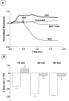
Effects of cAMP on nocodazole-induced breakdown in the barrier integrity. Exposure to 2 μM nocodazole led to a precipitous decrease in TER (A). Treatment with 10 μM forskolin alone enhances TER compared to control. Pretreatment with 10 μM forskolin for 30 min opposes the decrease in TER in response to nocodazole. (B) Histogram analysis of the representative experiments (n = 3) shown in (A). The % reduction in TER in response to nocodazole was significantly attenuated by pretreatment with forskolin 15 min onwards up to 1 hr. * indicates statistical significance at P < 0.001.
Fig. 10
Fig. 10
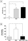
Influence of nocodazole on in vitro permeability. The changes in vitro permeability in response to nocodazole was ascertained by quantifying the flux of HRP across cells grown on culture inserts (A). Treatment with 2 μM nocodazole for 30 min significantly increases HRP flux compared to control (n = 5). Histamine (H) at the concentration of 100 μM for 10 min was used as a positive control. * indicates statistical significance at P < 0.001. The ability of elevated cAMP to oppose nocodazole-induced increase in paracellular permeability was quantified by the flux of FITC dextran across cells grown on porous culture inserts (B). Treatment with 5 μM nocodazole significantly increases permeability to FITC dextran in comparision to control. Pretreatment with 10 μM forskolin (FSK) in combination with 50 μM rolipram (ROL) for 1 hr significantly attenuates nocodazole-induced increase in permeability (n = 6). * indicates statistical significance at P < 0.01.
Fig. 11
Fig. 11
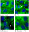
Effect of thrombin on microtubule organization. Immunostaining for microtubules in untreated cells (A). The characteristic uncondensed fibrillary extensions are evident. Treatment with 5 μM paclitaxel (PTX) alone for 1 hr (B) stabilizes microtubule assembly. Exposure to 2 U/ml thrombin for 2 min (C) induces microtubule condensation (shown by arrows). Pretreatment with 5 μM paclitaxel for 1 hr (D) opposes the thrombin response. The responses shown are typical of three independent trials.
Fig. 12
Fig. 12
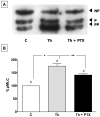
Effect of microtubule stabilization on thrombin-induced MLC phosphorylation. Treatment with 2 U/ml thrombin for 2 min induces increased MLC phosphorylation when compared to untreated cells. Pretreatment with 5 μM paclitaxel (PTX) for 1 hr (A) attenuates the thrombin (Th)-induced MLC phosphorylation. Summary of densitometric analysis (B) expressed as %pMLC of the representative experiments (n = 6) similar to that shown in (A). * indicates statistical significance at P < 0.001. ** indicates statistical significance at P < 0.01.
Fig. 13
Fig. 13
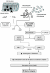
Cell signaling involved in the loss of barrier integrity secondary to microtubule disruption. RhoA-specific guanine nucleotide exchange factors (GEFs) on the microtubules are released upon disruption of microtubules, leading to activation of RhoA. Subsequently, Rho kinase inhibits MLC phosphatase (MLCP) by phosphorylating its MYPT1 subunit. The resulting increase in MLC phosphorylation induces actomyosin contraction, leading to disruption of the PAMR, which in turn causes breakdown in the barrier integrity. Activation of PKA, which prevents activation of RhoA, indirectly inhibits MLCP.

Similar articles

Cited by

References

    1. Ayala A, Warejcka DJ, et al. Corneal activation of prothrombin to form thrombin, independent of vascular injury. Invest Ophthalmol Vis Sci. 2007;48(1):134–43. - PubMed
    1. Banan A, Farhadi A, et al. The delta-isoform of protein kinase C causes inducible nitric-oxide synthase and nitric oxide up-regulation: key mechanism for oxidant-induced carbonylation, nitration, and disassembly of the microtubule cytoskeleton and hyperpermeability of barrier of intestinal epithelia. J Pharmacol Exp Ther. 2003;305(2):482–94. - PubMed
    1. Bereiter-Hahn J, Voth M, et al. Structural implications of mitochondrial dynamics. Biotechnol J. 2008;3(6):765–80. - PubMed
    1. Birukova AA, Adyshev D, et al. GEF-H1 is involved in agonist-induced human pulmonary endothelial barrier dysfunction. Am J Physiol Lung Cell Mol Physiol. 2006;290(3):L540–8. - PubMed
    1. Birukova AA, Birukov KG, et al. Involvement of microtubules and Rho pathway in TGF-beta1-induced lung vascular barrier dysfunction. J Cell Physiol. 2005;204(3):934–47. - PubMed

Publication types

MeSH terms