. 2009 Jul;7(7):749-755.e11.
doi: 10.1016/j.cgh.2009.03.022. Epub 2009 Apr 1.

A novel histologic scoring system to evaluate mucosal biopsies from patients with eosinophilic esophagitis

Affiliations

A novel histologic scoring system to evaluate mucosal biopsies from patients with eosinophilic esophagitis

Cheryl Protheroe et al. Clin Gastroenterol Hepatol. 2009 Jul.

Abstract

Background & aims: Eosinophilic esophagitis (EoE) is characterized by medically/surgically-resistant gastroesophageal reflux symptoms and dense squamous eosinophilia. Studies suggest that histologic assessment of esophageal eosinophilia alone cannot reliably separate patients with EoE from those with gastroesophageal reflux disease (GERD). Our goal was to develop an assay to identify EoE patients and perhaps differentiate EoE from other causes of esophageal eosinophilia.

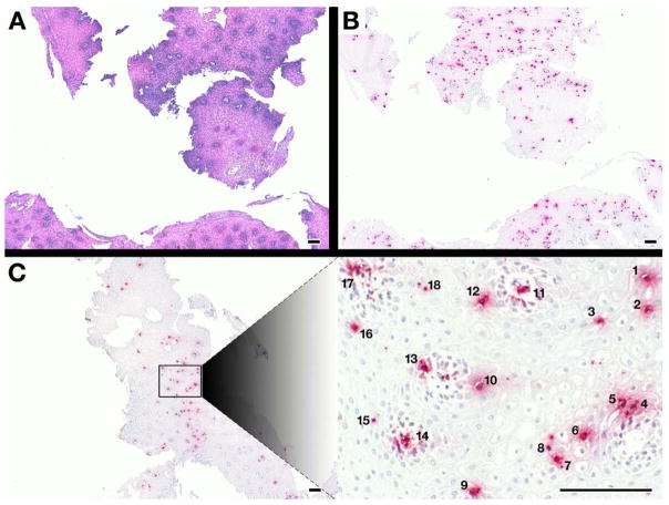
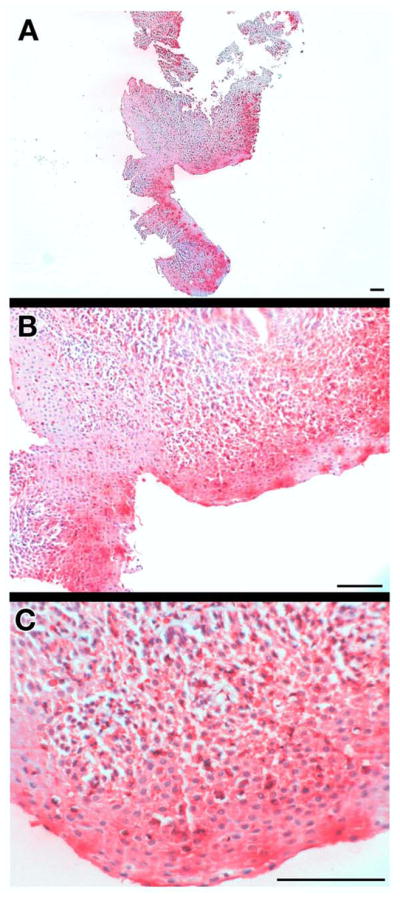
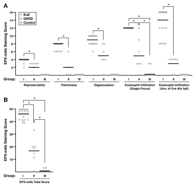
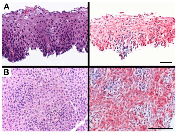
Methods: A monoclonal antibody specific for an eosinophil secondary granule protein (eosinophil peroxidase [EPX]) was developed and shown to specifically identify intact eosinophils and detect eosinophil degranulation in formalin-fixed specimens. A histopathologic scoring algorithm was developed to analyze data from patient evaluations; the utility of this algorithm was assessed by using archived esophageal tissues from patients with known diagnoses of EoE and GERD as well as controls from 2 tertiary care centers.

Results: Intraobserver/interobserver blinded evaluations demonstrated a significant difference (P < .001) between scores of samples taken from control subjects, from patients with esophageal eosinophilia who had a diagnosis of EoE, and from patients with GERD (P < .001). This algorithm also was able to identify patients whose clinical course was suggestive of a diagnosis of EoE, but that nonetheless failed to reach the critical threshold number of > or =15 eosinophils in a high-power (40x) microscopy field.

Conclusions: A novel immunohistochemical scoring system was developed to address an unmet medical need to differentiate histologic specimens from patients with EoE relative to those with GERD. The availability of a unique anti-EPX-specific monoclonal antibody, combined with the ease/rapidity of this staining method and scoring system, will provide a valuable strategy for the assessment of esophageal eosinophilia.


Figures

Figure 1
Figure 1. EPX-mAb based immunohistochemistry provides an efficient and rapid strategy to identify intact eosinophils infiltrating biopsies from eosinophil esophagitis patients
A comparison of low (5×, 20mm2 field of view) power microscopy of (A) hematoxylin-eosin stained sections and (B) serial sections of the same patient following EPX-mAb based immunohistochemistry demonstrated that this immunohistochemical strategy easily permits the identification of infiltrating eosinophils in multiple biopsies within the field. (C) EPX-mAb based immunohistochemistry permits a rapid evaluation for the presence of intact infiltrating eosinophils of entire esophageal biopsies and the location of focal areas of eosinophil accumulation. The insert photograph in this panel is a high (40×, 0.29mm2 field of view) power field that was quickly/efficiently identified as a focal area of eosinophil accumulation (identified eosinophils are numbered 1–18) without the need of laborious time-consuming cell differential analyses. Scale bar = 100 μm.
Figure 2
Figure 2. EPX-mAb based immunohistochemistry represents a novel strategy to detect eosinophil degranulation and the presence of released eosinophil peroxidase bound to tissue extracellular matrix
(A) Low (5×, 20mm2 field of view), (B) medium (16×, 1.8mm2 field of view), and (C) high (40×, 0.29mm2 field of view) microscopic fields of a proximal esophageal biopsy from an EoE patient demonstrate the utility of EPX-mAb based immunohistochemistry to detect eosinophil degranulation (i.e., EPX bound to extracellular matrix) in these biopsies. The results with this biopsy are representative of EoE patients which often display significant eosinophil degranulation. Scale bar = 100 μm.
Figure 3
Figure 3
EPX-mAb based immunohistochemistry provides a quantitatively significant strategy (Supplemental Table 2 (S-Table 2)) to distinguish EoE vs. GERD patients. (A) Examination of the scores for individual EPX-mAb based parameters associated with the EoE (Group I), GERD (Group II), and control patients (Group III) found in Supplemental Table 3 (S-Table 3) demonstrated statistical differences (*P<0.001) for each of the parameters comprising the EPX-mAb based algorithm. (B) Statistical assessments (ANOVA with Tukey) of the average total EPX-mAb based staining scores (means ± SEM) for the EoE, GERD, and control patients from Supplemental Table 3 (S-Table 3) demonstrated the utility of this algorithm to distinguish between these patient populations (*P<0.001).
Figure 4
Figure 4
EPX-mAb based immunohistochemistry permits a diagnosis of eosinophilic esophagitis among patients with appropriate clinical symptoms and borderline endoscopic/histological results but who fail to achieve the current guideline recommendations of at least a single focus of ≥15 eosinophils/40× hpf among the available biopsies. Serial sections of proximal esophageal biopsies from (A) patient #44 and (B) patient #46 (see Supplemental Tables 1 and 3 (S-Tables 1 and 3)) were either stained with hematoxylin/eosin (left panels) or subjected to EPX-mAb based immunohistochemistry (right panels) and photographed at high (40×, 0.29mm2 field of view) power. Although both patients had failed to meet traditional pathology guidelines for a diagnosis of EoE, EPX-mAb based immunohistochemistry detected the presence of extensive eosinophil degranulation in the absence of ≥15 intact eosinophils/40× hpf (0.29mm2 field of view), elevating their EPX-mAb based total score within the range indicating an EoE diagnosis. Scale bar = 50 μm.

References

    1. Furuta GT, Liacouras CA, Collins MH, et al. Eosinophilic esophagitis in children and adults: A systematic review and consensus recommendations for diagnosis and treatment. Gastroenterology. 2007;133:1342–63. - PubMed
    1. Collins MH. Histopathologic features of eosinophilic esophagitis. Gastrointest Endosc Clin N Am. 2008;18:59–71. viii–ix. - PubMed
    1. Rodrigo S, Abboud G, Oh D, et al. High intraepithelial eosinophil counts in esophageal squamous epithelium are not specific for eosinophilic esophagitis in adults. Am J Gastroenterol. 2008;103:435–42. - PubMed
    1. Odze RD. Pathology of eosinophilic esophagitis: What the clinician needs to know. Am J Gastroenterol. 2009;104:485–90. - PubMed
    1. Furuta GT, Nieuwenhuis EE, Karhausen J, et al. Eosinophils alter colonic epithelial barrier function: Role for major basic protein. Am J Physiol Gastrointest Liver Physiol. 2005;289:G890–7. - PubMed

Publication types

MeSH terms