. 2009 Aug 1;315(13):2215-30.
doi: 10.1016/j.yexcr.2009.03.020. Epub 2009 Apr 5.

Transport of mannose-6-phosphate receptors from the trans-Golgi network to endosomes requires Rab31

Affiliations

Transport of mannose-6-phosphate receptors from the trans-Golgi network to endosomes requires Rab31

A G Rodriguez-Gabin et al. Exp Cell Res. .

Abstract

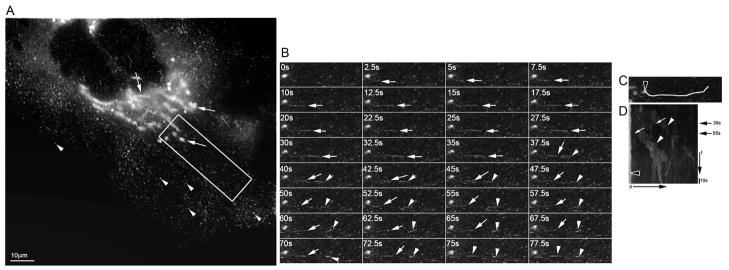
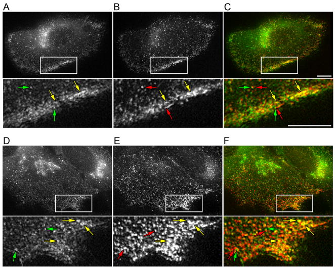
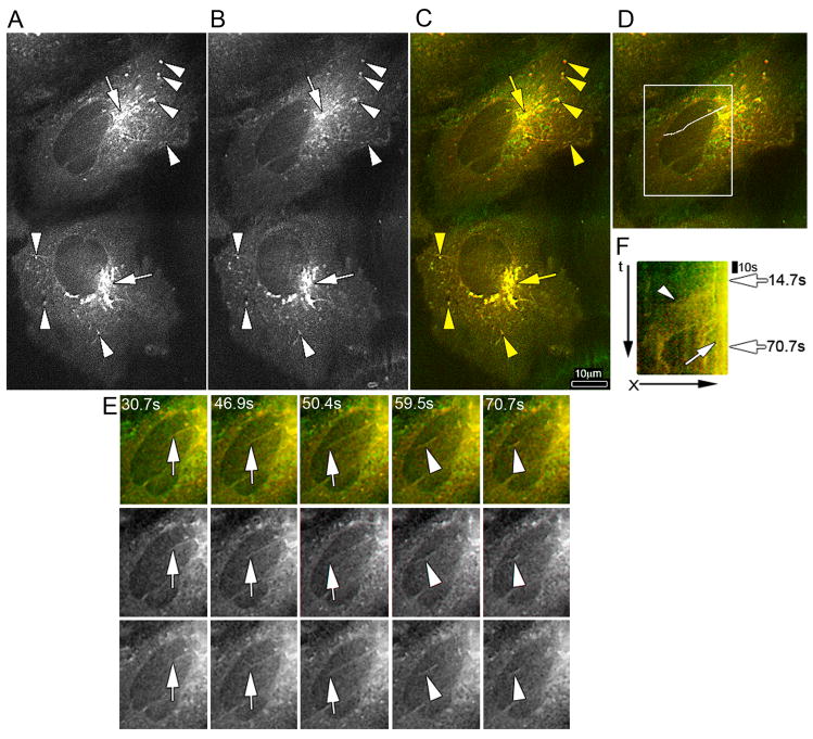
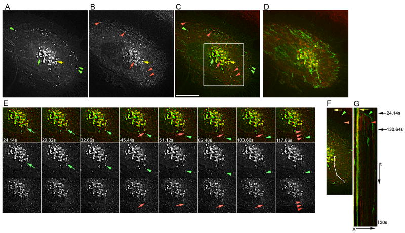
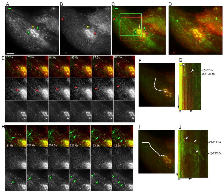
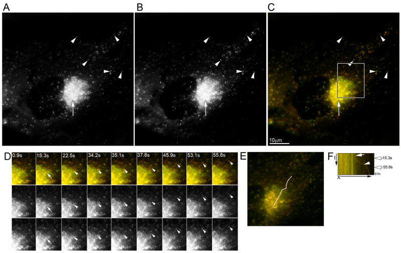
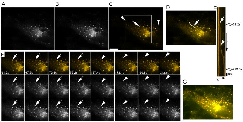
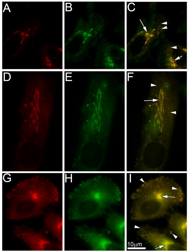
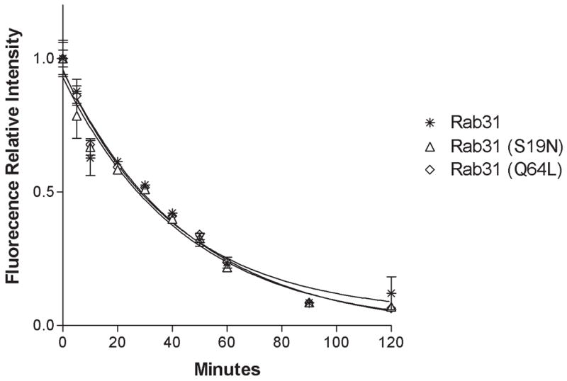
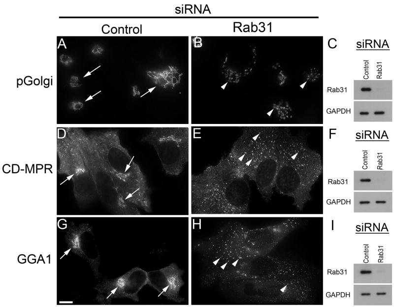
Rab31, a protein that we originally cloned from a rat oligodendrocyte cDNA library, localizes in the trans-Golgi network (TGN) and endosomes. However, its function has not yet been established. Here we show the involvement of Rab31 in the transport of mannose 6-phosphate receptors (MPRs) from TGN to endosomes. We demonstrate the specific sorting of cation-dependent-MPR (CD-MPR), but not CD63 and vesicular stomatitis virus G (VSVG) protein, to Rab31-containing trans-Golgi network carriers. CD-MPR and Rab31 containing carriers originate from extending TGN tubules that also contain clathrin and GGA1 coats. Expression of constitutively active Rab31 reduced the content of CD-MPR in the TGN relative to that of endosomes, while expression of dominant negative Rab31 triggered reciprocal changes in CD-MPR distribution. Expression of dominant negative Rab31 also inhibited the formation of carriers containing CD-MPR in the TGN, without affecting the exit of VSVG from this compartment. Importantly, siRNA-mediated depletion of endogenous Rab31 caused the collapse of the Golgi apparatus. Our observations demonstrate that Rab31 is required for transport of MPRs from TGN to endosomes and for the Golgi/TGN organization.


Figures

Figure 1
Figure 1. Dynamics of formation in the TGN of carriers containing Rab31
HeLa cells expressing Rab31-ECFP were transferred to recording medium, and the formation and the transport of post-Golgi carriers containing Rab31-ECFP at 32° C was monitored by time-lapse fluorescence microscopy. Images were captured at 0.5 sec intervals. A) Fluorescence pattern distribution. Rab31-ECFP localized in the TGN (arrow), and in small structures throughout the cytoplasm (arrowhead). B) Individual frames of the area boxed in (A). A TGN tubule extends (white arrow), and its tip breaks up to form a carrier (white arrowhead, 37.5 sec frame). The remaining tubule seems to retract, then again to extend and break up to form a second carrier (white arrowhead, 75 sec frame). Time in sec relative to the first image is shown. C) Area where the events in B occurred (carrier formation and movement to cell periphery) is indicated by a white lane. Black arrowhead shows carrier formation site. D) Kymograph analysis of the area defined in (C). A lack of movement of the TGN compartments results in a vertical trace (black arrowhead). Diagonal trace represents the formation of carrier and its movement through the area. Two white arrows indicate sequential breaking up of the TGN tubule to form two carriers (white arrowheads). The slope of the trace indicates speed of movement. Black arrows on the right indicate the time of budding in (B). X, distance in μm; t, time in sec.
Figure 2
Figure 2. Rab31 is present in endosomes
HeLa cells expressing Rab31-ECFP were incubated for 2 hours at 37° C in medium without FCS, but containing 0.2% BSA. Then cells were incubated with Texas Red transferrin (20 μg/ml) for 15–20 min. After incubation with fluorescent transferrin, cells were rinsed briefly in phosphate buffered saline (PBS) at 4°C and then fixed with 4 % paraformaldehyde in PBS at 4°C for 15 min. Cells were rinsed with PBS and analyzed by double fluorescence microscopy. A and D, Rab31-ECFP; B and E, Texas Red transferrin; C overlap of A and B, and F overlap of D and E. Texas Red Transferrin, Red; Rab31-ECFP, green; and overlapping of Texas Red Transferrin and Rab31-ECFP, yellow. Small panels are a 3.33 × magnification of the areas highlighted by white boxes. Compartments containing Rab31-ECFP (green arrows) are tightly associated with compartments containing Texas Red Transferrin (red arrows). Endosomal compartments containing both labels are present in the cell periphery (yellow arrows).
Figure 3
Figure 3. Rab31 is present in carriers that transport CD-MPR from TGN to endosomes
HeLa cells expressing CD-MPR-ECFP were transfected with a plasmid encoding Rab31-EYFP. Forty eight hours after transfection, cells were transferred to recording medium, and the formation and the transport of post-Golgi carriers were monitored at 32° C by time-lapse fluorescence microscopy. Images were collected every 0.70 sec. Fluorescence pattern distribution; A) CD-MPR-ECFP, B) Rab31-EYFP and C) overlapping of A and B, CD-MPR-ECFP (red), Rab31-EYFP (green), and overlapping of CD-MPR-ECFP and Rab31-EYFP (yellow). Rab31-EYFP and CD-MPR-ECFP co-localized in the TGN (arrow), and in small tubolo vesicular compartments throughout the cytoplasm (arrowhead). D) Area of the cells shown in E is marked by a white box. Area where the events in E occurred (carrier formation and movement to cell periphery) is indicated by a white lane. E) Individual frames showing a TGN tubule that extends (white arrow), and breaks up to form a carrier containing both CD-MPR-ECFP and Rab31-EYFP (white arrowhead). Rab31-EYFP, bottom panels; CD-MPR-ECFP, center panels and merged images top panels (color code, the same as in C). Time in sec relative to the first image is shown. F) Kymograph analysis of the area defined in (D). TGN compartments, vertical trace (white arrow). Carrier formation and its movement, diagonal trace (arrowhead). White arrows on the right indicate when the budding showed in (E) occurred. X, distance in μm; t, time in sec.
Figure 4
Figure 4. Rab31 and newly synthesized CD63 are sorted in the TGN into different carriers
HeLa cells expressing Rab31-ECFP were micro-injected intranuclearly with cDNAs encoding CD63-EYFP. Newly synthesized CD63-EYFP was trapped in the TGN and carrier formation was visualized by time-lapse double florescence microscopy. Images were collected every 1.42 sec. Fluorescence distribution pattern: A) CD63-EYFP, B) Rab31-ECFP and C) merged images. Green, CD63-EYFP; red, Rab31-ECFP; yellow, overlapping of CD63-EYFP; and Rab31-ECFP. Rab31-EYFP and CD63-ECFP co-localized in some TGN domains (yellow arrow), other TGN domains contain only CD63-EYFP (green arrow) or Rab31-ECFP (red arrow). Small vesicles present in the cytoplasm only contain Rab31-ECFP (red arrow heads) or CD63-EYFP (green arrow heads). D) Maximum pixel projection. Rab22b-ECFP and CD63-EYFP co-localized in the TGN but not in small vesicles present throughout the cytoplasm. E) Individual frames of the area marked with a box in (C). Top panels, merged images; center panels, CD63-EYFP and bottom panels, Rab31-ECFP. A TGN tubule containing CD63-EYFP extends (green arrow), and breaks up to form a carrier (green arrowhead). Then a long TGN tubule containing Rab31-ECFP extends along the same trajectory (red arrow), and simultaneously breaks in several regions to form three carriers (red arrowheads). F) Area where the events in E occurred (carrier formation and their movement to cell periphery) is indicated by a white lane. G) Kymograph analysis of the area defined in (F). TGN compartments, vertical traces (yellow arrow). The green trace that corresponds to the budding of carrier containing CD63-EYFP seen in (B) is indicated by a green arrowhead. The wide red trace corresponding to the formation of three carriers containing Rab31-ECFP is indicated by a red arrow. Black arrows on the right indicate the time when the budding occurred. The time in sec relative to the first image is shown. X, distance in μm; t, time in sec. Bar, 10 μm.
Figure 5
Figure 5. Rab31 and newly synthesized VSVG are sorted into different carriers in the TGN
HeLa cells expressing Rab31-ECFP were transfected with plasmid encoding VSVG-Venus, and then incubated at 40° C for 10–12 hrs. to accumulate VSVG-Venus in the ER. Transfected cells were then incubated at 20° C for 3 hrs, with cycloheximide (100 μg/ml) to trap VSVG-Venus in the TGN. The cells were transferred to recording media at 32 ° C to induce transport of VSVG from Golgi to PM. Time lapse photographs visualized the formation of carriers containing Rab31-ECFP and of carriers containing VSVG-Venus. Images were collected every 1.50 sec. Fluorescence distribution pattern: A) Rab31-ECFP, B) VSVG-Venus and C) merged images. Green, Rab31-ECFP; red, VSVG-Venus; yellow, overlapping of VSVG-Venus and Rab31-ECFP. Rab31-ECFP and VSVG-Venus co-localized in some TGN domains (yellow arrow), other TGN domains contain only Rab31-ECFP (green arrow). Small vesicles present in the cytoplasm only contain Rab31-ECFP (green arrow head) or VSVG-Venus (red arrow head). D) Maximum pixel projection. Rab22b-ECFP and VSVG-Venus co- localized in the TGN but not in small vesicles present throughout the cytoplasm. E) Individual frames of the area marked with red box in (C). Top panels, merged images; center panels, VSVG-Venus; and bottom panels, Rab31-ECFP. A TGN tubule containing VSVG-Venus extends (red arrow), and breaks up to form a carrier (red arrowhead). F) Area where the events in E occurred (carrier formation and movement to the cell periphery) is indicated by a white lane. G) Kymograph analysis of the area defined in (F). TGN compartments, vertical traces (white arrow). The red trace that corresponds to the budding of carrier containing VSVG-Venus seen in (E) is indicated by a white arrowhead. Arrows on the right indicate the time of budding in (E). H) Individual frames of the area marked with green box in (A). A TGN tubule extends (green arrow), and its tip breaks up to form a carrier (green arrowhead, 142.5 sec frame). The remaining tubule seems to retract, then again to extend and break up to form a second carrier (green arrowhead, 222.0 sec frame). Time in sec relative to the first image is shown. I) Area where the events in H occurred (carrier formation and movement to cell periphery) is indicated by a white lane. J) Kymograph analysis of the area defined in (G). TGN compartments, vertical trace (white arrow). Carrier formation and its movement, diagonal trace. Two white arrowheads indicate sequential breaking up of the TGN tubule to form two carriers. Arrows on the right indicate the time of budding in (H). X, distance in μm; t, time in sec. Bar, 10 μm.
Figure 6
Figure 6. Clathrin coats the carrier vesicles that contain Rab31
HeLa cells expressing clathrin-ECFP were transfected with a plasmid encoding Rab31-EYFP. Cells were transferred to recording medium, and the formation and the movement of post-Golgi carriers was monitored at 30° C by time-lapse fluorescence microscopy. Images were collected every 0.90 sec. Fluorescence pattern distribution; A) Rab31-EYFP, B) clathrin-ECFP and C) merged images. Clathrin-ECFP (red), Rab31-EYFP (green) and overlapping of clathrin-ECFP and Rab31-EYFP (yellow). Rab31-ECFP and clathrin-ECFP co-localize in the TGN (arrow), and in endosomes throughout the cytoplasm (arrowhead). D) Individual frames showing formation and movement of a carrier containing both clathrin-ECFP and Rab31-EYFP (white arrowhead). Top panels, merged images; middle panels, Rab31-EYFP; and bottom panels, clathrin-ECFP. Time in sec relative to the first image is shown. E) Area where the events in D (carrier formation and movement to cell periphery) occurred is indicated by a white lane. F) Kymograph analysis of the area defined in (E). TGN compartments, vertical yellow trace (white arrow). Carrier formation and its movement, diagonal yellow trace (arrowheads). White arrowhead identifies the trace that corresponds to the carrier budding shown in (D). Arrows on the right indicate the time when the budding in (D) occurred. X, distance in μm; t, time in sec. Bar, 10 μm.
Figure 7
Figure 7. Rab31 and GGA1 colocalize in carriers that bud from the TGN
HeLa cells expressing GGA1-ECFP were transfected with a plasmid encoding Rab31-EYFP. Cells were transferred to recording medium. Formation and the transport of post-Golgi carriers were monitored at 30° C by time-lapse fluorescence microscopy. Images were collected every 0.6 sec. Fluorescence pattern distribution; A) Rab31-EYFP, B) GGA1-ECFP and C) merged images. GGA1-ECFP (red), Rab31-EYFP (green) and overlapping of GGA1-ECFP and Rab31-EYFP (yellow). Rab31-ECFP and GGA1-ECFP co-localize in the TGN (arrow) (r = 0.912 ± 0.012), and in endosomes throughout the cytoplasm (arrowhead) (r = 0.914 ± 0.026). D) White lane marks the trajectory of a carrier formed in the TGN. Carrier formation and movement is shown in individual frames (F). E) Kymograph analysis of the trajectory mark in (D). TGN compartments, vertical yellow trace (white arrow). Carrier formation and its movement, diagonal yellow trace (arrowheads). White arrowhead identifies the trace that corresponds to carrier budding shown in (F). Arrows on the right indicate the time of budding shown in (F). X, distance in μm; t, time in sec. F) Individual frames showing a TGN tubule extending (white arrow), and breaking up to form a carrier containing both GGA1-ECFP and Rab31-EYFP (white arrowhead). Top panels, merged images; center panels, Rab31-EYFP; and bottom panels, GGA1-ECFP. Time in sec relative to the first image is shown. G) Maximum pixel projection. Rab22b-EYFP and GGA1-ECFP co- localized in the TGN and in small vesicles present throughout the cytoplasm. Bar, 10 μm.
Figure 8
Figure 8. Expression of two Rab31 mutants (S19N and Q64L) result in changes in the quantity of CD-MPR distributed between the TGN and endosomes
(A, B, C) HeLa cells expressing both CD-MPR-ECFP and wild type Rab31-EYFP. A) CD-MPR-ECFP (red), B) wild type Rab31-EYFP (green) and C) overlap of A and B, yellow compartments containing both wild type Rab31-EYFP and CD-MPR-ECFP (TGN, arrow and endosomes, arrowhead). Video 8 C shows that TGN tubules extend and break up to form carriers. (D, E, F) HeLa cells expressing both CD-MPR-ECFP and Rab31 (S19N)-EYFP. D) CD-MPR-ECFP (red); E) Rab31 (S19N)-EYFP (green); F) overlap of D and E, yellow compartments containing both Rab31(S19N)-EYFP and CD-MPR-ECFP (Golgi, arrow and endosomes, arrowhead). Notice the large size of the TGN (video 8 F shows that the long TGN tubules do not extent). (G, H, I) HeLa cells expressing both CD-MPR-ECFP and Rab31 (Q64L) mutant-EYFP. G) CD-MPR-ECFP (red), H) Rab31 (Q64L)-EYFP (green) and I) overlap of G and H, yellow compartments contain both Rab31(Q64L)-EYFP and CD-MPR-ECFP (Golgi, arrow and endosomes, arrowhead). Notice small size of the TGN and large amount of endosomes in the cell periphery (video 8 I shows that the long TGN tubules extend and break up to form carriers containing both proteins).
Figure 9
Figure 9. Rate of VSVG-Venus exit from Golgi
HeLa cells expressing either wild type Rab31 or Rab31 (S19N) mutant or Rab31 (Q64L) mutant were transfected with plasmid encoding VSVG-Venus, and maintained for 16 hours at 40° C. Newly synthesized VSVG-Venus was trapped in the Golgi by incubating the cells at 20° C for 3 hours with cycloheximide (100μg/ml). The transport of VSVG from Golgi to PM was induced by shifting the cells to 32° C. The amount of VSVG-Venus fluorescence present in the Golgi at different time periods was determined as described (Materials and Methods). The fluorescence intensities attributable to VSVG-Venus in the Golgi were plotted as a function of time for cells expressing: wild type Rab31 [*], Rab31(S19N) [Δ] and Rab31(Q64L) [◇]. The curves represent the optimized fit to a first order kinetic model. The rate constants obtained by non linear regression analysis were 0.024 ± 0.003 min−1, 0.022 ± 0.003 min−1, 0.021 ± 0.002 min−1 respectively; and the half resident times were 28.34 ± 3.27 min, 30.86 ± 3.00 min, 31.65 ± 2.5 min. The values are the means ± standard error for 10 measurements.
Figure 10
Figure 10. Depletion of endogenous Rab31 fragments Golgi/TGN structure
(A, D and G) HeLa cells were transfected with non targeting siRNA (control), or (B, E and H) siRNA that target a segment of the sequence of Rab31 present in exon 2 (5′-GGAUCACUUUGACCACCACAAC-3′). After 48 hrs, the level of Rab31 and GAPDH were determinate from immunoblots (C, F and I). The effect of RNAi depletion on the distribution of pGolgi-ECFP (A and B), CD-MPR-ECFP (D and E) or GGA1-ECFP (G and H) were analyzed by fluorescence microscopy. A, D and G; arrows indicate the position of Golgi/TGN in cells treated with non targeting siRNA. B, E and H; arrow heads indicate the position of Golgi/TGN fragments in Rab31 depleted cells. Bar, 10 μm.

Similar articles

Cited by

References

    1. Rohn WM, Rouille Y, Waguri S, Hoflack B. Bi-directional trafficking between the trans-Golgi network and the endosomal/lysosomal system. J Cell Sci. 2000;113 ( Pt 12):2093–2101. - PubMed
    1. Rothman JE. Mechanisms of intracellular protein transport. Nature. 1994;372:55–63. - PubMed
    1. Bonifacino JS, Glick BS. The mechanisms of vesicle budding and fusion. Cell. 2004;116:153–166. - PubMed
    1. Armstrong J. How do Rab proteins function in membrane traffic? Int J Biochem Cell Biol. 2000;32:303–307. - PubMed
    1. Plutner H, Cox AD, Pind S, Khosravi-Far R, Bourne JR, Schwaninger R, Der CJ, Balch WE. Rab1b regulates vesicular transport between the endoplasmic reticulum and successive Golgi compartments. J Cell Biol. 1991;115:31–43. - PMC - PubMed

Publication types

MeSH terms