

Nanodelivery of MRI contrast agent enhances sensitivity of detection of lung cancer metastases
- PMID: 19345904
- PMCID: PMC2689945
- DOI: 10.1016/j.acra.2008.12.002
Nanodelivery of MRI contrast agent enhances sensitivity of detection of lung cancer metastases
Abstract
Rationale and objectives: Early detection of lung cancer can be problematic. Although current imaging methods can identify lung cancers, they are limited in the size of detectable nodules. There is also lack of evidence that these methods can correctly classify nodules <7 mm as malignant because lung cancer can be mimicked in appearance by benign lesions that lower specificity. Therefore, there is a need for enhanced sensitivity/specificity of detection for small lung cancers.
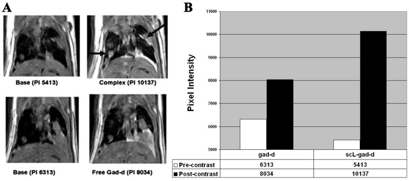
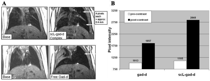
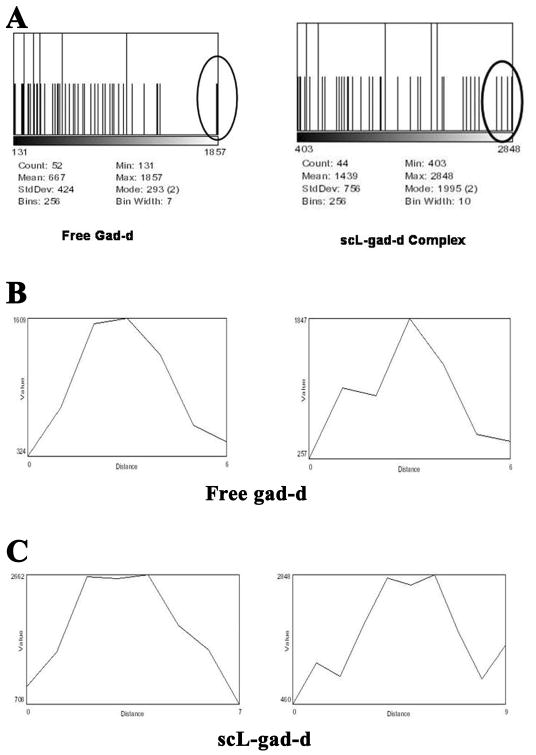
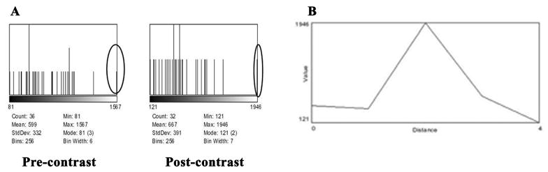
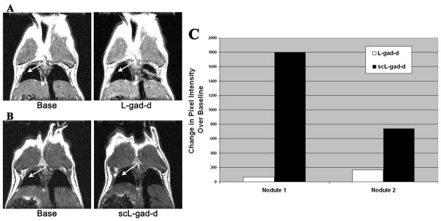
Materials and methods: We have developed a nanosized ( approximately 100 nm) immunoliposome complex for delivery of molecular medicines to tumors. In this complex, an anti-transferrin receptor single-chain antibody fragment (TfRscFv) decorates the surface of a cationic liposome encapsulating the payload. We have previously shown that this systemically administered complex (scL) selectively targets, and efficiently delivers its payload into, tumor cells. We have also encapsulated the magnetic resonance imaging (MRI) contrast agent gadopentetate dimeglumine ("gad-d") within this complex, resulting in increased resolution and image intensity in a mouse model of primary cancer. Here we examine the ability of the scL-gad-d complex to increase the sensitivity of detection of lung metastases.
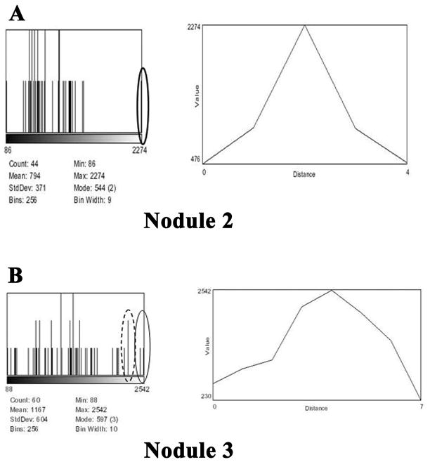
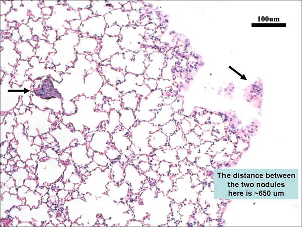
Results: These MRI studies show that the scL-gad-d nanocomplex is able to improve detection, and increase enhancement of, small lung cancers (400 microm and as small as 100 microm) compared to that of uncomplexed gad-d.
Conclusions: Because of its tumor targeting specificity, deliver of an MRI contrast agent via this nanocomplex has potential for use as an agent that can identify small lung cancers, thus improving early detection and possibly increasing survival.
Figures

Similar articles
-
A tumor-targeted nanodelivery system to improve early MRI detection of cancer.Mol Imaging. 2006 Jan-Mar;5(1):41-52. Mol Imaging. 2006. PMID: 16779969
-
Dynamic MRI of solitary pulmonary nodules: comparison of enhancement patterns of malignant and benign small peripheral lung lesions.AJR Am J Roentgenol. 2007 Jan;188(1):26-36. doi: 10.2214/AJR.05.1446. AJR Am J Roentgenol. 2007. PMID: 17179342
-
Primed infusion with delayed equilibrium of Gd.DTPA for enhanced imaging of small pulmonary metastases.PLoS One. 2013;8(1):e54903. doi: 10.1371/journal.pone.0054903. Epub 2013 Jan 31. PLoS One. 2013. PMID: 23382996 Free PMC article.
-
Detection of liver metastases on gadobenate dimeglumine-enhanced MRI: systematic review, meta-analysis, and similarities with gadoxetate-enhanced MRI.Eur Radiol. 2019 Oct;29(10):5205-5216. doi: 10.1007/s00330-019-06110-1. Epub 2019 Mar 26. Eur Radiol. 2019. PMID: 30915560
-
Alexa Fluor 680-labeled transferrin-cationic (NBD-labeled DOPE-DOTAP) liposome-encapsulated gadopentetate dimeglumine complex.2007 Oct 18 [updated 2007 Dec 17]. In: Molecular Imaging and Contrast Agent Database (MICAD) [Internet]. Bethesda (MD): National Center for Biotechnology Information (US); 2004–2013. 2007 Oct 18 [updated 2007 Dec 17]. In: Molecular Imaging and Contrast Agent Database (MICAD) [Internet]. Bethesda (MD): National Center for Biotechnology Information (US); 2004–2013. PMID: 20641669 Free Books & Documents. Review.
Cited by
-
A Phase l Study of a Tumor-targeted Systemic Nanodelivery System, SGT-94, in Genitourinary Cancers.Mol Ther. 2016 Aug;24(8):1484-91. doi: 10.1038/mt.2016.118. Epub 2016 Jun 13. Mol Ther. 2016. PMID: 27480598 Free PMC article. Clinical Trial.
-
Single-Chain Fragment Variable: Recent Progress in Cancer Diagnosis and Therapy.Cancers (Basel). 2022 Aug 30;14(17):4206. doi: 10.3390/cancers14174206. Cancers (Basel). 2022. PMID: 36077739 Free PMC article. Review.
-
Rational chemical design of the next generation of molecular imaging probes based on physics and biology: mixing modalities, colors and signals.Chem Soc Rev. 2011 Sep;40(9):4626-48. doi: 10.1039/c1cs15077d. Epub 2011 May 23. Chem Soc Rev. 2011. PMID: 21607237 Free PMC article. Review.
-
Safety and Efficacy in Advanced Solid Tumors of a Targeted Nanocomplex Carrying the p53 Gene Used in Combination with Docetaxel: A Phase 1b Study.Mol Ther. 2016 Sep;24(9):1697-706. doi: 10.1038/mt.2016.135. Epub 2016 Jun 30. Mol Ther. 2016. PMID: 27357628 Free PMC article. Clinical Trial.
-
The clinical potential of targeted nanomedicine: delivering to cancer stem-like cells.Mol Ther. 2014 Feb;22(2):278-291. doi: 10.1038/mt.2013.231. Epub 2013 Oct 11. Mol Ther. 2014. PMID: 24113515 Free PMC article.
References
-
- American Cancer Society. Cancer Facts and Figures 2008. http://www.cancer.org.
-
- Gohagan J, Marcus P, Fagerstrom R, et al. Writing Committee LSSRG. Baseline findings of a randomized feasibility trial of lung cancer screening with spiral CT scan vs chest radiograph: the Lung Screening Study of the National Cancer Institute. Chest. 2004;126:114–121. - PubMed
-
- Gohagan JK, Marcus PM, Fagerstrom RM, et al. Final results of the Lung Screening Study, a randomized feasibility study of spiral CT versus chest X-ray screening for lung cancer. Lung Cancer. 2005;47:9–15. - PubMed
-
- International Early Lung Cancer Action Program. Henschke CI, Yankelevitz DF, et al. Survival of patients with stage I lung cancer detected on CT screening. New England Journal of Medicine. 2006;355:1763–1771. - PubMed
-
- Stitik FP, Tockman MS. Radiographic screening in the early detection of lung cancer. Radiologic Clinics of North America. 1978;16:347–366. - PubMed
Publication types
MeSH terms
Substances
Grants and funding
LinkOut - more resources
Full Text Sources
Other Literature Sources
Medical
Research Materials
Miscellaneous
