. 2009 May 25;388(1):112-20.
doi: 10.1016/j.virol.2009.03.010. Epub 2009 Apr 5.

APOBEC3G cytidine deaminase association with coronavirus nucleocapsid protein

Affiliations

APOBEC3G cytidine deaminase association with coronavirus nucleocapsid protein

Shui-Mei Wang et al. Virology. .

Abstract

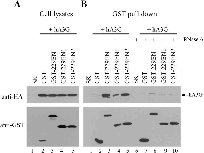
We previously reported that replacing HIV-1 nucleocapsid (NC) domain with SARS-CoV nucleocapsid (N) residues 2-213, 215-421, or 234-421 results in efficient virus-like particle (VLP) production at a level comparable to that of wild-type HIV-1. In this study we demonstrate that these chimeras are capable of packaging large amounts of human APOBEC3G (hA3G), and that an HIV-1 Gag chimera containing the carboxyl-terminal half of human coronavirus 229E (HCoV-229E) N as a substitute for NC is capable of directing VLP assembly and efficiently packaging hA3G. When co-expressed with SARS-CoV N and M (membrane) proteins, hA3G was efficiently incorporated into SARS-CoV VLPs. Data from GST pull-down assays suggest that the N sequence involved in N-hA3G interactions is located between residues 86 and 302. Like HIV-1 NC, the SARS-CoV or HCoV-229E N-associated with hA3G depends on the presence of RNA, with the first linker region essential for hA3G packaging into both HIV-1 and SARS-CoV VLPs. The results raise the possibility that hA3G is capable of associating with different species of viral structural proteins through a potentially common, RNA-mediated mechanism.


Figures

Fig. 1
Fig. 1
Schematic representation of chimeric constructs. Shown are mature HIV-1 Gag protein domain matrix (MA), capsid (CA), nucleocapsid (NC), p6, and amino acid residues in SP1-NC-SP2 junction. ΔNC has ten HIV-1 NC residues remaining in the deleted region; ΔPC has NC almost deleted, with SP1 partially removed. delNC has the two methionine residues (bracketed) in the SP1-NC junction removed. PCR-amplified fragments containing various portions of SARS-CoV N coding sequences were inserted into the deleted NC regions. Numbers denote codon positions at inserted SARS-CoV N protein sequence boundaries. Arrows indicate SP1-NC and NC-SP2 junction sites. Remaining HIV-1 NC residues in deleted regions are underlined. Altered or foreign amino acid residues inserted in juncture area are italicized. All constructs were expressed in HIVgptD25 (a HIV-1 PR-defective expression vector).
Fig. 2
Fig. 2
Incorporation of human APOBEC3G into VLPs. 293T cells were co-transfected with a human APOBEC3G (hA3G) expression vector and indicated plasmid. At 48–72 h post-transfection, cells and supernatant were collected and subjected to Western immunoblotting. hA3G plasmid DNA (2 or 8 μg) was used for co-transfection as indicated. ΔVif is a Vif-deficient mutation introduced into wt and NC(N2) (panel B, lanes 6–9). Wild-type Gag and chimeric proteins were probed with a monoclonal antibody directed against HIV-1 CA; an anti-HA monoclonal antibody was used to detect hA3G. Vif antiserum was used to detect Vif. Relative levels of VLP-associated hA3G are indicated along the bottom (panel C). hA3G, wt Gag, and chimeric proteins were quantified by scanning hA3G and p24CA-associated band densities from immunoblots. Ratios of hA3G to p24gag were determined and normalized to that of wt.
Fig. 3
Fig. 3
RNase A treatment significantly affected SARS-CoV N association with human APOBEC3G. (A) 293T cells were co-transfected with a human A3G (hA3G) expression vector and pBlueScript SK or indicated GST fusion construct. Cell lysates were subjected to Western immunoblotting 48 h post-transfection. (B). Equal amounts of cell lysates were treated with (lanes 10–18) or without (lanes 1–9) 0.2 mg/ml DNase-free RNase A for 30 min at 25 °C, followed by mixing with glutathione-agarose beads for 2 h at 4 °C. Complexes bound to the beads were pelleted, washed, and subjected to Western immunoblotting with anti-HA and anti-GST antibodies.
Fig. 4
Fig. 4
Incorporation of human APOBEC3G into SARS-CoV VLPs. (A) 293T cells were transfected with hA3G expression vector alone or together with plasmids encoding SARS-CoV M (membrane) and N, or with the indicated plasmids. At 48–72 h post-transfection, supernatant and cells were harvested, prepared, and subjected to Western immunoblotting. Levels of p24CA-, N-associated proteins and virus-associated hA3G in each sample were quantified by scanning immunoblot band densities. Ratios of VLP-associated hA3G versus p24CA-associated or N-associated protein levels were calculated for each sample and normalized to that of hA3G associated with wt VLPs in parallel experiments. (B) Sucrose density gradient fractionation analysis of SARS-CoV VLPs. 293T cells were co-transfected with hA3G, M and N expression vectors. At 48–72 h post-transfection, supernatants were collected and pelleted through 20% sucrose cushions. Viral pellets were resuspended in PBS buffer and centrifuged through a 20 to 60% sucrose gradient for 16 h. Fractions were collected from top to bottom, measured for density and analyzed for hA3G, M, and N proteins level by immunodetection. Densities of each fraction are indicated on the top.
Fig. 5
Fig. 5
Incorporation of human APOBEC3G into VLPs. (A) Schematic representatives of wt and human APOBEC3G (hA3G) expression constructs. Open boxes indicate the zinc-coordinating motif, HXE-X23–28-CX2–4C. The asterisk denotes the cytidine deaminase catalytic site. Domains that previously referred to the “linker” peptides are depicted by arrowheads. Numbers denote amino acid positions in hA3G. Indicated positions of deleted hA3G amino acid residue boundaries were used to designate mutant hA3G constructs. Note that each hA3G deletion mutant was tagged with a single copy of HA at the carboxyl terminus and that the wt hA3G was triple-tagged with HA. This may have caused a higher signal for the detected wt hA3G, thus leading to underestimates of the VLP-associated hA3G deletion mutants. (B–D) Incorporation of wt and mutant hA3G into VLPs. 293T cells were co-transfected with M and N, NC(N2) or an HIV-1 Gag expression vector (HIVgptD25) plus each of the hA3G expression vectors. To avoid effects from Vif on hA3G expression level, NC(N2) and HIVgptD25 were expressed in Vif-deficient (ΔVif) contexts. At 48–72 h post-transfection, supernatant and cells were harvested, prepared, and subjected to Western immunoblotting. M and N were probed with M antiserum and an anti-N monoclonal antibody. hA3G was probed with an anti-HA antibody and wt Gag and chimeric proteins were detected with an anti-p24CA antibody. Levels of p24CA- and N-associated proteins and virus-associated hA3G in each sample were quantified by scanning immunoblot band densities. Ratios of total hA3G versus p24CA-associated or N-associated protein levels were calculated for each sample and normalized to that of wt hA3G in parallel experiments. Dashes (–) denote ratios below 0.01. Similar results were observed in repeat and independent experiments. The immunoblots presented here were intentionally overexposed to allow for visualization of the individual hA3G mutants.
Fig. 6
Fig. 6
Incorporation of hA3G into chimeric VLPs. 293T (panel A) or 293 (panel B) cells were co-transfected with a human APOBEC3G (hA3G) expression vector and indicated plasmid. ΔNC(wtZiP) and ΔNC(KZiP) contained HIV-1 NC replacements consisting of wild-type or a mutated version of a leucine-zipper domain, respectively. NC(229EN2) contained the carboxyl-terminal half of a 229E human coronavirus nucleocapsid as a replacement for HIV-1 NC. At 48–72 h or 72 h (panel B) post-transfection, cells and supernatant were collected and subjected to Western immunoblotting. Wild-type Gag and chimeric proteins were probed with a monoclonal antibody directed against HIV-1 CA or SARS-CoV N; an anti-HA monoclonal antibody was used to detect hA3G.
Fig. 7
Fig. 7
Association of HCoV-229E nucleocapsid protein with hA3G. (A) 293T cells were co-transfected with a human A3G (hA3G) expression vector and pBlueScript SK, GST, or GST fusions encoding the full-length (GST-229EN), amino-terminal (GST-229EN1) or carboxyl-terminal (GST-229EN2) half of HCoV-229E nucleocapsid protein. Cell lysates were subjected to Western immunoblotting 48 h post-transfection. (B) Equal amounts of cell lysates were treated with (lanes 6–10) or without (lanes 1–5) 0.2 mg/ml DNase-free RNase A, followed by GST pull-down assay as described in the legend of Fig. 3.

Similar articles

Cited by

References

    1. Accola M.A., Strack B., Gottlinger H.G. Efficient particle production by minimal gag constructs which retain the carboxy-terminal domain of human immunodeficiency virus type 1 capsid-p2 and a late assembly domain. J. Virol. 2000;74(12):5395–5402. - PMC - PubMed
    1. Alce T.M., Popik W. APOBEC3G is incorporated into virus-like particles by a direct interaction with HIV-1 Gag nucleocapsid protein. J. Biol. Chem. 2004;279(33):34083–34086. - PubMed
    1. Bennett R.P., Nelle T.D., Wills J.W. Functional chimeras of the Rous sarcoma virus and human immunodeficiency virus gag proteins. J. Virol. 1993;67(11):6487–6498. - PMC - PubMed
    1. Bennett R.P., Presnyak V., Wedekind J.E., Smith H.C. Nuclear exclusion of the HIV-1 host defense factor APOBEC3G requires a novel cytoplasmic retention signal and is not dependent on RNA binding. J. Biol. Chem. 2008;283(12):7320–7327. - PubMed
    1. Berkowitz R.D., Luban J., Goff S.P. Specific binding of human immunodeficiency virus type 1 gag polyprotein and nucleocapsid protein to viral RNAs detected by RNA mobility shift assays. J. Virol. 1993;67(12):7190–7200. - PMC - PubMed

Publication types

MeSH terms