

Recovery of continence function following simulated birth trauma involves repair of muscle and nerves in the urethra in the female mouse
- PMID: 19346061
- PMCID: PMC4060426
- DOI: 10.1016/j.eururo.2009.03.020
Recovery of continence function following simulated birth trauma involves repair of muscle and nerves in the urethra in the female mouse
Abstract
Background: The natural history and the mechanisms behind the alteration of vaginal distension (VD) in a mouse model are not clear.
Objective: We examined the temporal sequelae of VD and pudendal nerve transection (PNT) on leak-point pressure (LPP) and the muscular and nerve components of the urethra in mice.
Design, setting, and participants: Seventy-two virgin female C57BL/6 mice were equally distributed into three groups. The VD group underwent VD for 1h. The PNT group received bilateral PNT. A control group underwent sham VD.
Intervention: Each group was divided into four subgroups of six mice for measurement of LPP at 0, 4, 10, and 20 d after VD or PNT.
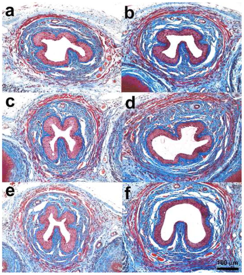
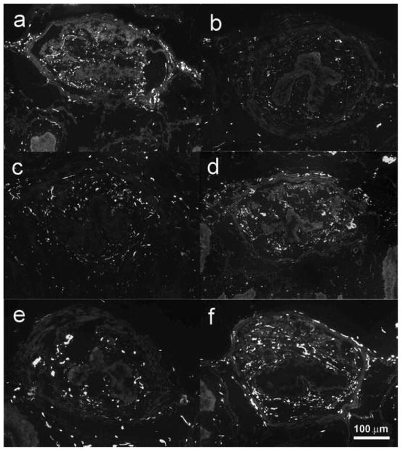
Measurements: LPP was measured. Morphology and neurofilament-immunoreactive nerve of the urethra were assessed.
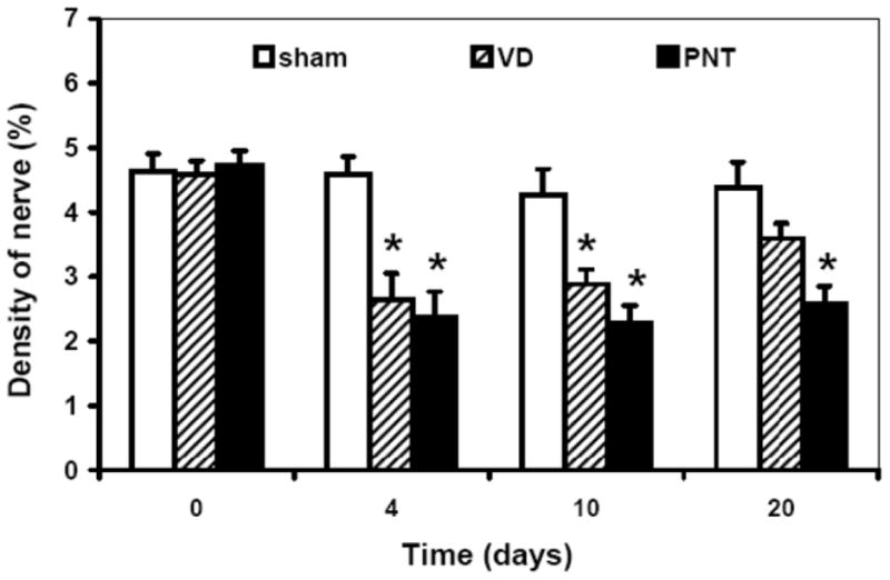
Results and limitations: LPP was decreased at 0, 4, and 10 d but not at 20 d after VD. Decreased LPP persisted to 20 d in the PNT group. The external urethral striated muscle appeared disrupted and/or wavy in two mice at 0 d, in three mice at 4 d, in one mouse at 10 d, and in one mouse in 20 d after VD. The density of neurofilament-immunoreactive nerve in the urethra was reduced at 4 and 10 d after VD, but not at 20 d, and at 4, 10, and 20 d after PNT compared with the corresponding values of the sham VD group. The limitation of this animal model is that the pelvic floor structure of the mouse is different from that of female humans. Therefore, results of this study should be carefully applied to human subjects.
Conclusions: VD causes reversible stress urinary incontinence in female mice. Recovery of continence function following VD is associated with repair of the external urethral sphincter and reinnervation of the urethra. This mouse model will be useful for mechanistic investigation and targeting of therapeutic intervention by taking advantage of genetic manipulation.
2009 European Association of Urology. Published by Elsevier B.V. All rights reserved.
Figures


Comment in
-
Editorial comment on: Recovery of continence function following simulated birth trauma involves repair of muscle and nerves in the urethra in the female mouse.Eur Urol. 2010 Mar;57(3):512-3. doi: 10.1016/j.eururo.2009.03.021. Epub 2009 Mar 13. Eur Urol. 2010. PMID: 19346062 No abstract available.
Similar articles
-
Stem cell homing factor, CCL7, expression in mouse models of stress urinary incontinence.Female Pelvic Med Reconstr Surg. 2013 Nov-Dec;19(6):356-61. doi: 10.1097/SPV.0b013e3182a331a9. Female Pelvic Med Reconstr Surg. 2013. PMID: 24165450 Free PMC article.
-
Editorial comment on: Recovery of continence function following simulated birth trauma involves repair of muscle and nerves in the urethra in the female mouse.Eur Urol. 2010 Mar;57(3):512-3. doi: 10.1016/j.eururo.2009.03.021. Epub 2009 Mar 13. Eur Urol. 2010. PMID: 19346062 No abstract available.
-
Dual simulated childbirth injuries result in slowed recovery of pudendal nerve and urethral function.Neurourol Urodyn. 2009;28(3):229-35. doi: 10.1002/nau.20632. Neurourol Urodyn. 2009. PMID: 18973146 Free PMC article.
-
Proteomic analysis related to stress urinary incontinence following vaginal trauma in female mice.Eur J Obstet Gynecol Reprod Biol. 2013 Nov;171(1):171-9. doi: 10.1016/j.ejogrb.2013.08.034. Epub 2013 Aug 29. Eur J Obstet Gynecol Reprod Biol. 2013. PMID: 24054828
-
Neurogenic aspects of stress urinary incontinence.Curr Opin Obstet Gynecol. 2010 Oct;22(5):425-9. doi: 10.1097/GCO.0b013e32833e499d. Curr Opin Obstet Gynecol. 2010. PMID: 20706117 Free PMC article. Review.
Cited by
-
Potential therapeutic role of punicalagin against mechanical-trauma-induced stress urinary incontinence via upregulation of Nrf2 and TGF-β1 signaling : Effect of punicalagin on mechanical trauma induced SUI.Int Urogynecol J. 2017 Jun;28(6):947-955. doi: 10.1007/s00192-017-3283-x. Epub 2017 Feb 6. Int Urogynecol J. 2017. PMID: 28168411 Free PMC article.
-
Salutary effect of gastric pentadecapeptide BPC 157 in two different stress urinary incontinence models in female rats.Med Sci Monit Basic Res. 2013 Mar 12;19:93-102. doi: 10.12659/MSMBR.883828. Med Sci Monit Basic Res. 2013. PMID: 23478678 Free PMC article.
-
Pelvic floor disorders following vaginal or cesarean delivery.Curr Opin Obstet Gynecol. 2012 Oct;24(5):349-54. doi: 10.1097/GCO.0b013e328357628b. Curr Opin Obstet Gynecol. 2012. PMID: 22907482 Free PMC article. Review.
-
Expression of monocyte chemotactic protein 3 following simulated birth trauma in a murine model of obesity.Urology. 2010 Dec;76(6):1517.e12-7. doi: 10.1016/j.urology.2010.07.466. Epub 2010 Oct 23. Urology. 2010. PMID: 20970834 Free PMC article.
-
Stem cell homing factor, CCL7, expression in mouse models of stress urinary incontinence.Female Pelvic Med Reconstr Surg. 2013 Nov-Dec;19(6):356-61. doi: 10.1097/SPV.0b013e3182a331a9. Female Pelvic Med Reconstr Surg. 2013. PMID: 24165450 Free PMC article.
References
-
- Retzky SS, Rogers RM., Jr Urinary incontinence in women. Clin Symp. 1995;47:2–32. - PubMed
-
- Altman D, Ekstrom A, Forsgren C, Nordenstam J, Zetterstrom J. Symptoms of anal and urinary incontinence following cesarean section or spontaneous vaginal delivery. Am J Obstet Gynecol. 2007;197:512–7. - PubMed
-
- Allen RE, Hosker GL, Smith AR, Warrell DW. Pelvic floor damage and childbirth: a neurophysiological study. Br J Obstet Gynaecol. 1990;97:770–9. - PubMed
-
- Rodriguez LV, Chen S, Jack GS, de Almeida F, Lee KW, Zhang R. New objective measures to quantify stress urinary incontinence in a novel durable animal model of intrinsic sphincter deficiency. Am J Physiol Regul Integr Comp Physiol. 2005;288:R1332–8. - PubMed
