Review
. 2009 Jun;30(6):1079-87.
doi: 10.3174/ajnr.A1579. Epub 2009 Apr 3.

Lyme neuroborreliosis: manifestations of a rapidly emerging zoonosis

Affiliations
Review

Lyme neuroborreliosis: manifestations of a rapidly emerging zoonosis

P Hildenbrand et al. AJNR Am J Neuroradiol. 2009 Jun.

Abstract

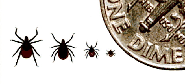
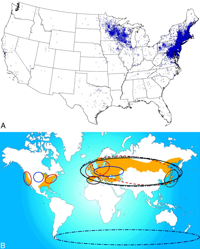
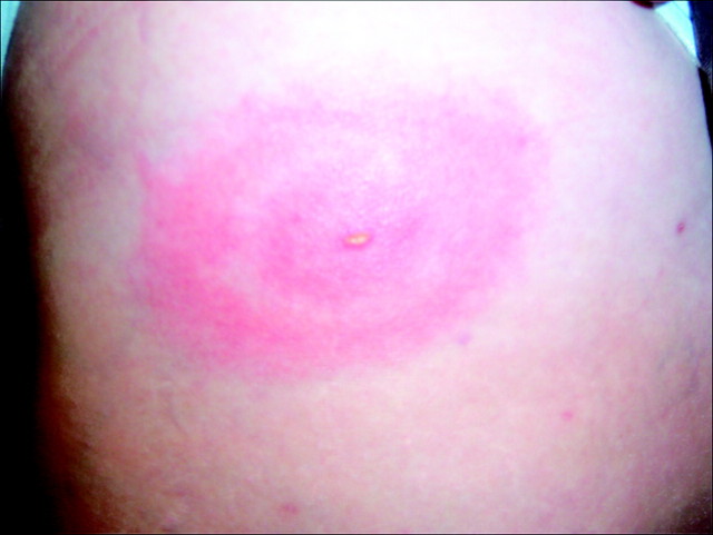
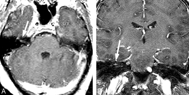
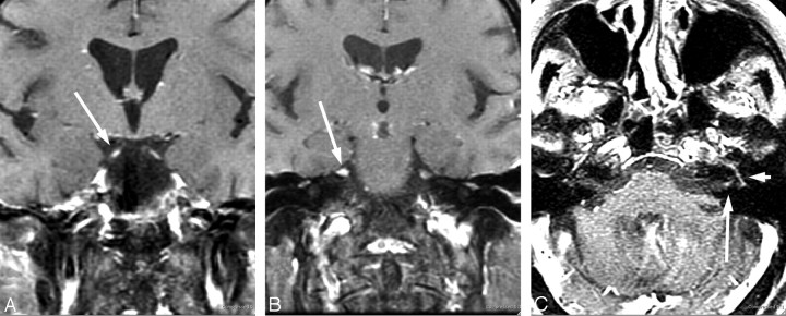
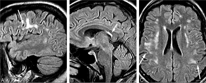
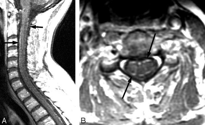
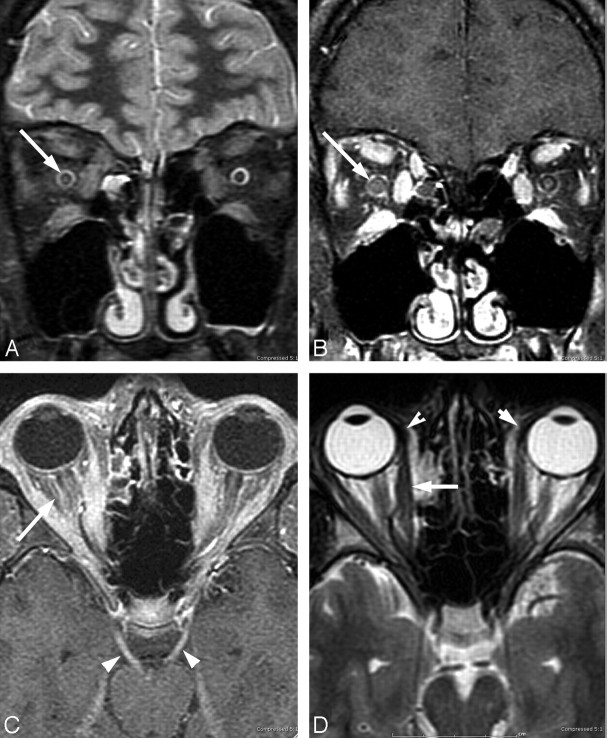
Lyme disease has a worldwide distribution and is the most common vector-borne disease in the United States. Incidence, clinical manifestations, and presentations vary by geography, season, and recreational habits. Lyme neuroborreliosis (LNB) is neurologic involvement secondary to systemic infection by the spirochete Borrelia burgdorferi in the United States and by Borrelia garinii or Borrelia afzelii species in Europe. Enhanced awareness of the clinical presentation of Lyme disease allows inclusion of LNB in the imaging differential diagnosis of facial neuritis, multiple enhancing cranial nerves, enhancing noncompressive radiculitis, and pediatric leptomeningitis with white matter hyperintensities on MR imaging. The MR imaging white matter appearance of successfully treated LNB and multiple sclerosis display sufficient similarity to suggest a common autoimmune pathogenesis for both. This review highlights differences in the epidemiology, clinical manifestations, diagnosis, and management of Lyme disease in the United States, Europe, and Asia, with an emphasis on neurologic manifestations and neuroimaging.


Figures

Fig 1.
Fig 1.
Blacklegged tick (I Scapularis): adult female, adult male, nymph, and larva. Reprinted with permission from the Centers for Disease Control and Prevention.
Fig 2.
Fig 2.
A, Summary of reported cases of Lyme disease in the United States. There is 1 dot within the county of residence for each reported case. Note the widespread diversity of cases correlating with the prevalence of infected Ixodes species in different geographic areas. Courtesy of the Centers for Disease Control. B, The beige-shaded areas indicate the geographic distribution of recorded clinical cases of Lyme borreliosis. The colored ellipses indicate the distribution of the various Borrelia subspecies. Fig 2B is reprinted with permission from Nature Publishing Group.
Fig 3.
Fig 3.
Erythema migrans rash with the typical target appearance that is virtually diagnostic of Lyme disease.
Fig 4.
Fig 4.
Facial neuritis. A 48-year-old woman with headache and peripheral right facial palsy. Prominent enhancement of the fundal tuft and labyrinthine segment of the seventh cranial nerve on postinfusion axial T1 (A) and coronal spoiled gradient-recalled sequences (B). The patient had CSF pleocytosis, Lyme-positive EIA, and Western blot (IgM and IgG) in serum and CSF. Resolution occurred with intravenous ceftriaxone therapy.
Fig 5.
Fig 5.
Evolving cranial neuritis. A 71-year-old woman with headache, malaise, fever, and diplopia. Initial coronal postcontrast T1 MR imaging (A and B) with enhancing bilateral third and fifth cranial nerves. C, Nine days later, she developed left facial palsy with enhancing fundal tuft and labyrinthine and tympanic segments of the seventh cranial nerve. The patient had CSF pleocytosis with positive Lyme-EIA and Western blot in both serum and CSF and CSF Lyme PCR-negative findings. Resolution occurred with intravenous ceftriaxone therapy.
Fig 6.
Fig 6.
A 50-year-old woman with a history of tick bite and erythema migrans rash treated with doxycycline, who had recurrent erythema migrans rash with headache, fever, nausea, and nuchal rigidity. The patient had CSF pleocytosis with positive Lyme serum EIA and IgM Western blot and negative Lyme antibodies in the CSF. Gradual symptomatic improvement occurred following intravenous ceftriaxone therapy. There has been stable MR imaging for 5 years. Sagittal (A and B) and axial (C) fluid-attenuated inversion recovery images show arcuate and confluent subcortical white matter involvement and callososeptal interface involvement remarkably similar to that in MS, but without involvement of the periventricular white matter.
Fig 7.
Fig 7.
A 74-year-old man with 2-year cognitive decline and memory loss. The patient had Lyme-positive serum EIA and Western blot (IgG and IgM) and CSF pleocytosis with CSF positive Lyme IgM and IgG antibodies. The patient improved with intravenous ceftriaxone therapy. The “dot-dash” callososeptal interface (A) and periventricular distribution of involvement (B) would be routinely ascribed to a demyelinating process.
Fig 8.
Fig 8.
A 56-year-old woman with neck, bilateral shoulder, and bilateral arm pain. In 2 weeks, she subsequently developed left facial palsy and positive serum EIA and Western blot (IgM and IgG) and CSF Lyme IgG and IgM antibodies. Complete resolution of symptoms occurred with oral doxycycline. Postcontrast sagittal and axial T1-weighted MR images show diffuse thin uniform cervical spinal cord leptomeningeal enhancement without apparent root or ganglion enhancement.
Fig 9.
Fig 9.
A 17-year-old boy with right papilledema and orbital pain and rule out pseudotumor. The patient had positive serum EIA and Western blot (IgM and IgG) and CSF Lyme IgM and IgG antibodies. Lyme PCR in the CSF was negative. Complete resolution of symptoms occurred after intravenous ceftriaxone therapy. Right optic nerve edema on fat-saturated T2-weighted fast spin-echo images (A) and right-greater-than-left optic nerve enhancement on coronal fat-saturated contrast-enhanced T1-weighted images (B). C, Bilateral third cranial nerve enhancement (arrowheads) and bilateral retrobulbar compartment congestion (arrow). Note the generalized extraocular muscle enlargement and enhancement, including the insertions. Additional imaging findings included enhancement of right fifth cranial nerve, optic chiasm, and intracanalicular right seventh nerve, all of which were occult to neurologic examination.

Similar articles

Cited by

References

    1. Aberer E. Lyme borreliosis: an update [in English, German]. J Dtsch Dermatol Ges 2007;5:406–14 - PubMed
    1. Jensenius M, Parola P, Raoult D. Threats to international travellers posed by tick-borne diseases. Travel Med Infect Dis 2006;4:4–13. Epub 2004 Dec 20 - PubMed
    1. Centers for Disease Control and Prevention (CDC). Lyme disease: United States, 2003–2005. MMWR Morb Mortal Wkly Rep 2007;56:573–76 - PubMed
    1. Mast WE, Burrows WM, Jr. Erythema chronicum migrans in the United States. JAMA 1976;236:859–60 - PubMed
    1. Mast WE, Burrows WM. Erythema chronicum migrans and “lyme arthritis.” JAMA 1976;236:2392 - PubMed