. 2009 Jun;75(11):3586-92.
doi: 10.1128/AEM.02683-08. Epub 2009 Apr 3.

Organoselenium coating on cellulose inhibits the formation of biofilms by Pseudomonas aeruginosa and Staphylococcus aureus

Affiliations

Organoselenium coating on cellulose inhibits the formation of biofilms by Pseudomonas aeruginosa and Staphylococcus aureus

Phat L Tran et al. Appl Environ Microbiol. 2009 Jun.

Abstract

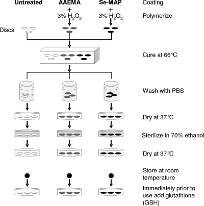
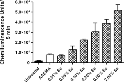
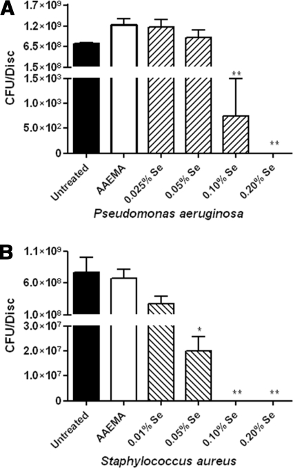
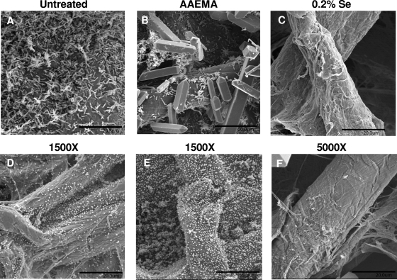
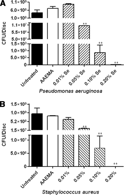
Among the most difficult bacterial infections encountered in treating patients are wound infections, which may occur in burn victims, patients with traumatic wounds, necrotic lesions in people with diabetes, and patients with surgical wounds. Within a wound, infecting bacteria frequently develop biofilms. Many current wound dressings are impregnated with antimicrobial agents, such as silver or antibiotics. Diffusion of the agent(s) from the dressing may damage or destroy nearby healthy tissue as well as compromise the effectiveness of the dressing. In contrast, the antimicrobial agent selenium can be covalently attached to the surfaces of a dressing, prolonging its effectiveness. We examined the effectiveness of an organoselenium coating on cellulose discs in inhibiting Pseudomonas aeruginosa and Staphylococcus aureus biofilm formation. Colony biofilm assays revealed that cellulose discs coated with organoselenium completely inhibited P. aeruginosa and S. aureus biofilm formation. Scanning electron microscopy of the cellulose discs confirmed these results. Additionally, the coating on the cellulose discs was stable and effective after a week of incubation in phosphate-buffered saline. These results demonstrate that 0.2% selenium in a coating on cellulose discs effectively inhibits bacterial attachment and biofilm formation and that, unlike other antimicrobial agents, longer periods of exposure to an aqueous environment do not compromise the effectiveness of the coating.


Figures

FIG. 1.
FIG. 1.
Schematic diagram for coating cellulose discs with AAEMA or Se-MAP. Se-MAP (22% [wt/wt] Se) was diluted in AAEMA to the desired concentrations and applied to 6-mm cellulose discs. Polymerization was initiated with 3% H2O2. Discs, including untreated discs, were cured at 66°C until dry and then washed twice with PBS (pH 7.4). Discs were dried at 37°C and sterilized in 70% ethanol. Discs were dried again at 37°C and stored at room temperature until used in the assays. Immediately prior to use in every assay, 300 μM GSH was added to each disc.
FIG. 2.
FIG. 2.
Selenium in Se-MAP on cellulose discs generates superoxide in a lucigenin-enhanced CL assay. Untreated, AAEMA-coated, or Se-MAP-coated (with various concentrations of selenium) cellulose discs were placed in a CL assay cocktail containing 1 μg/ml GSH and 1 μg/ml lucigenin in 0.05 M sodium phosphate buffer (pH 7.4), and CL was measured for 5 min. The final concentration of selenium present on the Se-MAP-coated discs is indicated on the graph. Values represent the means of triplicate experiments ± standard errors.
FIG. 3.
FIG. 3.
Selenium in Se-MAP inhibits P. aeruginosa and S. aureus biofilm formation. Untreated, AAEMA-coated, or Se-MAP-coated cellulose discs were prepared as described for Fig. 1 and inoculated with P. aeruginosa (A) or S. aureus (B). Biofilms were allowed to form for 24 h. The discs were gently washed twice in PBS to remove planktonic bacteria. Adherent bacteria (biofilm) were removed from the discs by vortexing in PBS, and CFU were determined by plating 10-fold serial dilutions on LB agar. The final concentrations of selenium present on the Se-MAP-coated discs are indicated on the graphs. Values represent the means of triplicate experiments ± standard errors. A one-way analysis of variance with Dunnett's multiple comparisons post test using the AAEMA-coated discs as the control was done to determine statistical significance. *, P < 0.05; **, P < 0.01.
FIG. 4.
FIG. 4.
SEM analysis of P. aeruginosa (A to C) and S. aureus (D to F) biofilm formation on untreated, AAEMA-coated, or Se-MAP-coated (0.2% selenium) cellulose discs. Biofilms were allowed to form as described for Fig. 3. After 24 h of incubation at 37°C, the discs were fixed, dried, affixed to aluminum mounts, and sputter coated with platinum and palladium. Observations were performed at 6 to 7 kV with a scanning electron microscope. Five fields of view were examined from randomly chosen areas from the optical surface of each sample at magnification of ×1,500 for untreated and AAEMA-coated discs and at ×5,000 for the 0.2% selenium Se-MAP-coated discs. Each experiment was conducted in triplicate. Representative fields of view are shown. Bars, 20 μm. Crystals visible in panels B and F are artifacts of fixation.
FIG. 5.
FIG. 5.
Comparison of the effectiveness of cellulose discs coated with Se-MAP in inhibiting the development of biofilms formed by the P. aeruginosa strain MPAO1 and its sodM mutant, PA4468ΔsodM. Biofilm development and analysis were conducted as described for Fig. 3. Values represent the means of triplicate experiments ± standard errors.
FIG. 6.
FIG. 6.
Se-MAP coating on cellulose discs remains stable for 1 week in aqueous solution. Cellulose discs were prepared and coated with AAEMA or Se-MAP in AAEMA (Fig. 1) and soaked for 1 week as described in Materials and Methods. The discs were then dried and tested for the ability to inhibit biofilm formation by P. aeruginosa (A) and S. aureus (B) as described for Fig. 3. The final concentrations of selenium present on the Se-MAP-coated discs are indicated on the graphs. Values represent the means of triplicate experiments ± standard errors. A one-way analysis of variance with Dunnett's multiple comparisons post test using the AAEMA-coated discs as the control was done to determine statistical significance. **, P < 0.01.

References

    1. Araujo, J. C., F. C. Teran, R. A. Oliveira, E. A. Nour, M. A. Montenegro, J. R. Campos, and R. F. Vazoller. 2003. Comparison of hexamethyldisilazane and critical point drying treatments for SEM analysis of anaerobic biofilms and granular sludge. J. Electron Microsc. (Tokyo) 52:429-433. - PubMed
    1. Bayle, C., E. Causse, and F. Couderc. 2004. Determination of aminothiols in body fluids, cells, and tissues by capillary electrophoresis. Electrophoresis 25:1457-1472. - PubMed
    1. Bebien, M., G. Lagniel, J. Garin, D. Touati, A. Vermeglio, and J. Labarre. 2002. Involvement of superoxide dismutases in the response of Escherichia coli to selenium oxides. J. Bacteriol. 184:1556-1564. - PMC - PubMed
    1. Braet, F., R. De Zanger, and E. Wisse. 1997. Drying cells for SEM, AFM and TEM by hexamethyldisilazane: a study on hepatic endothelial cells. J. Microsc. 186:84-87. - PubMed
    1. Cabiscol, E., J. Tamarit, and J. Ros. 2000. Oxidative stress in bacteria and protein damage by reactive oxygen species. Int. Microbiol. 3:3-8. - PubMed

Publication types

MeSH terms