

Cerebral malaria is associated with low levels of circulating endothelial progenitor cells in African children
- PMID: 19346372
- PMCID: PMC6043679
Cerebral malaria is associated with low levels of circulating endothelial progenitor cells in African children
Abstract
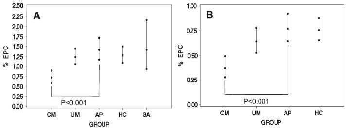
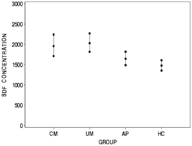
Damage to the cerebral microvasculature is a feature of cerebral malaria. Circulating endothelial progenitor cells are needed for microvascular repair. Based on this knowledge, we hypothesized that the failure to mobilize sufficient circulating endothelial progenitor cells to the cerebral microvasculature is a pathophysiologic feature of cerebral malaria. To test this hypothesis, we compared peripheral blood levels of CD34 (+)/VEGFR2(+) and CD34 (+)/CD133(+) cells and plasma levels of the chemokine stromal cell-derived growth factor 1 (SDF-1) in 214 children in Accra, Ghana. Children with cerebral malaria had lower levels of CD34 (+)/VEGFR2(+) and CD34 (+)/CD133(+) cells compared with those with uncomplicated malaria, asymptomatic parasitemia, or healthy controls. SDF-1 levels were higher in children with acute malaria compared with healthy controls. Together, these results uncover a potentially novel role for endothelial progenitor cell mobilization in the pathophysiology of cerebral malaria.
Figures
References
-
- Newton CR, Taylor TE, Whitten RO. Pathophysiology of fatal falciparum malaria in African children. Am J Trop Med Hyg. 1998;58:673–683. - PubMed
-
- Pongponratn E, Turner GD, Day NP, Phu NH, Simpson JA, Stepniewska K, Mai NT, Viriyavejakul P, Looareesuwan S, Hien TT, Ferguson DJ, White NJ. An ultrastructural study of the brain in fatal Plasmodium falciparum malaria. Am J Trop Med Hyg. 2003;69:345–359. - PubMed
-
- Taylor TE, Fu WJ, Carr RA, Whitten RO, Mueller JS, Fosiko NG, Lewallen S, Liomba NG, Molyneux ME. Differentiating the pathologies of cerebral malaria by postmortem parasite counts. Nat Med. 2004;10:143–145. - PubMed
Publication types
MeSH terms
Substances
Grants and funding
LinkOut - more resources
Full Text Sources
Medical
Research Materials