. 2009 Jun;296(6):L1076-84.
doi: 10.1152/ajplung.90332.2008. Epub 2009 Apr 3.

Francisella tularensis directly interacts with the endothelium and recruits neutrophils with a blunted inflammatory phenotype

Affiliations

Francisella tularensis directly interacts with the endothelium and recruits neutrophils with a blunted inflammatory phenotype

Jessica G Moreland et al. Am J Physiol Lung Cell Mol Physiol. 2009 Jun.

Abstract

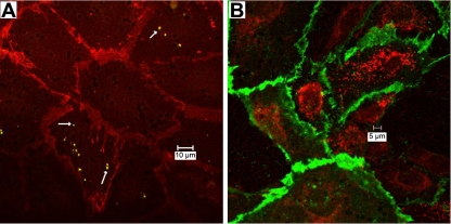
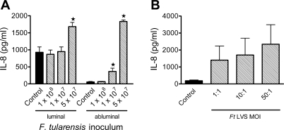
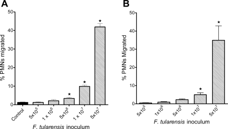
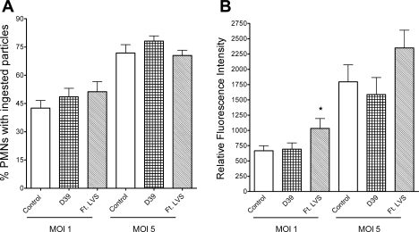
Francisella tularensis, the causative agent of tularemia, is a highly virulent organism, especially when exposure occurs by inhalation. Recent data suggest that Francisella interacts directly with alveolar epithelial cells. Although F. tularensis causes septicemia and can live extracellularly in a murine infection model, there is little information about the role of the vascular endothelium in the host response. We hypothesized that F. tularensis would interact with pulmonary endothelial cells as a prerequisite to the clinically observed recruitment of neutrophils to the lung. Using an in vitro Transwell model system, we studied interactions between F. tularensis live vaccine strain (Ft LVS) and a pulmonary microvascular endothelial cell (PMVEC) monolayer. Organisms invaded the endothelium and were visualized within individual endothelial cells by confocal microscopy. Although these bacteria-endothelial cell interactions did not elicit production of the proinflammatory chemokines, polymorphonuclear leukocytes (PMN) were stimulated to transmigrate across the endothelium in response to Ft LVS. Moreover, transendothelial migration altered the phenotype of recruited PMN; i.e., the capacity of these PMN to activate NADPH oxidase and release elastase in response to subsequent stimulation was reduced compared with PMN that traversed PMVEC in response to Streptococcus pneumoniae. The blunting of PMN responsiveness required PMN transendothelial migration but did not require PMN uptake of Ft LVS, was not dependent on the presence of serum-derived factors, and was not reproduced by Ft LVS-conditioned medium. We speculate that the capacity of Ft LVS-stimulated PMVEC to support transendothelial migration of PMN without triggering release of IL-8 and monocyte chemotactic protein-1 and to suppress the responsiveness of transmigrated PMN to subsequent stimulation could contribute to the dramatic virulence during inhalational challenge with Francisella.


Figures

Fig. 1.
Fig. 1.
Invasion of Francisella tularensis live vaccine strain (Ft LVS) into an intact pulmonary microvascular endothelial cell (PMVEC) monolayer. Ft LVS were incubated at the abluminal surface of the PMVEC monolayer [5 or 500 multiplicity of infection (MOI)] for 6 h; then monolayers were collected and fixed for microscopy. A: at ∼5 MOI, staining with rabbit anti-Ft LVS antibody (arrows) and secondary FITC reveals individual Ft LVS and staining with anti-platelet endothelial cell adhesion molecule (PECAM) antibody and secondary Texas Red reveals intercellular junctions. B: at 500 MOI, staining with rabbit anti-Ft LVS antibody and secondary Texas Red and staining with anti-PECAM antibody and secondary FITC reveal numerous intracellular bacteria. Images represent sections from 4 experiments.
Fig. 2.
Fig. 2.
IL-8 production elicited by Ft LVS-endothelial cell-PMN interactions. A: inocula of Ft LVS or no bacteria (control) were incubated at the abluminal surface of a PMVEC monolayer for 4 h; then PMN were added to the luminal surface, and medium was collected after 3 h (7 h after addition of bacteria) for determination of IL-8 levels. More IL-8 was secreted at the luminal than at the abluminal surface of the monolayer, consistent with the presence of PMN on that side of the monolayer. There was no significant increase in luminal levels of IL-8 above control levels in monolayers stimulated with up to 1 × 10⁷ Ft LVS. Coincubation of the monolayer with 5 × 10⁷ Ft LVS elicited a significant increase in IL-8 production above baseline levels in the luminal compartment. In the abluminal chamber, IL-8 levels were greater than control with inoculum of ≥1 × 10⁷/ml. Values are means ± SE (n = 5). *P < 0.05 vs. control for that compartment. B: PMN were incubated with Ft LVS at 1–50 MOI for 3 h before collection of supernatant for detection of IL-8 levels. Control PMN were incubated in the same medium; incubation of PMN with purified LPS from Escherichia coli was used as a positive control for IL-8 release. Ft LVS elicited release of IL-8 from PMN, although there was wide variation in the amount released. Values are means ± SE (n = 4).
Fig. 3.
Fig. 3.
PMN transendothelial migration in response to Ft LVS. Ft LVS elicited migration of PMN across PMVEC (A) and human dermal microvascular endothelial cells (HMVEC-D; B) in a concentration-dependent manner. Inocula of ≥5 × 10⁶ elicited PMN transmigration above baseline levels of migration in control, unstimulated monolayers (A). Values are means ± SE of 7 experiments done in triplicate. *P < 0.05 vs. control.
Fig. 4.
Fig. 4.
NADPH oxidase activity of transmigrated PMN as measured by lucigenin-enhanced chemiluminescence in relative luminescence units (RLUs). A: after transendothelial migration in response to Ft LVS, NADPH oxidase activity in PMN was significantly depressed in response to the phagocytic stimulus opsonized zymosan (OpZ) compared with control PMN or PMN that migrated in response to Streptococcus pneumoniae strain D39. Values are means ± SE (n = 5–8). *P < 0.05 vs. control and D39 (by 2-way ANOVA). B: response to PMA (10 ng/ml) was significantly increased in control PMN and PMN that migrated in response to D39 compared with PMN that migrated in response to Ft LVS. Values are means ± SE (n = 6). *P < 0.05 vs. control and D39. C and D: PMN incubated with bacteria-endothelial cell-conditioned medium for 1 and 3 h, respectively. There was no alteration in PMN NADPH oxidase activity in response to PMA after 1 h of incubation with bacteria-endothelial cell-conditioned medium from the luminal or abluminal compartment of monolayers exposed to Ft LVS (LVS) or S. pneumoniae (D39). After 3 h of incubation, NADPH oxidase activity was significantly enhanced in PMN exposed to D39-endothelial cell-conditioned medium compared with control PMN or PMN exposed to Ft LVS-endothelial cell-conditioned medium. Values are means ± SE (n = 3). *P < 0.05 vs. control and LVS.
Fig. 5.
Fig. 5.
Phagocytosis of OpZ by PMN after transendothelial migration. A: phagocytosis in control PMN and PMN after transendothelial migration in response to S. pneumoniae strain D39 or Ft LVS. At 1 and 5 MOI, percentage of PMN with ingested particles was similar in all 3 groups. B: mean fluorescence intensity was similar in control PMN and PMN after transendothelial migration in response to D39 when incubated with serum-opsonized zymosan particles at 1 and 5 MOI. Flow cytometry was used to determine mean fluorescence intensity per cell as a measure of the number of phagocytosed particles. Fluorescence was slightly enhanced in PMN after migration in response to Ft LVS at 1 MOI. Values are means ± SE (n = 5–6). *P < 0.05 vs. control.
Fig. 6.
Fig. 6.
Elastase release from PMN after transendothelial migration (post TM). A–C: fluorometric analysis of elastase release from control PMN and PMN after transendothelial migration in response to S. pneumoniae strain D39 or Ft LVS. Elastase release after pretreatment with dihydrocytochalasin B followed by stimulation with bacterial chemoattractant formyl-Met-Leu-Phe (fMLF; A), stimulation with fMLF alone (B), or stimulation with PMA (C) was significantly inhibited in PMN that migrated across the endothelium in response to Ft LVS compared with control PMN or PMN after migration in response to D39. Values are means ± SE (n = 5). *P < 0.05 vs. control. D: elastase release in response to fMLF alone was enhanced in PMN incubated with bacteria-endothelial cell-conditioned medium (cm) for 3 h. This significant increase in elastase release occurred in response to D39- and Ft LVS-endothelial cell-conditioned medium from luminal and abluminal surfaces of the endothelial monolayer. Values are means ± SE (n = 4). *P < 0.05 vs. control.

References

    1. Andrews S, Stephens LR, Hawkins PT. PI3K class IB pathway in neutrophils. Sci STKE 2007: CM3, 2007. - PubMed
    1. Boyum A Isolation of mononuclear cells and granulocytes from human blood. J Clin Lab Invest 21: 77–89, 1968. - PubMed
    1. Buckley CD, Ross EA, McGettrick HM, Osborne CE, Haworth O, Schmutz C, Stone PC, Salmon M, Matharu NM, Vohra RK, Nash GB, Rainger GE. Identification of a phenotypically and functionally distinct population of long-lived neutrophils in a model of reverse endothelial migration. J Leukoc Biol 79: 303–311, 2006. - PubMed
    1. Chiavolini D, Alroy J, King CA, Jorth P, Weir S, Madico G, Murphy JR, Wetzler LM. Identification of immunologic and pathologic parameters of death versus survival in respiratory tularemia. Infect Immun 76: 486–496, 2008. - PMC - PubMed
    1. Conlan JW, Chen W, Shen H, Webb A, KuoLee R. Experimental tularemia in mice challenged by aerosol or intradermally with virulent strains of Francisella tularensis: bacteriologic and histopathologic studies. Microb Pathog 34: 239–248, 2003. - PubMed

Publication types

MeSH terms