Comparative Study
. 2009 Jun;296(6):L947-58.
doi: 10.1152/ajplung.90288.2008. Epub 2009 Apr 3.

The contribution of Ca2+ signaling and Ca2+ sensitivity to the regulation of airway smooth muscle contraction is different in rats and mice

Affiliations
Comparative Study

The contribution of Ca2+ signaling and Ca2+ sensitivity to the regulation of airway smooth muscle contraction is different in rats and mice

Yan Bai et al. Am J Physiol Lung Cell Mol Physiol. 2009 Jun.

Abstract

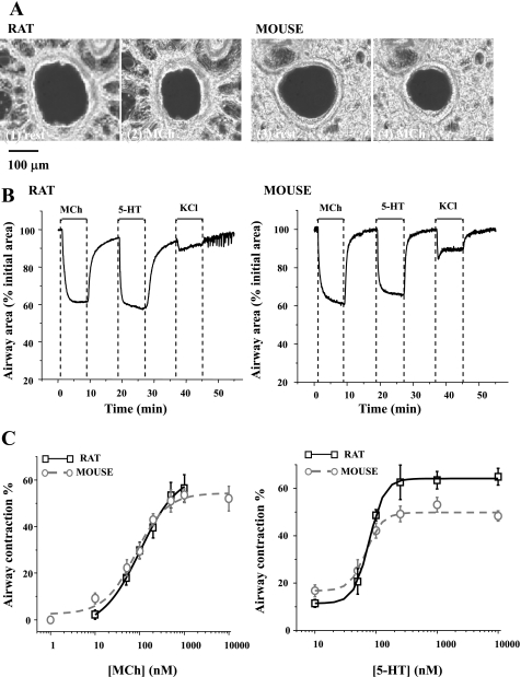
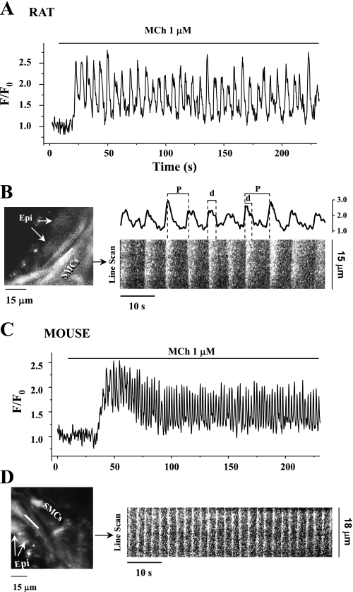
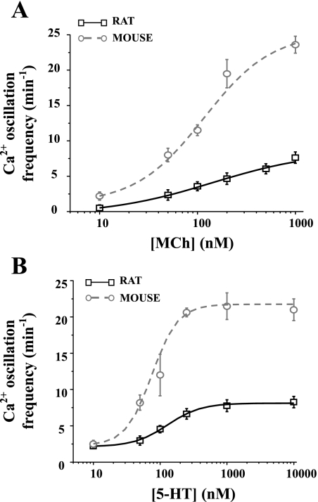
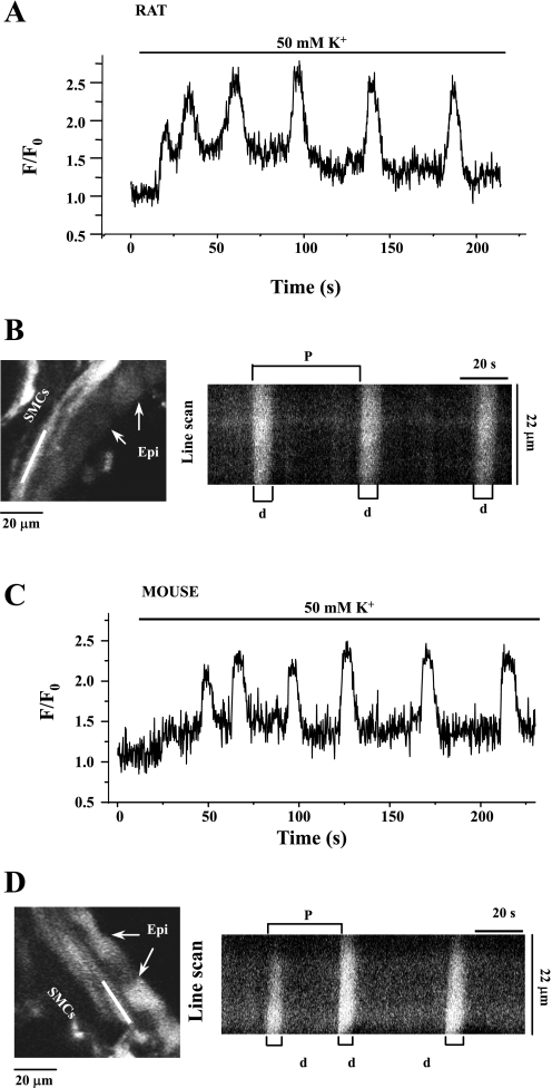
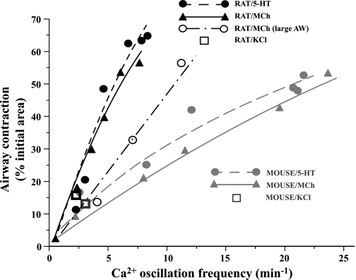
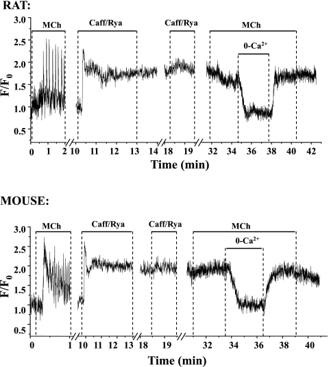
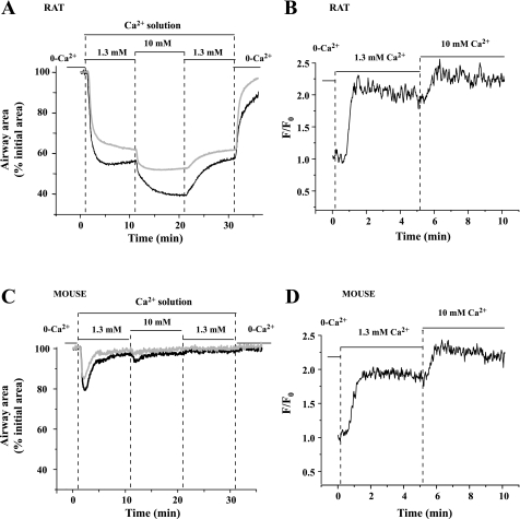
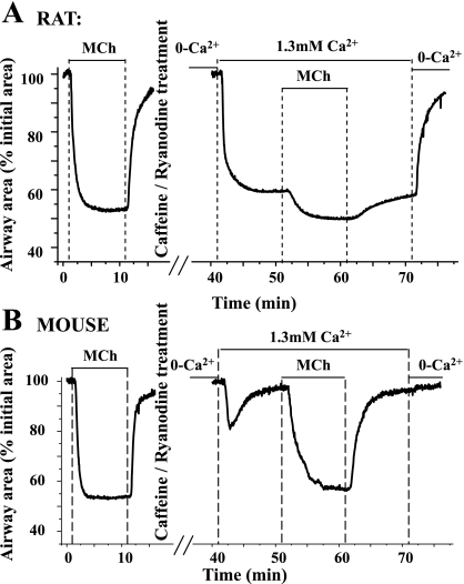
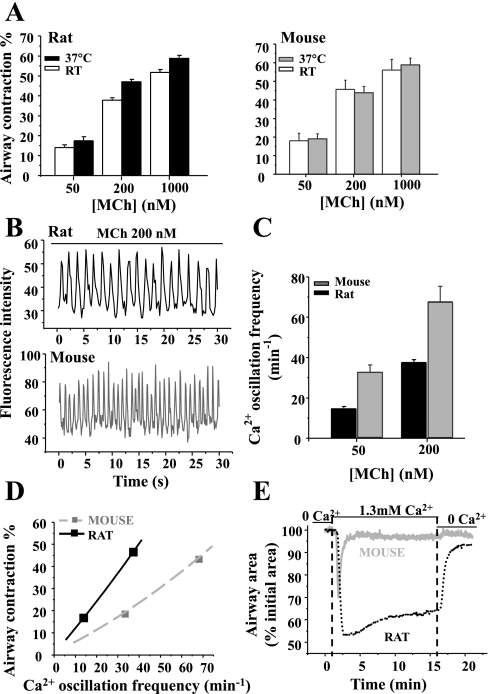
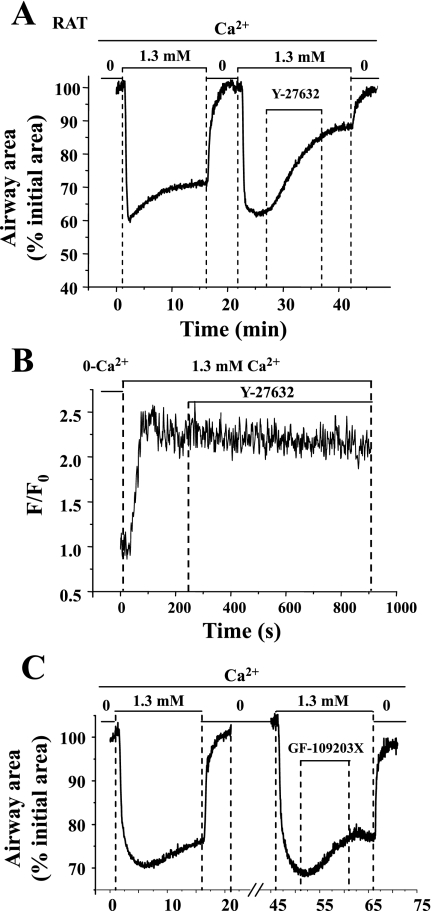
To determine the relative contributions of Ca(2+) signaling and Ca(2+) sensitivity to the contractility of airway smooth muscle cells (SMCs), we compared the contractile responses of mouse and rat airways with the lung slice technique. Airway contraction was measured by monitoring changes in airway lumen area with phase-contrast microscopy, whereas changes in intracellular calcium concentration ([Ca(2+)](i)) of the SMCs were recorded with laser scanning microscopy. In mice and rats, methacholine (MCh) or serotonin induced concentration-dependent airway contraction and Ca(2+) oscillations in the SMCs. However, rat airways demonstrated greater contraction compared with mice, in response to agonist-induced Ca(2+) oscillations of a similar frequency. Because this indicates that rat airway SMCs have a higher Ca(2+) sensitivity compared with mice, we examined Ca(2+) sensitivity with Ca(2+)-permeabilized airway SMCs in which the [Ca(2+)](i) was experimentally controlled. In the absence of agonists, high [Ca(2+)](i) induced a sustained contraction in rat airways but only a transient contraction in mouse airways. This sustained contraction of rat airways was relaxed by Y-23672, a Rho kinase inhibitor, but not affected by GF-109203X, a PKC inhibitor. The subsequent exposure of Ca(2+)-permeabilized airway SMCs, with high [Ca(2+)](i), to MCh elicited a further contraction of rat airways and initiated a sustained contraction of mouse airways, without changing the [Ca(2+)](i) of the SMCs. Collectively, these results indicate that airway SMCs of rats have a substantially higher innate Ca(2+) sensitivity than mice and that this strongly influences the transduction of the frequency of Ca(2+) oscillations into the contractility of airway SMCs.


Figures

Fig. 1.
Fig. 1.
The contractile responses of rat and mouse airways to methacholine (MCh), serotonin (5-HT), and KCl. A: typical phase-contrast images showing lung slices cut from rat (1 and 2) and mouse (3 and 4) in a resting state (1 and 3) and after 5 min of exposure to 200 nM MCh (2 and 4). B: representative experiments showing the change in airway lumen area of rat and mouse with respect to time in response to 200 nM MCh, 100 nM 5-HT, and 50 mM KCl. C: summary of MCh- (left) and 5-HT- (right) induced airway contraction in rats (black squares fitted with black solid line) and mice (gray circles fitted with gray dotted line). Contraction was measured as the decrease in lumen area after 5 min of agonist exposure. Each point represents the mean ± SE from at least 7 experiments from different slices from at least 4 rats and 5 experiments from 3 mice.
Fig. 2.
Fig. 2.
Oscillatory Ca2+ signaling of rat and mouse airway smooth muscle cells (SMCs) in response to MCh. Representative recordings are shown of the Ca2+ signaling in rat (A and B) and mouse (C and D) SMCs induced by 1 μM MCh. Changes in the fluorescence intensity representing changes in Ca2+ were collected either from regions of interest (ROI; 6 × 6 pixels) in single airway SMC and expressed as a fluorescence ratio (F/F0) (A and C) or from line scans along the SMCs with respect to time (B and D). Fluorescence confocal images showing part of rat (B, left) and mouse (D, left) airway indicate the position of the line scan (white line). Epi, epithelial cells. Line-scan plots were constructed by aligning the pixels along the white line from each fluorescence image from 120 to 180 s in rat (B, right, bottom) and mouse (D, right) airway SMCs. The Ca2+ oscillations of rat SMCs from ROI at right end of the scan line from 120 to 180 s are shown in B, right, top. The period (P) and duration (d) of Ca2+ oscillatory wave are shown. Ca2+ oscillation frequency = 1/period. Exposure to 1 μM MCh induced sustained Ca2+ oscillations in both the rat and mouse SMCs, but the Ca2+ oscillation frequency was higher in mouse (∼25 min−1) than in rat (∼10 min−1) airway SMCs. The duration of the Ca2+ oscillations was longer in the rat SMCs in response to 1 μM MCh.
Fig. 3.
Fig. 3.
Comparison of agonist-induced Ca2+ oscillations between rats and mice. The frequency of the Ca2+ oscillations after exposure for 5 min to MCh (A) or 5-HT (B) of rat (black squares fitted with black line) and mouse (gray circles fitted with gray line) airway SMCs. Rat airway SMCs displayed slower Ca2+ oscillations than those of mice. Each point represents the mean ± SE from at least 6 experiments from different slices from at least 3 rats and 5 experiments from at least 2 mice.
Fig. 4.
Fig. 4.
Comparison of KCl (50 mM)-induced Ca2+ oscillations in rat (A) and mouse (C) airway SMCs. Fluorescence images showing part of the rat (B, left) and mouse (D, left) airway and the line-scan plots (right) constructed by aligning pixels indicated by the white line along the SMCs from successive fluorescence images from 75 to 200 s. In both rats and mice, the Ca2+ oscillations induced by KCl were very slow (∼2 min−1) and had a long duration of Ca2+ elevation.
Fig. 5.
Fig. 5.
Relationship between Ca2+ oscillation frequency and airway contraction in mouse and rat. Contractile responses of rat airway (black symbols) and mouse airway (gray symbols) of a similar size in response to MCh (triangles, solid line), 5-HT (circles, dashed line), and 50 mM KCl (squares) or larger rat airways (AW) in response to MCh (black open circle, dot-dash line) were plotted with respect to the corresponding Ca2+ oscillation frequency and fitted with logistic functions. These data indicated that the same frequency of Ca2+ oscillations induced greater contraction in rat than in mouse airways irrespective of the airway generation.
Fig. 6.
Fig. 6.
MCh-induced Ca2+ signaling in rat and mouse airway SMCs before and after treatment with caffeine and ryanodine (Caff/Rya). These representative traces show that MCh induced Ca2+ oscillations before caffeine/ryanodine treatment. Exposure to caffeine/ryanodine to create a Ca2+-permeabilized lung slice induced a fast elevation of intracellular calcium concentration ([Ca2+]i) in the SMCs, which subsequently stabilized and remained at a high level when caffeine/ryanodine was removed. A 2nd exposure to caffeine/ryanodine or MCh had no further effect on the [Ca2+]i. However, [Ca2+]i dropped significantly when the extracellular Ca2+ was removed and was elevated to the previous level when extracellular Ca2+ was replaced. The responses were similar in rat and mouse airway SMCs.
Fig. 7.
Fig. 7.
The contraction and Ca2+ signaling of Ca2+-permeabilized rat and mouse airway SMCs in response to various extracellular Ca2+ concentration ([Ca2+]o). Representative traces show that in rat airway SMCs (top, black line), 1.3 mM Ca2+ [in HBSS supplemented with 20 mM HEPES buffer (sHBSS)] induced a sustained contractile response (A) and increase in [Ca2+]i (B). When the [Ca2+]o was increased to 10 mM, a greater contractile response (A) and higher level of [Ca2+]i (B) were observed. Ca2+-permeabilized large rat airways (top, gray line) had similar responses to the elevation of [Ca2+]o. In mouse airway SMCs (bottom, black line), 1.3 mM Ca2+ followed by 10 mM Ca2+ induced similar changes in [Ca2+]i (D) compared with the rat airway SMCs, but the contractile response consisted of a transient contraction followed by relaxation (C). The elevation of [Ca2+]i to 10 mM did not induce a further significant airway contraction in the mouse (C). Similar responses were observed in Ca2+-permeabilized large mouse airways (diameter: ∼220 μm; bottom, gray line).
Fig. 8.
Fig. 8.
Airway contraction in response to an elevation of [Ca2+]i, with and without agonist, in rat and mouse airways. MCh (200 nM) induced contractile responses (∼40%) in rat (top, left) and mouse (bottom, left). After Ca2+ permeabilization by caffeine/ryanodine treatment and exposure to 0 [Ca2+]o, the [Ca2+]i was low. Exposure to 1.3 mM Ca2+ induced ∼40% contraction in rat airways but only a transient contraction in mouse airways. Exposure to MCh induced an additional contraction in rat airway but a full contraction in the mouse airway. Compared with normal airways, 1 μM MCh induced ∼5% more contraction in the Ca2+-permeabilized rat airway but similar contraction in the Ca2+-permeabilized mouse airway.
Fig. 9.
Fig. 9.
The contractile and Ca2+ responses of airway SMCs at 37°C. A: summary of MCh-induced (50, 200, and 1,000 nM) contraction in rats (left) and mice (right) at room temperature (RT; white bars) and 37°C (filled bars) in the same airway. Each column represents the mean ± SE from 5 paired airways of 3 rats and mice. B: representative experiments showing the Ca2+ oscillations of rat (top) or mouse (bottom) airway SMCs in response to 200 nM MCh at 37°C. These oscillations are faster than those observed at RT. C: summary data of the frequency of the airway SMC Ca2+ oscillations after 5-min exposure to 50 and 200 nM MCh in rats (black bar) and mice (gray bar) at 37°C. Each column represents the mean ± SE from at least 5 experiments from different slices from 3 rats and 3 mice. D: MCh-induced airway contraction as a function of Ca2+ oscillation frequency of rat (black solid line) and mouse (gray dashed line) at 37°C. Although the slope of the relationship is reduced with increased temperature, an equivalent contraction of the rat and mouse airway is achieved by slower Ca2+ oscillations in the rat SMCs. E: representative traces showing the contractile responses of Ca2+-permeabilized rat (black dotted line) and mouse (gray solid line) airway SMCs to the elevation of [Ca2+]o in absence of agonists at 37°C; 1.3 mM Ca2+ induced a sustained contraction of rat airways and a transient contraction of mouse airways. These responses are similar to those obtained at RT.
Fig. 10.
Fig. 10.
The contraction and intracellular Ca2+ signaling of Ca2+-permeabilized rat airway SMCs in response to Y-27632, a Rho kinase inhibitor, and GF-109203X, a PKC inhibitor. A: a representative trace showing the relaxant effect of 10 μM Y-27632 on the Ca2+-induced contraction in the Ca2+-permeabilized rat airway SMCs. This relaxant response was not associated with significant change in [Ca2+]i (B). In comparison, 2 μM GF-109203X (C) had little effect on the Ca2+-permeabilized rat airway contraction induced by 1.3 mM Ca2+.

References

    1. Bai Y, Sanderson MJ. Modulation of the Ca2+ sensitivity of airway smooth muscle cells in murine lung slices. Am J Physiol Lung Cell Mol Physiol 291: L208–L221, 2006. - PubMed
    1. Bai Y, Zhang M, Sanderson MJ. Contractility and Ca2+ signaling of smooth muscle cells in different generations of mouse airways. Am J Respir Cell Mol Biol 36: 122–130, 2007. - PMC - PubMed
    1. Bergner A, Sanderson MJ. Airway contractility and smooth muscle Ca2+ signaling in lung slices from different mouse strains. J Appl Physiol 95: 1325–1332; discussion 1314, 2003. - PubMed
    1. Björck T, Gustafsson LE, Dahlén SE. Isolated bronchi from asthmatics are hyperresponsive to adenosine, which apparently acts indirectly by liberation of leukotrienes and histamine. Am Rev Respir Dis 145: 1087–1091, 1992. - PubMed
    1. Black JL Pharmacology of airway smooth muscle in chronic obstructive pulmonary disease and in asthma. Am Rev Respir Dis 143: 1177–1181, 1991. - PubMed

Publication types

MeSH terms