. 2009 Apr 21;106(16):6685-90.
doi: 10.1073/pnas.0901596106. Epub 2009 Apr 3.

Cells previously identified as retinal stem cells are pigmented ciliary epithelial cells

Affiliations

Cells previously identified as retinal stem cells are pigmented ciliary epithelial cells

Samantha A Cicero et al. Proc Natl Acad Sci U S A. .

Abstract

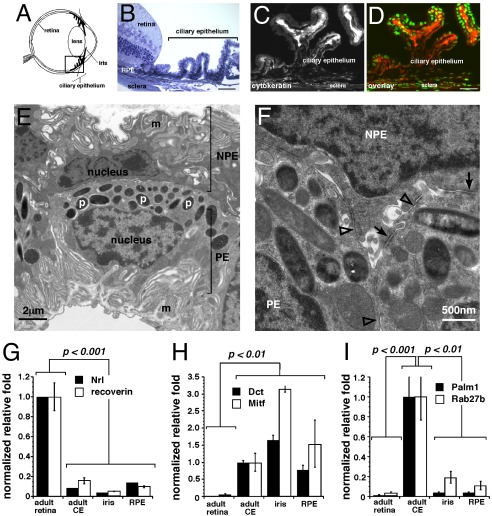
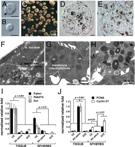
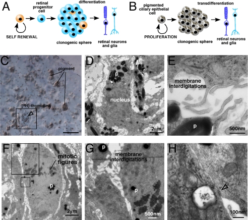
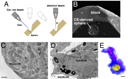
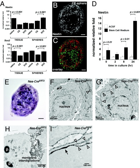
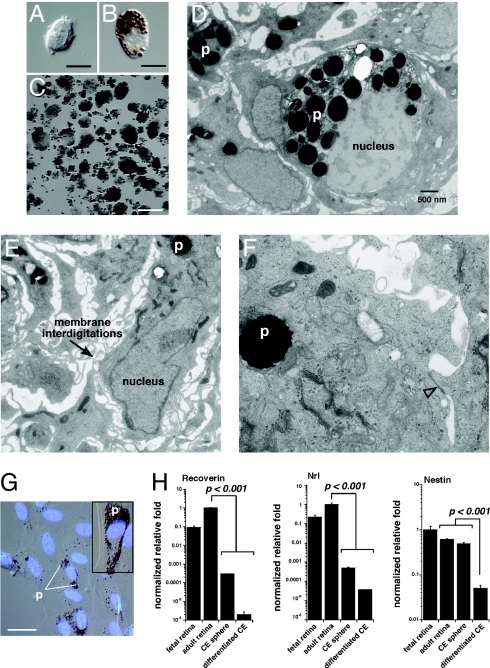
It was previously reported that the ciliary epithelium (CE) of the mammalian eye contains a rare population of cells that could produce clonogenic self-renewing pigmented spheres in culture. Based on their ability to up-regulate genes found in retinal neurons, it was concluded that these sphere-forming cells were retinal stem cells. This conclusion raised the possibility that CE-derived retinal stem cells could help to restore vision in the millions of people worldwide who suffer from blindness associated with retinal degeneration. We report here that human and mouse CE-derived spheres are made up of proliferating pigmented ciliary epithelial cells rather than retinal stem cells. All of the cells in the CE-derived spheres, including the proliferating cells, had molecular, cellular, and morphological features of differentiated pigmented CE cells. These differentiated cells ectopically expressed nestin when exposed to growth factors and low levels of pan-neuronal markers such as beta-III-tubulin. Although the cells aberrantly expressed neuronal markers, they retained their pigmented CE cell morphology and failed to differentiate into retinal neurons in vitro or in vivo. Our results provide an example of a differentiated cell type that can form clonogenic spheres in culture, self-renew, express progenitor cell markers, and initiate neuronal differentiation that is not a stem or progenitor cell. More importantly, our findings highlight the importance of shifting the focus away from studies on CE-derived spheres for cell-based therapies to restore vision in the degenerating retina and improving techniques for using ES cells or retinal precursor cells.


Conflict of interest statement

The authors declare no conflict of interest.

Figures

Fig. 1.
Fig. 1.
Isolation and characterization of the mouse ciliary epithelium. (A) Diagram of the location of the ciliary epithelium (CE) in the mammalian eye. (B) Toluidine blue stained 1-μm thick plastic section of the adult C57BL/6 mouse CE. (C and D) Cytokeratin immunofluorescense in the adult mouse CE (red fluorescence) alone (C) and overlaid with nuclear Sytox green stain D. (E and F) Transmission electron microscopy of the mouse CE showing membrane interdigitations (m), pigment containing melanosomes (p), and epithelial junctions found in the CE including tight junctions (open arrowheads) and gap junctions (arrows). (G–I) Real time RT-PCR analysis of mouse CE, retina, retinal pigment epithelium, and iris using probes for retinal specific genes (Nrl, recoverin), pigmented cells (Dct, Mitf), and CE (Palmdelphin, Rab27b). Each bar represents the mean and standard deviation from duplicate experiments for at least 3 independent samples for each piece of tissue. Abbreviations: RPE, retinal pigment epithelium; CE, ciliary epithelium; m, membrane interdigitations; p, pigmented melanosomes; NPE, nonpigmented ciliary epithelium; PE, pigmented epithelium. (Scale bars in B–D, 10 μm.)
Fig. 2.
Fig. 2.
CE-derived spheres are pigmented. (A and B) Representative DIC images of pigmented and nonpigmented dissociated mouse CE cells. (C) Brightfield image of CE-derived spheres after 7 days in culture from the mouse. (D and E) Cryosections of the mouse CE-derived spheres imaged using DIC optics. (F–H) TEM analysis of mouse CE-derived spheres emphasizing membrane interdigitations (F and G) and epithelial junctions (H) found in the normal CE (tight junctions, open arrowheads; gap junctions, arrow). (I) Real time RT-PCR analysis of CE-derived spheres using probes for genes found in the normal CE (Palm1, Rab27b, Dct). The source of retinal progenitor cells was P0 mouse retina, and the source of neural stem cells was adult SVZ. (J) Genes normally expressed in proliferating cells (PCNA, Cyclin D1) were also analyzed in these samples using real time RT-PCR. Each bar represents the mean and standard deviation from duplicate experiments for at least 3 independent samples for each piece of tissue. Abbreviations: m, mitochondria; p, pigment; CE, ciliary epithelium; SVZ, subventricular zone. (Scale bars in A and B, 5 μm; C, 100 μm; D and E, 10 μm.)
Fig. 3.
Fig. 3.
Pigmented CE cells are proliferating in CE-derived spheres. (A and B) Comparision of the retinal stem cell model and the transdifferentiation model. (C) S-phase cells were labeled by incubating Day 3 spheres with [3H]-tymidine for 1 h. Immediately after labeling, the spheres were fixed and a 1-μm thick section was collected from the center of the sphere. This section was stained with toluidine blue and overlaid with autoradiographic emulsion to detect the cells in S-phase (box, open arrowhead). (D and E) Immediately after the 1-μm section, 10 serial 50-nm sections were collected for transmission electron microscopy. These sections were imaged and aligned to the [3H]-tymidine–labeled section to characterize the morphology of the cells in S-phase. The cell shown in (C) is also shown in (D and E) by aligning the sections. (F–H) Cells in M-phase were identified by the presence of condensed chromosomes (mitotic figures). These cells also contained pigment, membrane interdigitations, and epithelial junctions (open arrowhead). Abbreviations: p, pigment. (Scale bar in C, 10 μm.)
Fig. 4.
Fig. 4.
Dual beam focused ion beam electron microscopic analysis of every cell in a CE-derived sphere. (A) Diagram of dual beam focused ion beam (FIB) technology. This procedure is based on alternating ion (Ga+) and electron beams. The ion beam mills the sample at 70 nm per round and the electron beam provides TEM quality images. (B) A scanning electron microscopic image of a sphere in a block halfway through the analysis. (C and D) Low power and high power images of representative sections in the sphere demonstrating the resolution of the analysis and pigment (p), membrane interdigitations (m), and epithelial junctions (open arrowhead). (E) Example of a 3D reconstruction of a single cell from the dual-beam FIB dataset showing the cytoplasm in blue, the pigment in pink, and the nucleus in yellow. Abbreviations: p, pigment; m, membrane interdigitations. (Scale bars in C, 10 μm; D and E, 1 μm.)
Fig. 5.
Fig. 5.
Expression of stem/progenitor cell markers in CE-derived spheres. (A) Real time RT-PCR for Nestin and Sox2 in CE-derived spheres and neural stem cells and retinal progenitor cells. (B and C) Immunofluorescence confirmed the expression of nestin (red fluorescence) in CE-derived spheres with green fluorescence overlay. (D) Timecourse of nestin induction in dissociated CE cells cultured in stem cell medium or artificial cerebrospinal fluid (ACSF). (E) Spheres derived from the Nes-CreERT2IREShPLAP strain expressed (AP) as detected with the NBT/BCIP substrate. (F–I) TEM analysis using lead citrate staining to detect the AP expressing (nestin+) cells. Using this method, lead citrate staining of AP expressing cells appears as a dark electron dense precipitate in TEM images along the membrane (open arrowheads). (H and I) These nestin-expressing cells in CE-derived spheres contained pigment, membrane interdigitations, and epithelial junctions (arrow). Abbreviations: SVZ, subventricular zone; ACSF, artificial cerebrospinal fluid; p, pigment. (Scale bars in B, C, and E, 25 μm.)
Fig. 6.
Fig. 6.
Differentiated mouse CE-derived spheres resemble pigmented CE cells. (A–D) CE-derived spheres were plated on laminin coated coverglass and grown in differentiation medium for 21 days. The spheres adhered to the coverglass and spread out from there. All of the cells appeared to be pigmented in DIC images (A and B) when compared with the pattern of nuclear staining (C and D). (E–G) TEM analysis of CE-derived sphere differentiation cultures. (H and I) Real time RT-PCR analysis of differentiated CE cells for genes found in retinal neurons or glia (H) and pigmented CE cells (I). (J–M) CE-derived spheres were injected into the subretinal space of newborn rats to study their transdifferentiation into retinal neurons and glia. Tissue was analyzed 21 days after injection. (J) In and around the site of injection, pigmented cells were readily detected in histological sections (arrows). (K–M) TEM analysis of these sections revealed that the cells remained differentiated as pigmented CE cells. The injected cells retained pigment, membrane interdigitations (microvilli), and epithelial junctions (arrow in M). Abbreviations: DIC, differential interference contrast microscopy; p, pigment; RPE, retinal pigment epithelium; ONL, outer nuclear layer. (Scale bars in A, B, and J, 10 μm.)
Fig. 7.
Fig. 7.
Human CE-derived spheres are made up of pigmented CE cells. (A and B) Postmortum human eyes were used as a source of CE cells for culture experiments. Dissociated human CE cell preparations were made up of pigmented and nonpigmented cells as for the mouse CE. (C) Clonogenic spheres from postmortum human CE samples. (D–F) TEM analysis of human CE-derived spheres showing pigment (p), membrane interdigitations, and epithelial junctions (open arrowhead). (G) Human CE-derived spheres were differentiated for 21 days coverslides as described for mouse CE-derived spheres. The cells spread out along the coverglass and took on the morphology of pigmented epithelial cells as for the mouse samples. (H) Real time RT-PCR analysis of human CE-differentiation cultures revealed that these cells did not express photoreceptor genes (recoverin, Nrl) or neural progenitor cell genes (nestin). Each bar represents the mean and standard deviation from duplicate experiments for at least 3 independent samples for each piece of tissue. Abbreviations: p, pigment. (Scale bars in A and B, 5 μm; C, 1 mm; G, 10 μm.)

References

    1. MacLaren RE, et al. Retinal repair by transplantation of photoreceptor precursors. Nature. 2006;444:203–207. - PubMed
    1. Lamba DA, Gust J, Reh TA. Transplantation of human embryonic stem cell-derived photoreceptors restores some visual function in crx-deficient mice. Cell Stem Cell. 2009;4:73–79. - PMC - PubMed
    1. Lamba DA, Karl MO, Ware CB, Reh TA. Efficient generation of retinal progenitor cells from human embryonic stem cells. Proc Natl Acad Sci USA. 2006;103:12769–12774. - PMC - PubMed
    1. Coles BL, et al. Facile isolation and the characterization of human retinal stem cells. Proc Natl Acad Sci USA. 2004;101:15772–15777. - PMC - PubMed
    1. Tropepe V, et al. Retinal stem cells in the adult mammalian eye. Science. 2000;287:2032–2036. - PubMed

Publication types