. 2009 Apr 21;106(16):6644-9.
doi: 10.1073/pnas.0810663106. Epub 2009 Apr 3.

Crystal structure of full-length KcsA in its closed conformation

Affiliations

Crystal structure of full-length KcsA in its closed conformation

Serdar Uysal et al. Proc Natl Acad Sci U S A. .

Abstract

KcsA is a proton-activated, voltage-modulated K(+) channel that has served as the archetype pore domain in the Kv channel superfamily. Here, we have used synthetic antigen-binding fragments (Fabs) as crystallographic chaperones to determine the structure of full-length KcsA at 3.8 A, as well as that of its isolated C-terminal domain at 2.6 A. The structure of the full-length KcsA-Fab complex reveals a well-defined, 4-helix bundle that projects approximately 70 A toward the cytoplasm. This bundle promotes a approximately 15 degree bending in the inner bundle gate, tightening its diameter and shifting the narrowest point 2 turns of helix below. Functional analysis of the full-length KcsA-Fab complex suggests that the C-terminal bundle remains whole during gating. We suggest that this structure likely represents the physiologically relevant closed conformation of KcsA.


Conflict of interest statement

The authors declare no conflict of interest.

Figures

Fig. 1.
Fig. 1.
Crystal structure of FL KcsA in complex with Fab2. (A) CDR sequences of the 3 Fabs selected against FL KcsA from a “reduced genetic code” phage display library. Numbering is according to the Kabat definition (36). Gly is green; Tyr, yellow, Ser, red; and nondiversified positions, gray. (B) Crystal packing of the KcsA–Fab2 complex at 3.8 Å. KcsA is in orange, and the light and heavy chains of the Fab are in cyan (light chain) and magenta (heavy chain). (C) Simulated annealing composite-omit 2Fo-Fc map (contoured at 1σ) of FL KcsA. The red trace shows the fitted model as Cα tracing. (D) The final model of the KcsA–Fab complex. Three regions distinguished by the level of symmetry are highlighted: the fourfold TM segments (blue color; residues 22–117), the twofold bulge helix (red color; residues 118–135), and the fourfold distal C-terminal bundle (gray color; residues 136–158). (E and F) Experimental (E) EPR mobility and (F) NiEdda accessibility parameters (11) from membrane-reconstituted FL KcsA, mapped on the crystal model of FL KcsA. The scales represent a linear increase in local dynamics (E) and accessibility to the aqueous media (F).
Fig. 2.
Fig. 2.
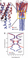
Influence of the C-terminal truncation on the conformation of the inner helix bundle gate. (A) Cα superposition of the high-resolution truncated KcsA structure (1K4C; red ribbons) with FL KcsA (blue ribbons). Inset highlights the splaying out of the inner helix bundle gate between residues 110 and 115, resulting in a 15° outward tilting. (B) Radius profile (calculated with the program HOLE; ref. 29) of truncated (red) and FL KcsA (blue).
Fig. 3.
Fig. 3.
Structure of the C-terminal bundle (residues 129–158) in complex with Fab4. (A) Top view of the CDKcsA structure as a tetramer with 4 Fab molecules bound. Each monomer of the CDKcsA is represented by different shades of orange. The heavy and light chains of Fab are shown in magenta and cyan, respectively. (B) Side chain packing of the CDKcsA core. Hydrophobic residues that constitute the core of the bundle are shown as green stick models. The distances between Cα–Cα are shown for A133 and L151. There is a 2-Å (15.4-11.4)/2 increase in the distance of Cα–Cα from the axis of symmetry at the N terminus of the CDKcsA relative to the C terminus. (C) Intersubunit electrostatic H-bonding interactions observed in the C-terminal 4-helix bundle. (Left) A top view of the network. (Right) A close-up, side view of the interactions between 2 subunits. A water molecule bridging several residues through H bonds is shown as a red sphere.
Fig. 4.
Fig. 4.
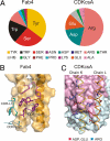
Side-chain interactions at the KcsA–Fab4 interface. (A) Pie chart representation of the amino acid composition of the binding interface of the Fab4–CDKcsA complex. The buried surface area on Fab or KcsA upon complex formation for each amino acid type is shown. Atomic structure of Fab–KcsA interface. (B) The helical cartoons with the transparent molecular surface representation in orange show CDKcsA. CDRs with residues within 4.5 Å of KcsA are shown as sticks in magenta for the heavy and in cyan for the light chain. (C) Fab4 is shown as molecular surfaces. Residues from CDKcsA contacting (closer than 4.5 Å) their respective antibody are shown as sticks for basic (blue) and for acidic (red) residues. The Cα traces of 2 of the KcsA helices are shown.
Fig. 5.
Fig. 5.
Functional effect of Fab4 binding on KcsA function and inner gate conformation. (A) I–V relationship in symmetric conditions (pH 4) of FL KcsA (black) and FL KcsA–Fab4 complex (red), as determined from patch-clamp experiments. (B) Change in FL KcsA–Fab4 single-channel activity in symmetric solutions (pH 4) upon a 150-mV jump. (C) The pH jump experiment at +150 mV (Left) and −150 mV (Right). (D) A model for FL KcsA activation gating. The channel (light gray) is shown in 2 conformations, both bound to the Fab4 (dark gray). The model assumes that opening of the inner bundle gate proceeds without dissociation of the distal C-terminal bundle, but implies a rearrangement of the “bulge” helix region to allow for a large TM2 movement.

Similar articles

Cited by

References

    1. Hille B. Ion Channels of Excitable Membranes. 3rd Ed. Sunderland, MA: Sinauer; 2003.
    1. Doyle DA, et al. The structure of the potassium channel: Molecular basis of K+ conduction and selectivity. Science. 1998;280:69–77. - PubMed
    1. Zhou Y, Morais-Cabral JH, Kaufman A, MacKinnon R. Chemistry of ion coordination and hydration revealed by a K+ channel-Fab complex at 2.0 A resolution. Nature. 2001;414:43–48. - PubMed
    1. Kelly BL, Gross A. Potassium channel gating observed with site-directed mass tagging. Nat Struct Biol. 2003;10:280–284. - PubMed
    1. Perozo E, Cortes DM, Cuello LG. Structural rearrangements underlying K+-channel activation gating. Science. 1999;285:73–78. - PubMed

Publication types