. 2009 Apr 21;106(16):6748-53.
doi: 10.1073/pnas.0900226106. Epub 2009 Apr 3.

Leishmania amazonensis promastigotes induce and are killed by neutrophil extracellular traps

Affiliations

Leishmania amazonensis promastigotes induce and are killed by neutrophil extracellular traps

Anderson B Guimarães-Costa et al. Proc Natl Acad Sci U S A. .

Abstract

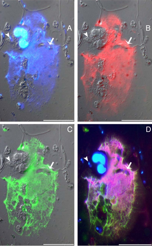
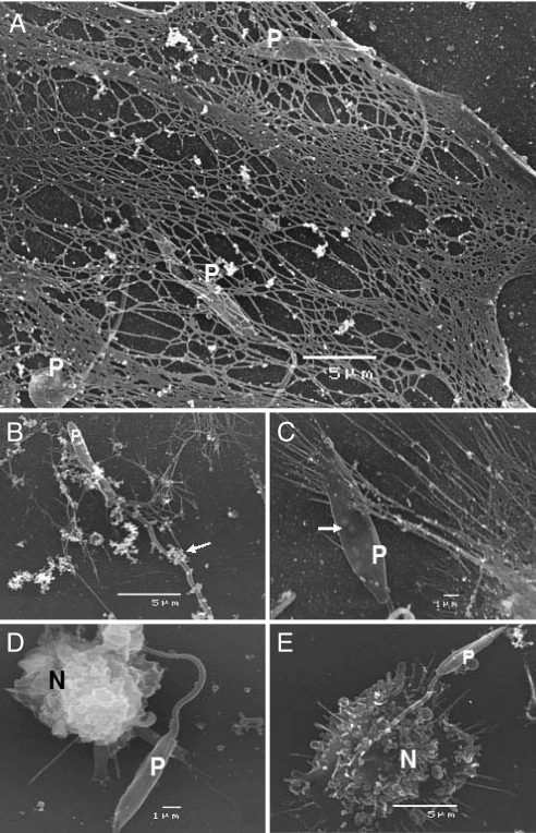
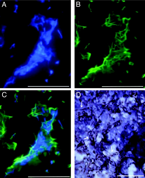
Neutrophils are short-lived leukocytes that die by apoptosis, necrosis, and NETosis. Upon death by NETosis, neutrophils release fibrous traps of DNA, histones, and granule proteins named neutrophil extracellular traps (NETs), which can kill bacteria and fungi. Inoculation of the protozoan Leishmania into the mammalian skin causes local inflammation with neutrophil recruitment. Here, we investigated the release of NETs by human neutrophils upon their interaction with Leishmania parasites and NETs' ability to kill this protozoan. The NET constituents DNA, elastase, and histones were detected in traps associated to promastigotes by immunofluorescence. Electron microscopy revealed that Leishmania was ensnared by NETs released by neutrophils. Moreover, Leishmania and its surface lipophosphoglycan induced NET release by neutrophils in a parasite number- and dose-dependent manner. Disruption of NETs by DNase treatment during Leishmania-neutrophil interaction increased parasite survival, evidencing NETs' leishmanicidal effect. Leishmania killing was also elicited by NET-rich supernatants from phorbol 12-myristate 13-acetate-activated neutrophils. Immunoneutralization of histone during Leishmania-neutrophil interaction partially reverted Leishmania killing, and purified histone killed the parasites. Meshes composed of DNA and elastase were evidenced in biopsies of human cutaneous leishmaniasis. NET is an innate response that might contribute to diminish parasite burden in the Leishmania inoculation site.


Conflict of interest statement

The authors declare no conflict of interest.

Figures

Fig. 1.
Fig. 1.
Killing of L. amazonensis by NETs from activated and naïve neutrophils. PMA-activated (A) or naïve (B) neutrophils were treated with CytD or DNase-1 and incubated with promastigotes (1 cell/0.1 parasite ratio) for 2 h at 35 °C. Schneider's complete medium was added to the cultures, and live parasites were counted after 2 days of incubation at 26 °C. Results of at least 7 independent experiments are shown as mean + SEM. PMA raw number: 8.2 × 10⁶ ± 0.7 × 10⁶ promastigotes; medium raw number: 3.2 × 10⁶ ± 0.7 × 10⁶ promastigotes. *, P < 0.002; **, P < 0.0001.
Fig. 2.
Fig. 2.
Immunostaining of NETs induced by Leishmania. (A–C) Naïve neutrophils were incubated with promastigotes (1:5 ratio) for 1 h at 35 °C. Cells were fixed, stained with DAPI (A), antihistone (B), or anti-elastase (C), the last 2 followed by Texas red-labeled (A) or FITC-labeled (B) secondary antibodies. Fluorescence staining images merged with differential interference contrast are shown. (D) Overlay of the fluorescence images. Arrow points to NET ensnared promastigote and arrowhead points to promastigote being phagocytosed. (Bars: 20 μm.)
Fig. 3.
Fig. 3.
Scanning electron microscopy of the interaction between naïve neutrophils and L. amazonensis. Naïve neutrophils were incubated with promastigotes (P) for 1 h at 35 °C. (A) An extended NET ensnares 3 promastigotes. (B) NET fibers and neutrophil granules (arrow) are attached to a promastigote. (C) NET threads trap a promastigote. (D and E) Neutrophils phagocytosing promastigotes. In A–C, a NET-ensnared promastigote presents a thin, flat body with protrusions (arrow in C), in contrast to swollen promastigotes (D and E) that were phagocytosed by neutrophils.
Fig. 4.
Fig. 4.
Leishmania and LPG stimulate NET release from naïve neutrophils. (A and B) Neutrophils were incubated with promastigotes (A) or amastigotes (B) of L. amazonensis at different cell ratios as indicated. Supernatants were recovered after 1 h at 35 °C and NETs were quantified. (C) Similar to A and B but using promastigotes of L. major or L. chagasi. Results of at least 3 independent experiments are shown. (A and B) P < 0.006. (C) *, P < 0.0001. (D) Neutrophils were incubated at the indicated concentrations of purified LPG for 1 h at 35 °C, supernatants were recovered, and NETs were quantified. Results of at least 6 independent experiments are shown. *, P < 0.05.
Fig. 5.
Fig. 5.
Histone kills Leishmania. (A) Killing of Leishmania by NETs is inhibited by antihistone antibody. Neutrophils treated with PMA and CytD were exposed to 5 μg/mL of anti-H2A histone or IgG isotypic antibodies, and then incubated with promastigotes (1 cell/0.1 parasite ratio) for 2 h at 35 °C. Schneider's complete medium was added to the cultures and live parasites were counted after 2 days incubation at 26 °C. Results of 4 independent experiments are shown as mean + SEM. PMA raw number: 6.6 × 10⁵ + 0.8 × 10⁵ promastigotes. *, P < 0.005. (B) Supernatants from activated neutrophils are toxic to Leishmania. Supernatants from neutrophils treated with PMA and DNase were added to 107 promastigotes in the presence or absence (Ctrl) of different concentrations of antihistone antibody. After 30 min, live parasites were counted. Results from 4 independent experiments are shown as mean + SEM. Control raw number: 1.2 × 10⁶ ± 0.1 × 10⁶ promastigotes. P = 0.05. (C) Histone H2A is toxic to Leishmania. Purified histone H2A was added at different concentrations to 5 × 10⁶ promastigotes. After 30-min incubation at 35 °C, live parasites were counted. Sodium acetate buffer (Na-acetate, histone's diluent), was used as a control. Results from 3 independent experiments are shown as mean + SEM. *, P < 0.0001.
Fig. 6.
Fig. 6.
Analysis of tissue sections from lesions of cutaneous leishmaniasis. (A and B) Immunofluorescence staining with DAPI (A) reveals nuclear and extracellular localization of DNA, which largely overlaps with staining for histone (B). (C) Overlay of the images in A and B. (D) Staining for elastase reveals fibrous extracellular material (arrows). Tissue sections are of 6-μm thickness. (Bars: 50 μM.)

Similar articles

Cited by

References

    1. Nathan C. Neutrophils and immunity: Challenges and opportunities. Nat Rev Immunol. 2006;6:173–182. - PubMed
    1. Brinkmann V, et al. Neutrophil extracellular traps kill bacteria. Science. 2004;303:1532–1535. - PubMed
    1. Brinkmann V, Zychlinsky A. Beneficial suicide: Why neutrophils die to make NETs. Nat Rev Microbiol. 2007;5:577–582. - PubMed
    1. Urban CF, Lourido S, Zychlinsky A. How do microbes evade neutrophil killing? Cell Microbiol. 2006;8:1687–1696. - PubMed
    1. Urban CF, Reichard U, Brinkmann V, Zychlinsky A. Neutrophil extracellular traps capture and kill Candida albicans yeast and hyphal forms. Cell Microbiol. 2006;8:668–676. - PubMed

Publication types

MeSH terms

LinkOut - more resources